All posts by vmcimchiefs

Neurologic sarcoidosis

We discussed a case about a middle-aged woman with history of cluster migraines, presenting with chronic headaches & acute left eye optic neuritis and left CN V1-3 palsy, found to have neurologic sarcoidosis.

Sarcoidosis is a granulomatous disease that can affect all organ systems. It causes non-necrotizing (non-caseating) granulomas.

Epidemiology

  • African Americans 2-3x > Caucasians
  • Females 2x > males
  • Young adults

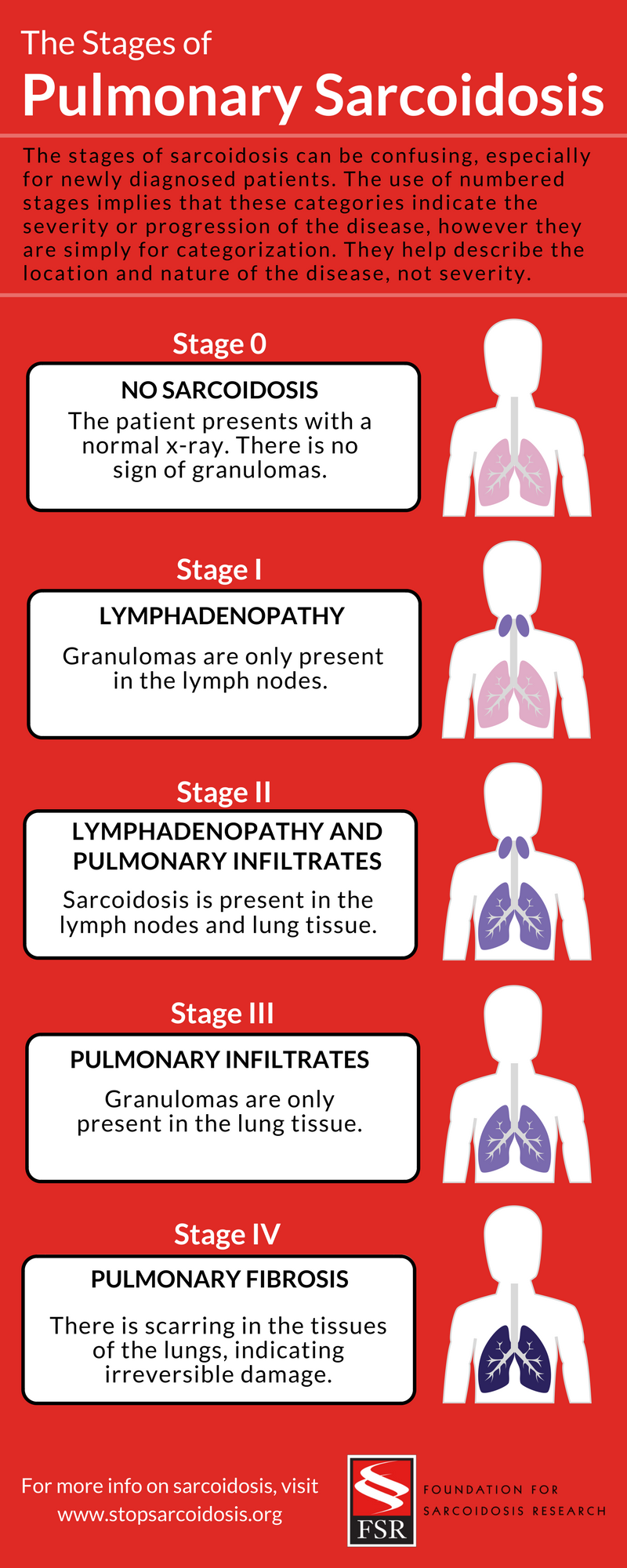
Pulmonary sarcoidosis
Chest radiographic findings are organized into stages. This gives an anatomic guide to lung involvement, but does not reflect disease activity or functional deficits.

  • Stage I:
    • Bilateral hilar adenopathy
  • Stage II:
    • Bilateral hilar adenopathy and parenchymal involvement (most commonly reticulonodular opacities)
  • Stage III:
    • Parenchymal involvement without adenopathy
  • Stage IV:
    • Fibrosis

Extrapulmonary sarcoidosis

  • Sarcoidosis can affect all organs
  • Most common extrapulmonary involvment
    • Skin
    • Eyes
    • RES
    • MSK
    • Exocrine glands
    • Heart
    • Kidney
    • CNS

Ocular Sarcoidosis

  • Affects up to 25% of pts with sarcoidosis
  • Females > males
  • More common in African American and Japanese populations
  • Intraocular: uveitis
  • Extraocular: lacrimal glands, conjunctiva, extraocular muscles, optic sheath

Lofgren syndrome
A specific acute presentation of sarcoidosis.
Classic triad of:

  • Bilateral hilar lymphadenopathy
  • Erythema nodosum
  • Arthropathy

Treatment

  • Initial: glucocorticoids
  • Refractory / intolerant to steroids: immunosuppression agents (MTX, azathioprine, leflunomide, or TNFα inhibitors

Nephrotic Syndrome due to Membranous Nephropathy secondary to Syphilis

We discussed a middle-aged man with risk factors including homelessness and high risk sexual behavior with idiopathic hyperkeratosis presenting with acute to subacute anasarca secondary to nephrotic syndrome ( proteinuria (>20 g per day) with hypoalbuminemia, protein gap and anasarca). He was diagnosed on kidney biopsy with Membranous Nephropathy due to Secondary Syphilis. 

Ddx for nephrotic syndrome 

  • FSGS:
    • Secondary to Infectious (HIV, Parvovirus, CMV, COVID-19); Drugs (Bisphosphonates/Heroin); Metabolic Diseases (DM, HTN, Obesity); Adaptive FSGS from hyperinfiltration injury after nephrectomy 
  • Membranous Nephropathy:
    • Subset have Phospholipase A2 Receptor on podocytes (+Ab PLA R2 is linked with MN)
    • Secondary to colon/breast/lung/hepatitis B and syphilis 
  • Minimal Change Disease
    • Malignancy (Hodgkins), drug induced (NSAID use) and mostly idiopathic MCD
  • Other
    • DM, Medications, Infection, Malignancy, SLE, Amyloid, Pre-Eclampsia 

Tests to order: ANA, dsDNA, DM, SPEP/UPEP/SPIE; HIV/CMV/Parvovirus/Syphilis; PLA-2R

Syphilis testing

  • The traditional screening algorithm
    You start off with a non treponemal test (RPR); if reactive, you confirm with a treponemal test. If the treponemal test is also reactive, you have a diagnosis of syphilis. If it is negative, you do not have syphilis or it is SO early in the infection, that you have not made antibodies. 
  • Reverse Screening algorithm
    You may start off with a treponemal test (such as EIA) for screening. If this is positive, then you reflex to a non-treponemal test (non-specific test like VDRL or RPR) to confirm syphilis. However, if you have a negative non-treponemal test, there may be 2 scenarios at play:
    1. Non treponemal tests can often seroconvert to inactive (RPR negative), so you may have late latent syphilis
    2. Early syphilis where RPR or VDRL hasn’t yet turned positive. In these cases, you order a treponemal test (like TPPA) to be a tie-breaker. If reactive, you have diagnosed syphilis. If negative, you consider your first treponemal test to be a false positive. 

Nontreponemal tests

  • Rapid plasma reagin (RPR)
  • Venereal Disease Research Laboratory (VDRL)

Treponemal tests

  • Fluorescent treponemal antibody absorption (FTA-ABS)
  • Treponema pallidum particle agglutination (TPPA)
  • T. pallidum enzyme immunoassay (TP-EIA)

Acute Promyelocytic Leukemia (APL)

We discussed a case about a young man who was previously healthy, presenting with subacute-chronic fevers + chills + malaise + unintentional weight loss + night sweats + bruising and acute bleeding from the mouth, found to have acute promyelocytic leukemia.

APL is a variant of AML

  • Adults (20-50s, w/ median in 40s)
  • Hallmark is atypical promyelocytes in blood of bone marrow
  • t(15;17) translocation & PML-RARA gene fusion → blocks myeloid differentiation, so myeloid cells are stuck at the promyelocyte stage- increased risk for DIC & hyperfibrinolysis → hemorrhage (esp brain bleeds), so it is important to do a good neuro exam!
  • Treatment: ATRA, even if only suspect APL (don’t need to wait for confirmatory test results)

Nocardia pneumonia

We discussed a case about an elderly woman with PMH of ESRD s/p renal transplant (within the year) on immunosuppressants & recent history of CMV colitis, who presented with acute fevers, malaise, cough, found to have Pulmonary Nocardia. We learned about:

Main risk factor: immunocompromised state

  • Patients on glucocorticoid therapy
  • Malignancy
  • Organ transplant recipients on immunosuppressant therapy
  • AIDS

Sites of infection: Pulmonary, CNS, cutaneous, disseminated

Definitive diagnosis: isolation & identification of the organism
Partially acid-fast w/ modified (Kinyoun stain)

Treatment: dependent on susceptibilities

  • 1st line: TMP-SMX
  • Alt: linezolid, clarithromycin, amikacin, cephalosporins, carbapenems
  • Duration: prolonged course depending on severity (e.g ~3-6 months vs 6-12+ months)

Still’s disease

We discussed a middle aged man who presented with acute painful knee with daily fevers, leukocytosis, elevated inflammatory markers (ESR, CRP, Ferritin) and transaminitis found to have adult onset still’s disease. Our patient had a debridement in the OR due to concerns of septic arthritis however no pus was seen and joint fluid aspiration showed inflammation (not infection). Due to daily fevers in the hospital and leukocytosis, rheumatology was consulted and found a ferritin >17k. Patient met Yamaguchi criteria (see below) and was diagnosed with Still’s Disease. Patient was started on high dose steroids and his fever, transaminitis, arthritis improved. 

Still’s Disease Yamaguchi criteria

  • Major
    1. Fever>39, >1 week (patient met this)
    2. Arthritis >2 weeks (possible?)
    3. Rash (our patient had rash after starting abx) – not characteristic rash seen in stills 
    4. Leukocytosis (patient met this)
  • Minor
    1. Sore throat (patient does not have)
    2. LAD (does not have)
    3. Hepatomegaly, Splenomegaly (patient does not have)
    4. High AST/ALT/AlkPhos (has this)
    5. Negative ANA and RF (has this)

Ddx for painful joints

  • Infections
    1. Disseminated gonorrhea (mono or oligoarticular)
    2. S. Aureus (monoarticular)
    3. Strep Pneumoniae
    4. Beta hemolytic strep
    5. Fungi like candida, cocci, blaso, cryptoneoformans
    6. TB (pott’s disease)
    7. Other (Nocardia, Brucella)
    8. Syphilis and Chlamydia
  • Inflammation
    1. Spondyloarthritis (Reactive arthritis, Ankylosing Spondylitis, Psoriatic Arthritis)
    2. Lofgrens Syndrome (Sarcoid)
    3. HSP
    4. Cryoglobulin – oligoarticular, HCV, purpura 
    5. RA
    6. SLE
    7. Still’s Disease
    8. DM/PM
  • Crystals
    1. Gout (monosodium urate crystals)- oligo/polyarticular
    2. Pseudogout (CPPD) – oligo/polyarticular
  • Tumor
    1. Chondroma, Giant cell tumor, osteoma
  • Blood
    1. Clotting disorder
    2. Trauma

Cardiac tamponade

We discussed a case about a middle aged woman with subacute-chronic SOB, fatigue, pleuritic CP, and productive cough, found to have a large pericardial effusion with features of cardiac tamponade. We learned about various features of cardiac tamponade:

  •  Exam: Beck’s triad, pulsus paradoxus
  • EKG: low voltage QRS, tachycardia, electrical alternans
  • TTE / POCUS
    • Cardiac chamber collapse
    • Dilated IVC
    • Reciprocal respiratory variations in ventricular volumes
  • Pericardial vs pleural effusion on POCUS peristernal long axis (PLAX)
    • Pericardial: fluid ANTERIOR to the descending aorta
    • Pleural: fluid POSTERIOR to the descending aorta

Dermatomyositis

We discussed a young female with facial swelling, subacute onset generalized weakness without laboratory evidence of myositis (CK normal) and cutaneous findings of periorbital edema, heliotrope rash, gottron’s papules and mechanics hands diagnosed with amyopathic dermatomyositis with MDA-5+. Dermatomyositis is the autoimmune inflammation of perimysium and cutaneous findings; average age ~40 with female predominance. Cutaneous findings include heliotrope rash, periorbital and facial swelling, and violaceous erythema of face that INVOLVES nasolabial fold (unlike SLE). Patient may complain of difficulty combing hair, rising from a chair. Remember that in patients with DM, you should send the mymarker panel which surveys for many types of antibodies. Our patient was MDA-5+ which gives rise to ILD. Other antibodies include Anti-Mi2 which is seen in 30% of DM; ANA which is seen in 80% of DM; and finally antiJo-1 antibody which is seen in DM with Antisynthetase syndrome (ILD, Raynaud, arthritis, mechanics hands). Remember to survey for cancers (12x increased malignancy risk compared to age matched population). 

Myopathy ddx

  1. Autoimmune
    • Primary
      • Dermatomyositis
      • Polymyositis
      • Inclusion body myositis
    • Secondary
      • Vasculitis
  2. Non-inflammatory
    • Meds: Statin, Steroids
    • Toxins: EtOH
    • Critical Illness

Wolff-Parkinson-White (WPW) syndrome

We discussed a case about a previously healthy young man who presented with acute dizziness, SOB, and palpitations due to tachyarrhythmia from Wolff-Parkinson-White Syndrome.

WPW pattern: preexcitation and absence of symptomatic arrhythmia-

WPW syndrome: preexcitation + symptomatic arrhythmia involving the accessory pathway 

The accessory pathway can conduct bidirectionally (anterograde or retrograde)

EKG: delta wave (slurred and broad upstroking of the QRS complex)

Several tachyarrhythmias can be related to WPW (e.g. afib, AVRT)

  • Orthodromic: anterograde via AV node; retrograde via accessory pathway
    • Treatment: synchronized cardioversion (if unstable); start w/ vagal maneuvers & adenosine (if stable)
  • Antidromic: anterograde via accessory pathway; retrograde via AV node
    • Treatment: synchronized cardioversion (if unstable); start w/ vagal maneuvers & consider procainamide (if stable)
    • Unmitigated conduction via anterograde accessory pathway can cause unstable rhythm that degenerates to vtach

If chronic and stable: 1st line therapy is ablation

Disseminated histoplasmosis

Young female with high-risk exposure to HIV presenting with constitutional symptoms (fever and malaise), GI sx (diarrhea, vomiting) and intermittent confusion found to have oral thrush, cholestatic liver injury diagnosed with HIV/AIDS with 1) CMV colitis with viremia; 2) disseminated histoplasmosis (blood, bone marrow, colon) likely leading to HLH vs macrophage activation syndrome. Her ferritin on admission was >100,000 which improved to normal with treatment of underlying disease. Bone marrow biopsy showed hemophagocytosis and histoplasmosis. She was treated with 2 weeks Ambisome IV to treat histoplasmosis before starting abacavir 600 mg/dolutegravir 50 mg/lamivudine 300 mg therapy. Discharged with PO itraconazole 200 BID. Treatment with valganciclovir and then foscarnet for CMV colitis. Atovaquone for PJP ppx. 

Opportunistic infection by CD4 count

Disseminated histoplasmosis
Our patient worked in a food factory with live chickens. Remember, it starts off as mold in soil from animal droppings (birds) and turns into yeast in the host. Aerosolized particles are inhaled and spread though our lymph nodes. HIV/AIDS is a risk factor to develop disseminated histoplasmosis. The most common complication is GI tract involvement . Histoplasmosis itself is shown to induce HLH. Treatment of disseminated histoplasmosis is 14 days of IV Amphotericin followed by PO itraconazole. 

Starting HAART
Start HAART therapy as soon as possible except if there is CMV retinitis, cryptococcal meningitis, TB meningitis, new pulmonary TB or disseminated MAC. 

Rhino-orbital-cerebral mucormycosis

We discussed a case of a middle aged man with poorly controlled T2DM, medication non-adherence, presenting with acute/subacute headache, worsening L facial numbness & L Bell’s palsy, L eye swelling and pain, necrotic nasal and palate wound due to rhino-orbital-cerebral mucormycosis.

Rhino-orbital-cerebral mucormycosis

  • Risk factors: T2DM / DKA, immunocompromised
  • Symptoms: Fever, acute sinusitis, nasal congestion, purulent nasal discharge, headache, sinus pain, necrotic wounds
  • Infiltration of disease -> CN palsies, cavernous venous sinus thrombosis, carotid artery involvement
  • Can affect various systems: Pulmonary, GI, Cutaneous, Renal, Isolated CNS, Disseminated disease

Diagnosis

  • Histopathology w/ culture confirmation
  • CT (bony erosions) & MRI (intracranial / orbit / cavernous sinus)
  • 1,3 beta-D-glucan (Fungitell) & Galactomannan: negative because not part of mucormycosis cell structure

Treatment

  • Combo surgical + antifungal treatment
  • Initial: Amphotericin B
  • Step-down therapy: Posaconazole & Isavuconazole (PO or IV)