Category Archives: Morning Report

Legionnaires’ disease

We discussed a case about an older man with chronic HCV (treated ~2 years ago) and 10-pack-year smoking history, who presented with acute fevers, rigors, SOB, cough, and was admitted for acute hypoxic respiratory failure due to Legionella pneumonia (Legionnaires’ disease).

Severity of CAP

There are several severity scoring systems for CAP. Most common systems are the PSI / POST Score and CURB-65. The IDSA / ATS also has a criteria for defining severe CAP.

Highlights about the clinical utility of MRSA nares screening to rule out MRSA pneumonia

Legionella pneumonia (Legionnaires’ disease)

  • Accounts for ~1-10% of CAP
  • Patients can present as severe hypoxic respiratory failure. There are case series that report up to 44% of Legionnaires’ disease require ICU admission.
  • In addition to outbreaks from aerosolized contaminated water systems, sporadic cases are quite common (causing the majority of cases) and the source of infection is often unknown.
  • Older age, smokers / chronic lung disease, other chronic diseases (CVD, renal), and immunocompromised state are risk factors.
  • Consider testing for Legionella in…
    • All patients with moderate to severe CAP
    • Patients with CAP who require hospitalization
    • Any patient with CAP or nosocomial pneumonia who has a known or possible exposure to Legionella
    • Immunocompromised patient (given high risk for Legionella and severe disease)
  • Diagnosis of Legionella
    • PCR of lower respiratory tract samples (ie BAL) is preferred because of high diagnostic accuracy and detects all Legionella spp (there are >60 Legionella spp!).
    • Urine Ag is also acceptable. This only detects Legionella pneumophila serotype 1 (which is prevalent in the US).
  • Treatment of Legionella
    • Macrolides and fluoroquinolones
    • Duration: ~5-10 days and do not stop therapy until patient is clinically stable and afebrile for at least 48 hrs
    • Procalcitonin-guided deescalation is not well established in Legionnaires’ disease. Procal levels do rise in Legionnaires’ disease, but may not rise to the same degree as with pneumonia caused by other organisms such as strep pneumoniae.

Diverticulitis and CMV colitis

We discussed a case of an immunocompetent middle-aged man with HTN, HLD, history of diverticulitis, who presented with subacute diarrhea, LLQ to diffuse abdominal pain, nausea + vomiting, decreased PO intake, found to have diverticulitis and CMV colitis.

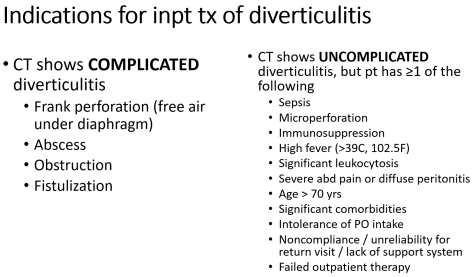
When should a patient be admitted and treated for diverticulitis?

Risk factors for diverticulitis

  • A diet low in fiber, high in fat, and high in red meat
  • Physical inactivity
  • Obesity
  • Other medical problems eg Ehler-Danlos, Marfan’s, Williams-Beuren syndromes, HIV, patients undergoing chemotherapy
  • Current smokers


    One topic that is brought up when talking about risk factors for diverticulitis is seeds and nuts in one’s diet. These are not associated with an increase in risk of diverticulosis, diverticulitis, or diverticular bleeding.

Workup

  • Acute diverticulitis is often a clinical diagnosis and does not require imaging to confirm the diagnosis.
  • CT Abd/Pelv W Con can help differentiate uncomplicated vs complicated diverticulitis.
  • Endoscopic evaluation has no role in establishing the diagnosis of acute diverticulitis. In fact, endoscopy should be avoided in the acute setting due to risk of perforation or exacerbation of existing inflammation.

Highlights in the Management of diverticulitis

  • Outpatient treatment includes antibiotics that cover GI organisms (GNR and anaerobes). Some common antibiotics regimen include: ciprofloxacin + metronidazole, augmentin, levofloxacin + metronidazole.
    We should expect improvement in symptoms within a few days. Patients should follow-up during this time (in 2-3 days) to ensure symptoms are not progressing, which may warrant inpatient management.
  • There is some controversy regarding the use of antibiotics in acute uncomplicated diverticulitis. Recommendations from European experts / guidelines tend to be against management with antibiotics, while American experts / guidelines tend to favor the use of antibiotics.
    Current evidence suggests antibiotics do not shorten the clinical course of diverticulitis, though some sources suggest antibiotics may help prevent complications (e.g. abscesses) and future episodes of diverticulitis.
  • Colon cancer can manifest similarly to acute diverticulitis (colicky abdominal pain, LLQ pain, fevers, relief with flatus or BMs).
    Therefore, especially in patients who have not undergone recent (w/in 3 years) screening colonoscopies, colonoscopy should be done after resolution of acute diverticulitis (4-8 weeks after) to exclude colon malignancy.

CMV colitis

  • Rare in immunocompetent.
    In immunosuppressed patients, CMV infection tends to be reactivation of latent disease.
    While in immunocompetent patients, CMV infection can also be due to primary infection.
  • In an immunocompetent patient, serologic testing may help with diagnosis of primary infection either with detection of…
    • CMV-specific IgM or
    • 4x rise in CMV-specific IgG
  • Treatment consists of antiviral therapy e.g. ganciclovir and valganciclovir

References

AIDS & PJP pneumonia

We discussed a case of a younger man with HIV, who presented with subacute fevers, B symptoms, fatigue, progressively worsening SOB with dry cough, found to have diffuse bilateral GGO on CT chest and diagnosed with AIDS and hypoxemic respiratory failure due to Pneumocystis pneumonia.

We reviewed the CD4 count thresholds for antibiotic prophylaxis against a few significant opportunistic infections (OI) in patients with HIV:
One recent update to antibiotic prophylaxis guidelines is for MAC. Primary prophylaxis against MAC is NOT recommended for patients with HIV who immediately initiate ART. Otherwise, for people not on effective ART, the CD4 cutoff to prophylax against MAC is <50 . Read more about this guideline here!

We also discussed the different clinical manifestations between HIV+ PJP pulmonary infection and HIV- PJP pulmonary infection:

Severity of PJP pulmonary infection is dependent on degree of hypoxia:

  • Mild
    • A-a gradient <35
    • PaO2 > 70 mmHg
  • Moderate
    • A-a gradient 35-45
    • PaO2 > 70 mmHg
  • Severe
    • A-a gradient >45
    • PaO2 50-70 mmHg

Besides TMP-SMX for treatment, adjunctive glucocorticoids should be used is specific situations:

  • Moderate to severe PJP infection

References:

Cerebral venous thrombosis

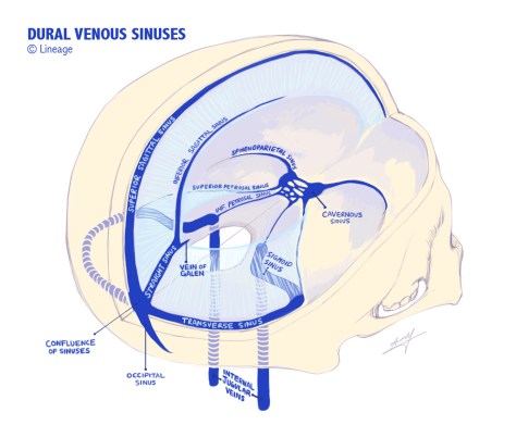
We discussed a case of a middle-aged man with uncontrolled diabetes, who presented with acute-subacute fevers, headache, L neck pain, L retroauricular pain, and was found to have L ophthalmoplegia, acute L eye swelling due to MRSA bacteremia, left skull base MRSA osteomyelitis, and septic cavernous sinus thrombosis.

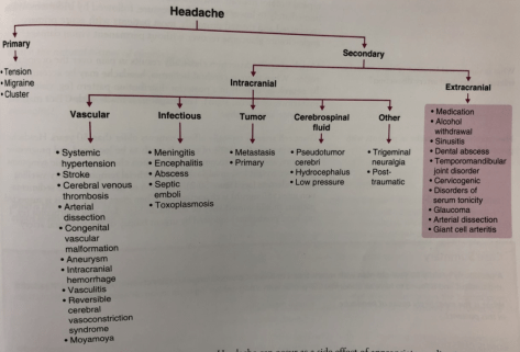
We reviewed various frameworks for secondary headache.
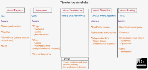
Thunderclap headache = Abrupt onset and reaches its max intensity within 1 minute or less of onset

Cerebral venous thrombosis is a rare (1%) cause of strokes. It usually affects younger patients (20-50 y/o) with a female > male predominance.

In septic cerebral vein thrombosis, thrombosis most commonly arises from contiguous spread of infection (nasal, sinus, dental) into the veins/sinuses.

Risk factors for cerebral venous thrombosis include:

  • Thrombophilia e.g. hx of prior VTE, oral contraceptives, obesity, pregnancy, autoimmune diseases (lupus, IBD, antiphospholipid), nephrotic syndrome
  • Head and neck infections e.g. mastoiditis, sinusitis, otitis media, meningitis, cerebral abscess, Lemierre’s syndrome
  • Mechanical causes e.g. trauma (at the skull base or jugular foramen), NSGY procedures, jugular vein procedures

Clinical manifestations to definitely watch out for include:

  • Headache: acute to subacute, can be thunderclap headache
  • Fevers
  • Focal neuro symptoms: CN III, IV, VI palsies, paresis, dysarthria, diplopia…depending on where the clot is
  • Seizure
  • Encephalopathy

Treatment goals:

  • Recanalize the occluded vein
  • Prevent thrombus propagation
  • Treat the underlying prothrombotic state

Treatment:

  • Heparin (unfractionated or LMWH)
  • Intracerebral hemorrhage is NOT a contraindication to anticoagulation.

References:

3rd degree heart block

We discussed a case about an older man with smoking history, who presented with acute exertional dizziness + fatigue + diaphoresis + nausea + presyncope, found to have symptomatic bradycardia due to 3rd degree heart block.

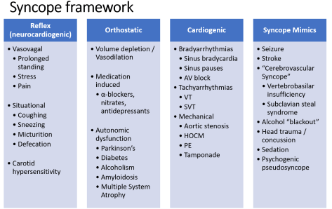
SYNCOPE FRAMEWORK

ACLS ADULT BRADYCARDIA ALGORITHM

  • In 2020, there was an update that recommended the 1st dose of Atropine given be 1mg (instead of 0.5mg) q3-5 min for a max of 3 mg.

PERMANENT PACEMAKER PLACEMENT INDICATIONS
FIRST, RULE OUT ALL REVERSIBLE CAUSES!

  1. Symptomatic bradycardia due to sinus node dysfunction
  2. Permanent afib & symptomatic bradycardia
  3. Alternating bundle branch blocks
  4. Asymptomatic high-degree AV block, 2nd degree Mobitz type 2 block, or 3rd degree block

REFERENCES

Pancreatic adenocarcinoma

Today we discussed a case about an older woman who presented after subacute painless jaundice and chronic weight loss + fatigue + nausea, found to have new onset DM, mixed cholestatic and hepatocellular liver injury, and a large pancreatic head mass. She was diagnosed with pancreatic adenocarcinoma.

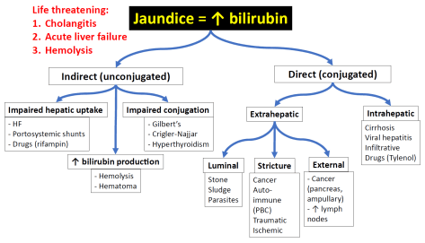
JAUNDICE FRAMEWORK

LIVER INJURY

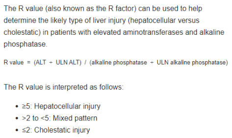
  • The R factor (or R value) could be useful in helping distinguish cholestatic vs hepatocellular liver injury.

PANCREATIC ADENOCARCINOMA

  • Definitely be concerned in patients presenting with painless jaundice. But patients with pancreatic cancer can also present with abdominal pain.
  • Of the patient diagnosed with pancreatic cancer, up to 2/3 can develop new onset DM in the 36 months surrounding the diagnosis of pancreatic cancer.
  • Your illness script should include risk factors such as
    • Age > 50
    • Hx chronic pancreatitis
    • Obesity
    • T2DM
    • High red meat consumption
    • EtOH use disorder
    • Tobacco use disorder
    • Mucinous cystic lesions of the pancreas
    • Inherited conditions (e.g. Peutz-Jeghers syndrome, BRCA2 germline mutations, hereditary pancreatitis, Lynch syndrome)
  • CA 19-9 is the most sensitive (70-92%) and specific (68-92%) tumor marker with clinical utility.
  • Serial monitoring of CA 19-9 levels can be useful in following patients after potentially curative surgeries and for those receiving chemotherapy for advanced disease.
    Rising CA 19-9 levels usually precede the radiographic appearance of recurrent disease.

RESOURCES

TEN

We presented an an elderly male presenting after recent travel and new medication exposure with ill-defined coalescing plaques with dusky centers as well as diffuse erythema over trunk, scrotum, UE/LE with areas of skin sloughing associated with pain and fever, + Nikolsky sign diagnosed with TEN.

SJS and TEN are a spectrum of disease, TEN being more severe and SJS less severe. TEN covers >30% of total body surface area and SJS covers <10%. If between 10 and 30 percent of TBSA is covered by the rash, it is termed SJS/TEN overlap syndrome. sure SJS/TEN is provoked by exposure to an inciting drug. Complete lists of drugs with reported association with this condition can be found online, but the most notable categories include allopurinol, antiepileptics, antibacterial sulfas, nevirapine (NNRTI), and NSAIDs ending in “-oxicam.” Patients with HIV have a particularly increased risk of developing SJS/TEN. The typical exposure period before reaction is 4 days to 4 weeks, but risk continued for 8 weeks after treatment with a new drug. 

The classic description and progression of SJS/TEN on physical exam are as follows. First, a flu-like prodrome may occur (malaise, myalgias, arthralgias, and fever) 1-3 days before the onset of rash. When the rash begins, it is extremely painful, ill-defined, coalescing macules with purpuric centers on the face and thorax that spread diffusely and symmetrically. These lesions are typically termed “dusky.” It usually spares the scalp, palms, and soles. Soon vesicles and bullae form and within days, the skin sloughsMucosal surfaces are involved in >90% of patients, which may cause symptoms such as photophobia, conjunctival itching/burning, odynophagia, dysuria, and painful defecation. Nikolsky sign is positive in these patients (the skin sloughs when tangential pressure is applied). Other diseases with a positive Nikolsky sign include pemphigus vulgaris and staphylococcal scalded skin syndrome. The “wet newspaper sign” is also classic for SJS/TEN, the shiny and lighter tone of exposed skin after a layer of affected epidermis sloughs off. The SCORTEN score can be used to determine prognosis and severity of the condition. 

Treating SJS/TEN is mainly supportive, including removing the offending agent, IV fluids, wound care, nutrition, and electrolyte repletion, but a multidisciplinary approach is necessary. Consults to dermatology, burn surgery, ophthalmology, OBGYN for a vaginal exam if the patient is female, and possible urology consult for males should occur immediately. High suspicion for infection and low threshold to start antibiotics are also beneficial.

There are multiple adjunctive treatments your dermatology consultants may recommend, but additional research is needed on most of them. Cyclosporine, anti-TNF drugs, steroids, IVIG, and plasmapheresis are a few of them.

Mucormycosis

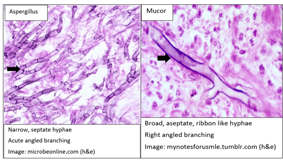
Elderly female with history of uncontrolled DM presenting with DKA and found to have a cavitary lung lesion found to have biopsy proving broad, aseptate, ribbon like hyphae consistent with mucormycosis. Mucormycosis is a invasive fungal infection (common species include Rhizopus, Mucor species); can cause necrosis of host tissue from invading vasculature. The main risk factor for developing mucormycosis: DM with DKA, steroids, underlying malignancy, AIDS and malnutrition. Most commo presentation of Mucor is rhino-orbital-cerebral mucor and pulmonary mucor. Treatment for our patient included lobectomy and Amphtericin IV prior and post surgery.

Differential for cavitary mass:

  • Infection
    • Lung abscess
    • Mycobacteria
    • S. Milleri
    • S. Aureus
    • S. Pyogenes
    • Klebsiella
    • Pseudomonas
    • Tricuspid Endocarditis
    • Lemierre Syndrome
    • Cryptococcus
    • Endemic Mycosis
    • Molds- Mucormycosis
    • Parasites-Paragonimiasis, Entamoeba
  • Malignancy
    • Squamous cell carcinoma
    • Mets from GI/GU tumors
    • Lymphoma, Kaposi Sarcoma
  • Autoimmune
    • GPA
    • RA
  • PE and infarcts

Tonsillar Syphilis

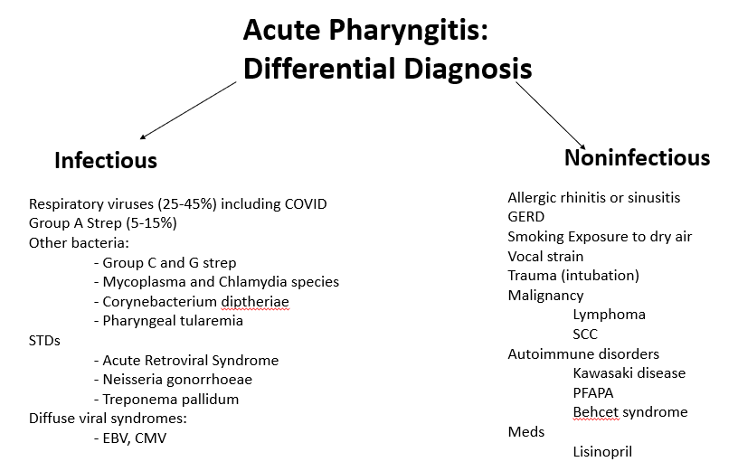
We discussed an interesting case of a middle aged female with high risk sexual behavior, active smoker with alcohol use disorder who presents with 3 weeks of globus sensation and odynophagia likely a presentation of acute pharyngitis.

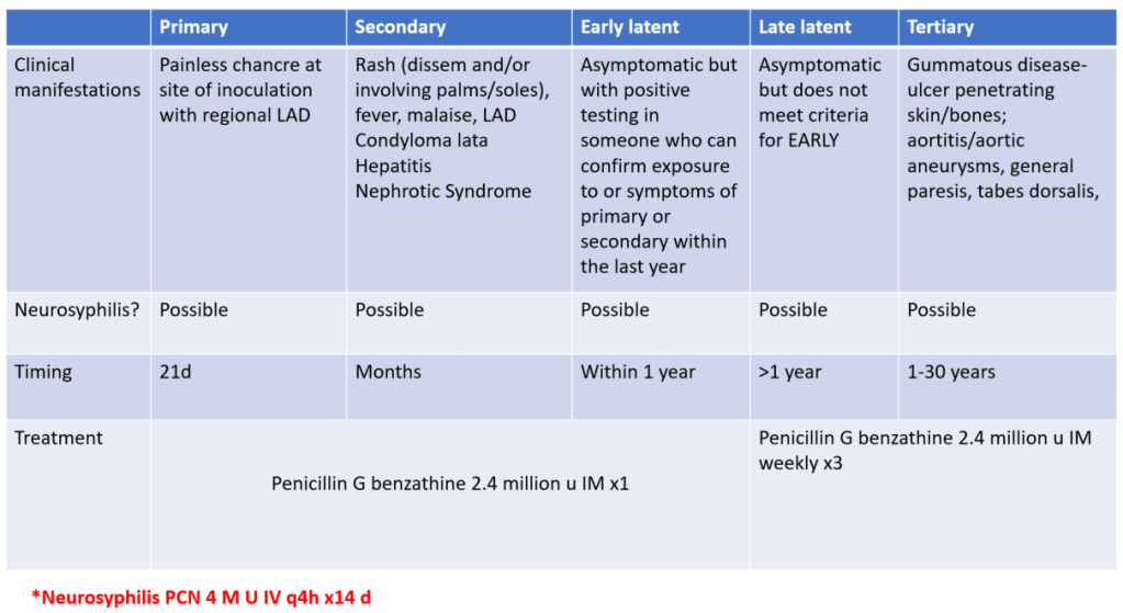
Remember to always take a sexual history in an adult with acute pharyngitis. On physical exam, make sure to look for signs of muffled voice, drooling, stridor, tripod positioning, crepitus and trismus when evaluating an adult with acute pharyngitis. Patient received a biopsy of her tonsil that resulted in IHC stain positive for Treponema Pallidum; RPR 1:65; Syphilis EIA positive. Patient was diagnosed with tonsillar syphilis- likely a primary syphilis presentation.

Immune Mediated Necrotizing Myositis due to paraneoplastic process of thymoma with associated myasthenia gravis

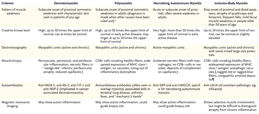
We discussed a middle aged female with uncontrolled diabetes that presents with profound subacute (5 week history) development of proximal over distal weakness and associated muscle pain. Other symptoms included 1 week of dysphagia to solid>liquid food. Overall, she presented with proximal muscle weakness with absent Babinski sign and 2+ reflexes bilaterally. Her medication included Simvastatin 20qhs. EMG shows myopathic process; ANA 1:160; HMGcoAR Antibody negative, Anti-SRP negative; biopsy showing fibrocellular variation with necrotic fibers within clusters of inflammation most consistent with necrotizing myopathy. CTCAP showed a large mediastinal mass which are diagnosed to be a invasive thymoma. Due to symptoms of dysphagia and probably bulbar involvement, patient was checked for associated myasthenia gravis. Ach Receptor Ab 159 mmol/L and 1:2560 striated muscle antibody IGg titer. Anti MUSK negative. Patient was treated with thymectomy and IVIG followed by rituximab.

We know that this patient was not dealing with a UMN or LMN process but rather a muscular weakness. We discussed framework of proximal muscle weakness, particularly in regards to muscle weakness and neuromuscular weakness.

Differential for muscle weakness:

  • Medication
  • Toxins
  • Electrolyte disturbances
  • Deconditioning
  • Endocrinopathy
  • Critical illness
  • Rhabdomyolysis
  • Infection
  • Inflammatory Myopathy (DM, PM, Inclusion body myositis)
  • Muscular Dystrophy
  • Metabolic Myopathy