Category Archives: Morning Report

AM Report 9/22/2016 – Cauda Equina Syndrome

Cauda Equina  Syndrome:

Cauda Equina: set of intradural nerves that provide sensory/motor innervation from L2-S5

Nerve Roots of Cauda Equina:

  • Hip muscles
  • Lower extremity muscles
  • Urinary and anal sphincters
  • Sexual organ muscles

Causes of Cauda Equina:

  • Disc herniation (MOST COMMON)
  • Lumbar stenosis (OA)
  • Trauma
  • Infection/abscess
  • Metastatsis/neoplasma (Metastasis most common, ependymoma or schwannoma possible)
  • Inflammatory disease (CIPD, Paget’s, ankylosing spondylitis)
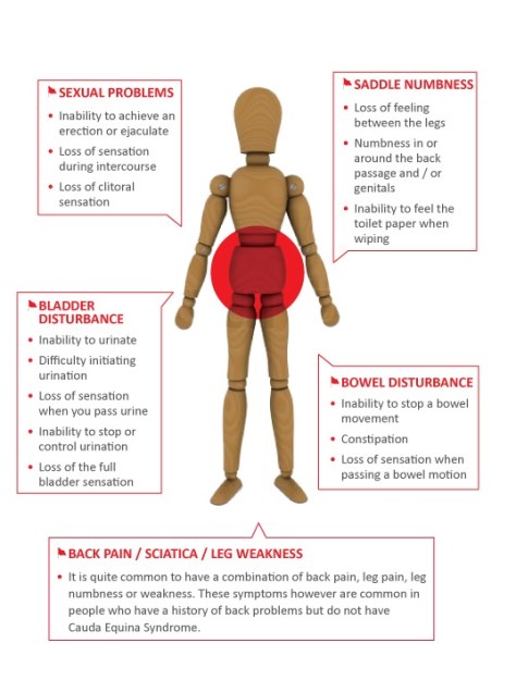
Symptomsbased on level 
Low back/radiating LE pain with at least one below

  • Urinary incontinence/retention
  • Fecal incontinence/retention
  • Loss of anal sphincter tone
  • Sexual dysfunction
  • Saddle anesthesia/hypoesthesia

new-cesa-dl-back-21-5-13

Treatment:
– urgent neurosurgical evaluation to reduce side/eliminate mass
– eradicate causative organisms

Most common organisms:
– staph (63%)
– strep (9%)
– gram negative anaerobes (16%)

Empiric antibiotic regimen: vancomycin and cephalosporin +/- nafcillin

Steroids indications:
– within 8 hours of traumatic spinal cord injury
– metastatic disease (followed by surgery/radiation)

9/20/2016 AM Report – Cannabinoid Hyperemesis Syndrome

Cyclic Vomiting Syndrome
Define:

  • Stereotypical episodes of vomiting <1 week
  • 3 episodes in past year, 2 in past 6 months, at least one week apart
  • Absence of vomiting between episodes

3 Phases – Interepisodic, Prodromal, and Vomiting

1.jpeg

Epidemiology:

  • More common in children
  • More frequent bouts in children (12/year) vs. adults (4/year)
  • Broad age of onset for adults
  • Symptoms longer for adults prior to diagnosis
  • Nausea between episodes is more common in adults

Differential diagnosis:

ACUTE:

  • Acute gastroenteritis (rotovirus, enteric aderovirus, norovirus)
  • Post-operative nausea/vomiting
  • Vestibular neuritis
  • Chemotherpy/Drugs

CHRONIC:

  • Pregnancy
  • Chronic nausea/vomiting syndrome
  • Gastroparesis
  • Gastric outlet obstruction
  • CVS
  • Rumination syndrome

Cannabinoid Hyperemesis Syndrome
Define:

  • Stereotypical episodic vomiting similar to CVS (onset, duration, frequency)
  • Presentation after prolonged/excessive cannabis use
  • Relief of symptoms with sustained (1 week) cessation of cannabis*

* often associated with pathologic bathing behavior (prolonged hot baths/showers) – considered pathognomonic for CHS

9/7/2016 AM Report – IgA Nephropathy

Nephrotic Syndrome:
Define:
– Proteinuria (>3.5 g/24h or urine/creatinine > 3.5 mg/mg)
– Hypoalbuminemia
– Clinical evidence of edema
* Other clinical manifestations (hyperlipidemia, coagulation abnormalities) are related to losses of proteins other than albumin or caused by increased protein synthesis.

Nephrotic Range Proteinuria: > 3 g/24h without other findings

Nephritic Syndrome:
Characterized by: proteinuria, hematuria, pyuria, and often decreased kidney function and hypertension
– due to injury or inflammation within the glomerulus that allows the passage of protein, erythrocytes, and leukocytes into the renal tubule.
– resulting damage can lead to decreased GFR, kidney function, and abnormal blood pressure

IgA Nephropathy:
– most common primary glomerulonephritis
– an immune complex disease in which IgA antigen-antibody complexes are depositied primarily in the mesangium
– may be primarily involve the kidney or may be secondarily associated with other conditions

Epidemiology:
occurs at any age, but usually found in men (20-30 yo) who are white or Asian
– develops to ESRD in approximately 20-30% of patients within 10-20 years of onset

Risk factors for progression:
younger age at onset, hypertension, proteinuria > 1 g/24h, elevated creatinine, and low GFR

Clinical manifestations:

– typically asymptomatic at presentation with only microscopic hematuria and proteinuria on urine evaluation
– common presentation: recurrent gross hematuria occuring synchronously with an episode of respiratory or GI infection

Diagnosis:
confirmed by kidney biopsy

Management: ACE inhibitors or ARBs, with goal urine protein excretion < 1 g/24h

Simplified Approach to GN:
Step 1: determine the broad cause of the acute or progressive renal failure
– Pre-renal, intrinsic renal, or post-renal
– FENa (if oliguric to differentiate pre-renal from ATN), UA (evaluated for sediment), and renal US (rule out obstruction)
– If INTRINSIC renal than the differential is either: Glomeruli, Tubules, Interstitum, Vessels

Step 2: If a glomerular cause – differentiate between GLOMERULONEPHRITIS pattern from a NEPHROTIC SYNDROME pattern
– UA, renal US, 24 hour urine protein (urine protein:creatinine ratio)
– Look for active sediment (UA with protein and RBCs, dysmorphic RBCs, and/or RBC casts)
1.jpg

Step 3:
Use the serum complement levels to differentiate cause of GN
Low Complement GN:
Systemic: SLE, endocarditis, cyroglobulinemia
Isolated Renal: post-infectious GN, MPGN

Normal Complement GN:
Systemic: HSP, ANCA-associated, Goodpasture’s syndrome, hypersensitivity vasculitis
Isolated Renal: IgA nephropathy, anti-GBM disease, RPGN

09/12/16 AM Report: Mesenteric Ischemia

Remember to distinguish ACUTE mesenteric ischemia from CHRONIC mesenteric ischemia, can sometimes see Acute on Chronic ischemia. 


Acute
: SUDDEN onset abdominal pain due to intestinal hypoperfusion ~50 % mortality if not treated

Chronic: gradual chronic abdominal pain due to intestinal hypoperfusion (intestinal angina)

Etiology (Acute): Arterial embolism (eg: dislodged thrombus from LA/LV/cardiac valve, septic emboli from IE, hx of A.fib not on anticoagulation), or arterial thrombosis.

Etiology (Chronic):Most common cause of chronic mesenteric ischemia is atherosclerosis, commonly affecting SMA or celiac arteries

Risk factors: History of atrial fibrillation, atherosclerotic disease (CAD, MI, PAD, smoking, DMII), for chronic mesenteric ischemia, age>60, female 3x>male, smoking, 50 % of patients with CAD, CVA, PAD

Acute mesenteric ischemia S&S: pain out of proportion to exam, severe pain, guarding (no time to form collaterals!), nausea/vomiting

Chronic mesenteric ischemia S&S: recurrent episodes of acute abdominal pain after eating, can lead to weight loss: Triad includes postprandial pain,food phobia , and weight loss. 

Diagnosis: CTA has high sensitivity/specificity, can also use Duplex ultrasound but more variability in sensitivity as operator dependent

For chronic mesenteric ischemia, must see high grade mesenteric vascular stenoses in at least 2 major vessels (usually one vessel alone will

Treatment of ACUTE mesenteric ischemia: broad spectrum abx, anticoagulation (heparin), open embolectomy, aorto-mesenteric bypass, laparotomy to remove necrosed bowel, and use of Vasodilators if non-occlusive.
Treatment of CHRONIC mesenteric ischemia: supportive care, if severe symptoms, can do revascularization with angioplasty +- stenting, aorto-mesenteric bypass

Comparing Mesenteric Ischemia and Ischemic Colitis 
mesenteric ischemia.JPG


09/08/16 AM Report-Amyloidosis

Etiologies for Restrictive Cardiomyopathy
                                             

Idiopathic
Endomyocardial fibrosis, Loeffler eosinophilic endomyocardial fibrosis

Secondary restrictive cardiomyopathy
Infiltrative-Hemochromatosis, Amyloidosis (most common cause of RCM in the USA), Sarcoidosis
OtherRadiation induced, Anthracyclines (eg: Doxorubicin), Malignancy, Glycogen storage diseases, Systemic sclerosis

Etiologies for Constrictive Pericarditis

Idiopathic, viral pericarditis, Tb (most common worldwide), connective tissue disorders, uremia, neoplasms, drugs (eg:Procainamide/Hydralazine)

Restrictive Cardiomyopathy vs. Constrictive Pericarditis

cm-restrictive

Amyloidosis-remember that it is a systemic disease!

  • RENAL (# 1 MC): proteinuria (usually nephrotic range)
  • CV (#2 MC): cardiomyopathy (restrictive), arrhythmias, angina, low voltage on EKG with pseudo-infarct pattern
  • GI: Hepatosplenomegaly, gastroparesis
  • NEURO: Peripheral/autonomic neuropathy, median nerve compression (carpal tunnel), orthostatic hypotension
  • MSK: pseudo hypertrophy of muscles, macroglossia with lateral scalloping, amyloid arthropathy (thickening of synovial membrane)
  • HEMATOLOGIC: Decreased Factor X activity (increased risk of bleeding)
  • PULM: Tracheobronchial infiltration most common, pleural effusion, rare to have Pulm. HTN
  • SKIN: easy bruising (ecchymosis), periorbital ecchymosis (“Racoon Eyes”) that is worse with Valsalva maneuvers, nail dystrophy, Alopecia,


    Making the diagnosis

  • Start with fad pad biopsy due to low risk of bleeding but if negative, may need to biopsy affected organs to make diagnosis. Stain with Congo Red stain and look for Apple-Green birefringence under polarized light to make diagnosis
  • Most common cause of Amyloid in the USA is AL Amyloid-clonal expansion of plasma cells in the bone marrow that secrete a clonal Ig Lambda chain that deposits as amyloid fibrils into tissues, can occur with or without multiple myeloma, including with non-Hodgkins lymphoma and Waldensrom’s macroglobulinemia 
  • Check immunofixation and free light chain assay (SPEP/UPEP can be falsely negative)


09/06/16 AM Report: PCP Pneumonia in HIV patients

Pneumocystis jirovecii (previously Pneumocystis carinii)

Problem script 

Epi: immunocompromised (eg: HIV with CD4<200 or not on Bactrim PPX, hematologic malignancy s/p BMT)
Timing: Sub-acute (weeks)-contrast with acute bacterial PNA which is more acute!
S&S: Fever (>80 %), progressive exertional dyspnea (95 %), non-productive cough, CXR usually with bilateral infiltrates but can also cause PTX, can also see GGO on CT scan

Labs to support diagnosis but does not make the diagnosis

Elevated LDH (sensitive but not specific)
ABG-check degree of hypoxia and AA gradient
Elevated 1,3 beta-d-glucan-part of cell wall (sensitive but not specific)
HIV with CD4 count <200 (risk factor)

Imaging

CXR-classically bilateral interstitial/alveolar infiltrates but can be variable. Suspect PCP if spontaneous PTX in patient with PNA and risk factors for PCP!
CTexcellent sensitivity, can see bilateral patchy/nodular ground glass opacities (not necessary but can order CT if unclear diagnosis)

Making the diagnosis– must VISUALIZE cystic/trophic forms, PCP cannot be cultured 

Step 1: Sputum Induction (order specifically for PCP!), specificity of ~100 % but variable sensitivity of 55-90 %
 If negative and high suspicion for PCP, proceed to Step 2

Step 2: Bronchoalveolar Lavage, 90-100 % sensitivity, ~100 % specificity

Treatment

First line is Bactrim 15-20 mg/kg oral or IV TID/QID x 21d (remember Bactrim has good bioavailability!)-No need to hold treatment before making diagnosis if high suspicion!
Treat with adjunctive steroids if PaO2<70 or A-A gradient >35
                     Dosing: Prednisone 40 mg PO BID x 5d, 40 mg PO daily x 5d, 20 mg PO daily x 11d.

Remember that it can take several days to see response in treatment, can get worse before getting better!

Primary Prophylaxis in HIV patients

capture


09/01/16 AM report: Sim Session

It can be scary to go into a RRT or Code Blue.Check your own pulse and remember the tips below!

Important tips we reviewed today:

Introduce yourself when you come into the room and identify one person as the RRT or Code Blue Leader
Avoid AIR orders-tell specific people what you want done and have them confirm when it is actually done. “John, please hang one liter of NS wide open and let me know when it is done”
-In Code Blue, press the code blue button in the room and put a backboard under the patient!
-The Team Leader should stand at the head of the bed so he/she can see everyone
SHOCK Vfib or pulseless Vtach, then can resume CPR unless waiting for pads or charging-know which side of the ACLS algorithm you are on (the crash carts have the card hanging if you don’t have it!)
-Don’t forget about maintaining adequate IV access, cycling vitals, and giving fluid or O2 as needed.

Assign specific roles to each person

1)Airway (Respiratory Therapist/Anesthesia)- Remember that intubation is NOT an emergency if able to get good bag-valve ventilation and CPR needs to be interrupted. It is more important to avoid interrupting CPR, and intubation can be done during a pulse check.
2)Medications (RN)- Avoid air orders!
3)Time Recorder and Charting (RN)- Ask him/her to alert you every 2 minutes and when epinephrine or amiodarone is due
4)CPR (anyone who is available)- Don’t forget to have at least 2-3 people who can rotate through CPR as it can be very tiring!

==========================================================================
After ROSC, check a blood pressure and assess neurologic status (is this patient a candidate for therapeutic hypothermia or simply avoiding hyperthermia)?

Case 1 (AMS with hypoglycemia)-take home points 

1)Always consider hypoglycemia on the differential for altered mental status and check finger stick for any patient who is unresponsive
2)Review recent medications to see if contributing to AMS
3)For on-going hypoglycemia that is refractory to glucose ampules push, start D5W or D10W drip and consider glucagon

Case 2 (COPD exacerbation)-take home points

1)Know the contraindications to Bipap for COPD exacerbations
2)Avoid excessive ventilation in patient with COPD as can lead to auto-peep (air trapping) so set RR low. Otherwise risk of causing PTX and PEA arrest. Remember these patients may have some chronic hypercapnia so do not correct their CO2 all the way to “normal” range.
3)If inadequate ventilation, patient may need heavy sedation and/or use of paralytic agents.


08/30/16 AM report: Thrombocytopenia

Think about thrombocytopenia in three broad categories after ruling out pseudothrombocytopenia

Pseudothrombocytopenia (low platelet count due to clumping in EDTA tube, repeat with heparin or citrate top to confirm)

Underproduction (anything in the BM that prevents production, usually affects other lines as well)

eg: marrow failure (aplastic anemia), nutritional deficiency (B12/folate/copper)
eg:marrow invasion-leukemia, tumor, fibrosis, granulomatous diseases like sarcoidosis)
eg:marrow injury-drugs (long list), radiation, infection (Hep C, viral, HIV), sepsis
eg:congenital (Wiskott Aldrich, Bernard Soulier)

-Splenic sequestration (anything that can cause splenomegaly. eg: cirrhosis)

-Peripheral destruction (can be broken up into immune mediated and non-immune mediated)

Etiologies of thrombocytopenias that are life-threatening and can cause thromboses in addition to bleeding-does your patient have one of these?

HITT-need to discontinue heparin and treat with non-heparin anticoagulant (eg: direct thrombin inhibitor, Argatroban), can cause arterial and venous thromboses
APLS– associated with both arterial and venous thromboses
DIC-risk of bleeding and thromboses (usually venous)
-HUS-TTP-can be life-threatening if plasmapheresis is not initiated

ITP

-Acquired thrombocytopenia due to auto-antibodies against platelet antigens
-Usually ISOLATED thrombocytopenia without anemia or leukopenia
-Can be Primary or Secondary

Secondary etiologies: HIV, Hep B,C, CLL, SLE, APLS, H Pylori, thyroid disease

Treatment: 

1)Asymptomatic and platelet count >30-40k: no treatment!
2)Symptomatic: Glucocorticoids/IVIG
3)Refractory: Rituximab, TPO agonist (Eltromabag, Romiplostim), splenectomy

Comparing ITP vs. TTP vs. DIC (http://emedicine.medscape.com/article/206598-overview)

ITP vs DIC vs TTP

8/29/16 AM Report: Hyperthyroidism

Recognize the physical exam findings of hyperthyroidism! 

Skin: warm moist skin, pretibial myxedema(Graves disease), thyroid acropachy, clubbing (Graves)
Eyes: Lid lag, stare, Proptosis (Graves disease), extra-ocular impairment (Graves disease)
CV: arrhythmias (sinus tachycardia, atrial fibrillation), high output CHF
Neuro: fine tremor, hyperreflexia
Psych: anxiety, agitation, psychosis, depression, insomnia, mania
Thyroid: diffuse thyromegaly, thyroid bruit (specific for Graves)

See the Stanford 25 website on how to do a thyroid exam 

High radioactive iodine uptake on RAIU scan in hyperthyroidism (de novo synthesis of thyroid hormone) 

1)Graves disease-can also diagnose with TSI-high sensitivity/specificity
2)Toxic adenoma
3)Multinodular Toxic Goiter
4)Iodine deficiency (rare in the USA)

Low radioactive iodine uptake on RAIU scan in hyperthyroidism (inflammation and destruction of thyroid tissue with release of pre-formed hormone OR extra-thyroidal source of thyroid hormone)

1)Thyroiditis (subacute granulomatous thyroiditis (De Quervains), painless, post-partum, suppurative/infectious thyroiditis)
2)Exogenous thyroid hormone administration
3)Struma Ovarii
4)Drug induced (eg: Amiodarone)
5)IV contrast load

Treatment

-Methimazole and beta blockers (eg:Propranolol) for hyper-adrenergic symptoms
-AVOID radio-iodine ablation if Graves’ ophthalmopathy, as can worsen eye involvement but
-Monitor for risk of agranulocytosis and hepatotoxicity with Methimazole!

See picture below for symptoms of hyperthyroidism! (copyright Nursing Education Consultants Inc) *Note that hyperdefecation commonly occurs, not diarrhea.

hyperthyroidism


8/25/16 AM Report – VZV Encephalitis

CSF Study Interpretation:

CSF Studies:
Normal Bacterial Meningitis Viral Meningitis Fungal/TB
Opening Pressure (cm H2O) < 15 ↑↑ Normal/mild ↑ Variable (very elevated with crypto)
Protein (mg/dL) 15-45 Normal/mild ↑
Glucose (mg/dL) 40-70 Normal
WBC 0-8 ↑↑ (>1000) ↑ (5-300)
WBC differential Even >80% PMNs >50% Lymphs, <20% PMNs 50-80% Lymphs
Gram Stain Normal 60-90 % + N/A 37-87% AFB +

CT Before LP:
1) Altered Mental Status / Fecal Deficits
2) Immunocompromised
3) History of CNS disease – mass lesion, stroke, etc.
4) Seizure within 1 week of presentation
5) Papilledema

Bad causes of headache to always think about:
– Meningitis (or other CNS infection)
– Tumor/Space occupying lesion
– SAH
– Temporal arteritis

Headaches: POUNDPulsatile, One day, Unilateral, Nausea, Disabling
Migraine:
Epi: 3x more common in adult females; peaks in the 5th decade of life
S/S: unilateral, throbbing headache, +/- photophobia/phonophobi/nausea

Tension-Type Headache
:
Epi: stress/sleep disruption are common triggers
S/S: bilateral, steady, mild-moderate discomfort

Cluster Headache:
Epi: male sex/tobacco use are risk factors; typically occurs within a few hours of falling asleep
S/S: periorbital/temporal intense pain usually ipsilateral; can develop autonomic features such as ptosis, lacrimation, conjunctival injection, and rhinorrhea

Clinical Manifestations of VZV:
Rash: erythematous papules progressing to grouped vesicles; usually limited to one dermatome, but can affect 2-3 neighboring dermatomes. Thoracic/Lumbar dermatomes are most commonly involved.
Acute neuritis: most common symptom; 75% have prodrome of pain – usually described as “burning, throbbing, stabbing.”

HZ Ophthalmicus:
serious, sight-threatening condition caused by VZV reaction within trigeminal ganglion. Usually preceded by prodrome of headache, malaise, fever.
2
Hutchinson’s Sign:
vesicles on the tip of the nose – associated with HZ opthalmicus.
1
Ramsey-Hunt Syndrome: major otologic complication of VZV reactivation.
Triad (typicially) 1) Ipisilateral facial paralysis
2) Ear pain
3) Vesicles in auditory canal