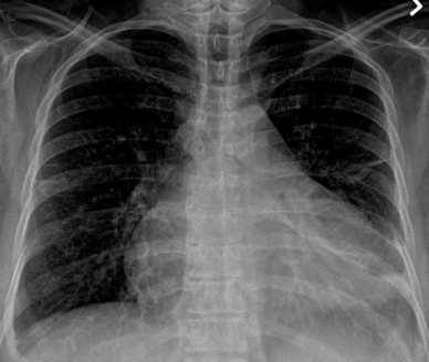
Stable cardiac tamponade you say?? That’s right! Today, our esteemed Dr. Whitney Chew presented a fascinating case of a middle-aged woman with a remote history of malignancy who presented with a month of progressive fatigue, shortness of breath, and pleuritic chest pain, found to have a large pericardial effusion with compression of the right atrium and ventricle on echocardiogram concerning for cardiac tamponade. Vital signs were stable (BP 131/71 without tachycardia) and the patient was reported to be in no acute distress on documented physical exams.
Clinical Pearl: Cardiac tamponade can be acute OR subacute. When acute, the fluid accumulates quickly and the pericardium has no time to stretch or allow the body to compensate for the decreased diastolic filling. Hemodynamic collapse occurs quickly in this case. In subacute cardiac tamponade, usually from renal failure or malignancy, the fluid accumulates slowly, allowing the pericardial compliance to increase gradually. In this case, as much as 2L of fluid can accumulate in the pericardium before hemodynamic collapse occurs.


CXR of pericardial effusion: Above are images of pericardial effusion on chest X-ray. On the left, cardiomegaly with a slight water bottle appearance can be appreciated. In addition, a retrocardiac air-fluid level with the air lower than the fluid despite the upright position of the patient is indicative of a pericardial effusion. The fluid is contained within the pericardium and sitting atop some aerated lung, creating this reverse air-fluid level. On the right, the later film shows the “Oreo Cookie Sign” of a vertical opaque line between 2 radiolucent vertical lines indicative of pericardial fluid between the pericardial and epicardial fat. With large pericardial effusions, widening of the subcarinal angle may also be seen.
