All posts by vmcimchiefs

AM Report 2/8/17: Guillain-Barré Syndrome

GBS Variants:

-AIDP (Acute inflammatory demyelinating polyneuropathy)- 85-95 % of GBS
-Miller Fisher Variant (5 % US but up to 25 % in Japan)
-AMAN (Acute motor axonal neuropathy)
-AMSAN (Acute sensorimotor axonal neuropathy)

Timing:

Acute (<4 weeks)

Pathophysiology:

Due to molecular mimicry from immune response to preceding infection that cross reacts with components of the peripheral nerve.

Diagnostic Criteria:

  1. Progressive weakness of ≥ 2 limbs due to neuropathy
  2. Areflexia
  3. Disease course < 4 weeks
  4. Exclusion of other causes

Supportive criteria:

  1. Symmetric weakness
  2. Mild sensory involvement
  3. Absence of fever
  4. Typical CSF profile
  5. EMG evidence of demylination

Most common infectious triggers:
-Campylobacter Jejuni (most common), CMV, EBV, HIV, VZV, Mycoplasma, and even Zika virus

Most sensitive physical exam findings in GBS

-Absent/Depressed DTR (90 %)
-Ascending extremity weakness (90 %)
-Paresthesias (80 %)
-Dysautonomia (70 %)
-Facial weakness or bulbar signs (55 %)
-Back/extremity pain, respiratory failure, oculomotor weakness

What do you see on LP?

Albumino-cytologic dissociation (normal WBC with high protein)

Miller Fisher variant TRIAD:

-Ophthalmoplegia
-Ataxia
-Areflexia
Antibodies against GQ1b (anti-ganglioside) present in 85-90 % of patients

Treatment

-Supportive treatment
-IVIG
-Plasma Exchange

Remember that steroids are not effective!

AM Report 1/23/17: Endocrinology Board Review

Surreptitious Insulin Use:
– Exogenous insulin intake is associated with increased insulin and low C-peptide levels

Whipple’s Triad:
1.Symptoms consistent with hypoglycemia
2.Documented low plasma glucose when symptoms are present
3.Relief of symptoms following resolution of hypoglycemia

– Consider a psychiatric evaluation in patient’s suspected of intentional exogenous insulin use

Picture1.png
– Endogenous insulin is formed as two insulin chains (A&B) linked by C-peptide.  Measurement of C-peptide can help distinguish from endogenous versus exogenous insulin

Insulinoma:
Diagnosis:
– Clinical + measurement of insulin (normal or elevated), proinsulin (normal or elevated), and C-peptide (normal or elevated)
– Localization of tumor with imaging; start with CT of the abdomen

Glucose Insulin Proinsulin C-peptide BHB
Exogenous Insulin
Insulinoma, NIPHS, PGHS ↑/NL ↑/NL ↑/NL
Sulfonylurea Ingestion

– Use a urine sulfonylurea toxicity screen to distinguish insulinoma from sulfonylurea ingestion
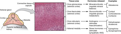
Pheochromocytoma:

– always think about pheo when the clinical case includes episodes of hypertension and headache

– Pheochromocytomas are tumors composed of chromaffin cells of the adrenal gland

– Pheochromocytomas almost always realease catecholamines

Classic Triad:

– Diaphoresis

– Headache

– Tachycardia

Genetic disorder associated with pheochromocytoma:

– MEN 2A and 2B

– Neurofibromatosis type 1

– Von Hippel-Lindau syndrome

Diagnosis: – Plasma / Urine catecholamines

– plasma high sensitivity (96-100%), but lower specificity (85-89%); 24 hour urine sensitivity/specificity (91-98%)

– Plasma will exclude a pheo when negative, but need to confirm if positive

– Following biochemical diagnosis – radiographic localization is needed

Preoperative management:

– Alpha-blocker – typically with phenoxybenzamine – is first-line therapy; followed by B-blockers (metoprolol, propranolol) to treat reflex tachycardia

MEN 1, 2A, and 2B disorders:

MEN 1: Pituitary adenoma, Parathyroid hyperplasia, Pancreatic tumors

MEN 2A: Medullary thyroid cancer, Parathyroid hyperplasia, Pheochromcytoma

MEN 2B: Medullary thyroid cancer, Marfanoid habitus/Mucosal neuroma, Pheochromocytoma

picture3

Cushing Syndrome:
– Bright purple abdominal striae always, always, always are associated with Cushing syndrome – striae larger than 1 cm in width are highly specific for hypercortisolism
picture5
Other classic stigmata:
– cervicodorsal fat pad aka “buffalo hump,” acne, hirsutism, moon facies, plethora, easy bruising, hypertension, insulin resistance, hypokalemic metabolic alkalosis, osteoporosis

Picture4.png
Cause:
– Elevated cortisol
– Most common cause is exogenous glucocorticoid therapy for another cause

3 biochemical testing options for CS:
– 1 mg (low dose) dexamethasone suppression test – failure to suppress AM cortisol indicates true Cushing syndrome
– 24 hour urine free cortisol (UFC) – excludes CS when not elevated
– Late-night salivary cortisol
Never measure a random cortisol as part of the work up

After confirming CS biochemically, measure ACTH to establish etiology
– differentiate between ACTH-dependent/Cushing Disease (usually > 20 pg/mL) and ACTH-independent (usually < 5 pg/mL)

If ACTH-dependent CS: order pituitary MRI
If ACTH-independent CS: adrenal gland imaging (either CT or MRI)

ACTH-secreting pituitary adenoma is known as Cushing disease
Pearl: pituitary adenomas do not suppress with low-dose dex, but do suppress with high dose

Other possible ACTH-secreting tumors (but very rare):
– An ectopic ACTH-secreting tumor
– Usually these are primary lung cancers or a carcinoid tumor
– The clinical presentation of an ectopic lung tumor usually is less cushingoid (presenting with only HTN and metabolic abnormalities) due to the rapid growth of these tumors


AM Report 1/9/17: Infectious Disease

Pneumocystis jiroveci pneumonia (PJP):
– opportunistic infections are the most common etiologies of infection in patients 1-6 months after solid organ transplant.
Common signs/symptoms of PJP:
– Progressive exertional dyspnea (95%)
– Fever (90%)
– Non-productive cough (90%)
Pearl: walk patients with suspected PCP to reveal hypoxemia!

Diagnosis:
– Direct fluorescent antibody stain (DFA stain)
– Gomori methenamine silver stain (GMS stain)
*Must visualize the cystic/trophic forms directly
picture1

Treatment: TMP/SMX for 21 days
Add steroids for pO2 ≤ 70 or A-a gradient ≥ 35

Toxic Shock Syndrome:
– expect a TSS board question to present as overwhelming sepsis in the context of a menstruating female or a post-surgical wound infection
– toxins (called super antigens) stimulate cytokine production, resulting in systemic signs of shock

Triad of TSS:
Shock
Fever
Rash – diffuse macular rash with subsequent desquamation, especially on the palms and soles
*Along with involvement of at least 3 organ systems
picture2

Organisms often involved:
– S. aureus
– S. pyogenes

Treatment:
S. pyogenes: penicillin plus clindamycin
MSSA: Nafcillin or oxcillin plus clindamycin
MRSA: Vancomycin plus clindamycin
* add clindamycin to suppress protein synthesis and, therefore, toxin production

Scarlet Fever:
Key features:
– “circumoral pallor” – pale area around mouth
– “Pastia lines” – petechial lines in the skin creases
– desquemation

Most common organism: Group A Streptococcus

5 “S'” of Scarlet Fever:
– Streptococci (causative organism)
– Sorethorat
– Swollen tonsils
– Strawberry tongue
– Sandpaper rash

Treatment: Oral penicillin V; amoxicillin, 1st generation cephalosporins, and IM PCN G are alternatives


Lyme Disease:

– Erythema migrans is the associated rash
picture3
– Borelia burgdorferi is the causitive organism and Ixodes tick is the vector
– > 95% of cases occur in regions where the Ixodes tick is abundant
picture4
Other infections spread by Ixodes tick:
– Babesiosis
– Anaplasmosis

Signs/Symptoms of different stages of Lyme disease:
Localized: erythema migrans (target lesion at site of tick attachment ~ 60-80%), fever/other systemic symptoms are rarely present
Early disseminated: erythema migrans at multiple sites, febrile illness with constitutional symptoms (myalgia, arthralgia, and headache)
Cardiac: asymptomatic PR prolongation → complete heart block
Neurologic: facial nerve palsy (most common) either unilateral or bilateral
Late disseminated: oligoarticular inflammatory arthritis involving large joints (i.e. knee)

Rocky Mountain Spotted Fever:
Look for a history of exposure to ticks in endemic areas (southeast / south central US) and features of:
– Pancytopenia (esp thrombocytopenia)
– Hyponatremia
– No evidence of DIC (normal PT/PTT)
– ↑ transaminases

picture6
Rash of RMSF:
– >85% of patients by 1 week
– May lag behind other symptoms (~50% by day 3)
– Typically starts at the distal extremities and progress centrally; involves the palms/soles in >30% and typicially spares the face

Treatment:
– Doxycycline; choloramphenicol is an alternative option in pregnancy

Ehrlichiosis:
Think of Ehrlichia as “Rocky Moutain spotless fever”
– presents similarly to Anaplasmosis
– endemic to the southcentral and southeastern US
– spread via the Lone Star tick

Symptoms in order of frequency:
Fever (~90%) > headache > myalgia > arthralgia > meningismus

– Blood smear can help with visualization of intracytoplasmic inclusions in WBCs; only present in ~30%

Treatment:
– Doxycycline

Coccidioides Infection (Valley Fever):
– Clues to be aware of: Arizona/New Mexico and erythema nodosum
– Endemic to SW US (Arizona, New Mexico, Texas, and central valley of California)
– Route of infection: inhalation of fungal particles found in the sand
– Arthralgia of multiple joints “desert rheumatism” is common.
picture7
Diagnosis confirmed on fungal stains
– Thick walled spherules (10-80 uM) with endospores are seen in tissue
picture8
No treatment for mild disease; use itraconazole or fluconazole for severe illness

Histoplasmosis:
Exposure history is key here; think of histo with any of the following exposures in the SE/SC US:
– Bats (or guano)
– “Spelunking” (cave exposure)
– Farm buildings / bird-roosting locations
picture9
Most infections are subclinical (~95%); can see mucocutaneous lesions
Antigen detection in urine great for disseminated infections (>85%)

Blastomycosis:
– Endemic to Ohio and Mississippi/ River valleys
picture10
– Primarily a pulmonary infection, may disseminate to the skin and bone
– Well demarcated skin lesion is most common manifestation of disseminated disease
picture11
– Appears as a broad based budding pattern at 37 C
picture12

AM Report 2/6/17: Neurology Board Review

Normal Pressure Hydrocephalus:
Crude mnemonic: Wet (incontinence), Wobbly (ataxia), and Wacky (dementia)
Abnormal gait typically develops first!
Magnetic gait” – is the characteristic gait of NPH – the patient’s walk appears as if his/her feet are stuck to the floor.
The gait is wide based, with slow, small steps, and reduced foot-to-floor clearance

Visualize hydrocephalus on CT/MRI – specifically ventriculomegaly that is disproportionate to corticol atrophy
picture1

LP: Normal (or slighty elevated) opening pressure and cell count

Standard of care: Ventriculoperitoneal (VP) shut – can perform large volume (30-50 mL) LP prior to placement to verify benefit

picture2

Cluster Headache:
Presentation: Shorter duration (usually < 3 hours), ipsilateral congestion and lacrimation with headache, nocturnal attacks
Epidemiology: Young or middle aged, male sex, history of cigarette smoking
Common trigger: Alcohol

First-line therapy: Inhalation of 100% oxygen and/or SQ or intranasal sumatriptans
Prevention medication: Verapamil

Transverse Myelitis:
TM is thought to be due to an autoimmune inflammation
~50% of the time it is preceded by an infection

Diagnosis requires:
– Presence of clinical features
– Evidence of inflammation (either leukocytosis in CSF or contrast enhancement on MRI)
– Exclusion of other potential causes

Treatment: IV methylprednisolone; plasmapheresis or cyclophosphamide for refractory disease

Alzheimer Disease:
Presentation: >60 years old, decline is insidious and progressive, definite impairment in 2 or more domains of cognition that impacts daily living

Treatment options:
– Cholinesterase inhibitors: Donepezil, Rivastigmine, Galantamine
– N-methyl-D-asapartate receptor antagonists: Memantine
*mild improvements in measured cognition and performance of some activities; no clinically significant outcome

Frontotemproal Dementia:
– 3rd most common type of neurodegenerative disorder (2nd in those < 65 years old); average age of onset is 50-60
– Social behavior / personality changes are variable
– Genetics of FTD are heterogenous ~40% of patients with FTD have at least one family member with a neurodegenerative disorder

Difference with Lewy Body Dementia – Lack of significant psychosis – especially visual hallucinations

Parkinson Disease:
4 main features:
Tremor: “pill-rolling” and always gets better with action
Bradykinesia
Rigidity
Gait/Postural impairment: “festinating gait” – meaning hurried small shuffles, often on tippy toes
* diagnostic criteria require the presence of bradykinesia with at least one other cardinal feature
* MRI is recommended to rule out vascular lesions and hydrocephalus when suspected

Treatment options:
– Non-pharmacologic: rigorous daily exercise
– Pharmacologic: Dopamine substrate (levodopa), Decarboxylase inhibitors (carbidopa), Dopamine agonists (Ropinirole, Bromocriptine, Pergolide), Catechol-O-methyltransferase inhibitors (Entacapone, Tolcapone), Monoamine oxidase type B inhibitors (Selegiline, Rasagiline), Glutamate antagonist (Amatadine), Anticholinergic agents (Benztropine)
– Surgical: Deep brain stimulation (DBS) – consider in patients who have sustained motor benefit from medication, but are limited by adverse effect of medications

Myasthenia Gravis:
Cause: autoimmune disease directed against post-synaptic NM junction
Labs:
Anti-acetylchoine receptor antibodies (~90% sensitive)
– Anti-muscle-specific kinase (MuSK) antibodies
EMG: repetitive nerve stimulation shows decremental response
Imaging: CT or MRI of mediastinum to screen for thymoma (~15% present, 85% hyperplasia)

Treatment:
– Cholinesterase inhibitors: pyridostigmine
– Glucocorticoids
– Immunosuppressive agents: azathioprine, cyclosporine, etc.

Crisis Treatment:
– Plasmapheresis or IVIG

Lambert-Eaton Syndrome:
Cause: autoantibodies against voltage-gated calcium channels located at the presynaptic NM junction
Associations: small cell lung cancer – can occur before tumor has been discovered; found in as many as 60% of patients

Differentiate from MG:
Repetitive contraction IMPROVES muscle strength
– Hyporeflexia and dysautonomia are also present

Treatment:
– Remove the malignancy
– IVIG +/- immunomodulators

AM report 02/01/16 Melioidosis

Melioidosis (Whitmore’s disease)

-Caused by the Gram (-) organism Burkholderia pseudomallei
burkholderia

Epidemiology

–Predominantly in Southeast Asia, Northern Australia, and China (most commonly from Thailand, Malaysia, Singapore)

Transmission

–Percutaneous inoculation (soil/contaminated water)
-Inhalation (more common during severe weather events, eg:Tsunami/Hurricane)
-Aspiration
-Rarely Ingestion

Risk factors

Occupational exposure (Eg: Rice farming)
Immunocompromised
–DMII
–Alcohol use
–CKD
–Chronic lung disease
–Thalassemia

However, can occur in healthy individuals as well! 

Clinical Manifestations

1)Localized Infection-skin ulcers/abscesses
2)Pulmonary Infection->50 % of patients, with >25 % having cavitary pulmonary nodules 
3)Bloodstream infection-More than half of patients have bacteremia on presentation and septic shock develops in >20 % of patients 
4)Disseminated infection-Septic Arthritis, osteomyelitis, but can form parenchymal abscesses in ANY organ (eg: Spleen, Kidney, Prostate, Brain amongst others)

Can MIMIC Tb and Malignancy-Consider Melioidosis in any patient with cavitary nodules and skin findings! 

Treatment 

Remember that Burkholderia is inherently RESISTANT to penicillin, ampicillin, 1st/2nd gen cephalosporins amongst others-Treatment is very prolonged!

Intensive phase (10-14d): IV Ceftazidime, Meropenem, or Imipenem
Oral Eradication Therapy (3-6 months): TMP-SMX (preferred over doxycycline)

Source Control! Search and Treat internal-organ abscesses 

AM Report 1/31/17: PD Peritonitis and Shock Liver

Steps to an Acid/Base Problem:

  1. Step 1: Check for internal consistency[H+] = 24 x pC02/HCO3
  2. Step 2: Use pH to determine primary disorder
  3. Step 3: Calculate the AGAG = [Na+] – [Cl- + HCO3]
  4. Step 4: Determine the presence of additional disordersCorrected HCO3 = measured HCO3 + [AG – 12]
  5. Step 5: Calculate the expected pC02 for metabolic acidosis (Winter’s Formula)Expected PC02 = 1.5 (HCO3) +8 +/- 2

5 Etiologies with AST/ALT elevations > 1000:

  • Drugs/Toxins (acetaminophen, toxic mushrooms, etc.)
  • Hepatic ischemia
  • Acute viral hepatitis
  • Acute autoimmune hepatitis
  • Wilson disease

3 components that define acute liver injury:

  • Elevated aminotransferases
  • Hepatic encephalopathy
  • Elevated PT/INR

*No preexisting cirrhosis or liver disease; <26 weeks

Acetaminophen Toxicity:

  • 4 grams is the daily maximum recommended dose
  • N-acetylcysteine is the treatment of choice in suspected acetaminophen toxicity -nmay be given IV, using a 20 hour protocol, or orally, using a 72 hour protocol
  • Use the Rumack-Matthew nomogram to elevate for poisoning severity

picture14

Options for renal replacement therapy (RRT):

  • Hemodialysis
  • Peritoneal dialysis
  • Transplantation
  • Non-dialytic management – manage symptoms and focus on QOL

Peritoneal dialysis – a dialysate solution is intermittently instilled into the peritoneal cavity via an indwelling catheter, and excess water/solutes are removed by osmosis and diffusion across the peritoneal membrane into the dialysis solution.

picture15picture16

Two different options for PD exist:

  1. Continuous ambulatory peritoneal dialysis (CAPD)
  2. Automated peritoneal dialysis (APD)

    *CAPD does not require a machine, while APD relies on a “cycler” to fill and empty your belly 3-5 times during the night

    *There is no difference in clinical outcome between PD and HD – although there is a trend toward superior outcomes for PD patients with new-onset ESRD who have residual kidney function

Potential complications with PD:

  • Peritonitis – from frequent access via the catheter or from perforation or microperforation of bowel
  • Hernias at catheter site
  • Subcutaneous edema
  • Difficulty draining catheter (often due to constipation)
  • Pleural effusions
  • Sclerosing peritonitis: scarring/fibrosis from recurrent peritonitis – can lead to bowel constriction.  Prognosis is extremely poor (often fatal) and treatment requires surgical stripping.

Diagnosis of peritonitis requires 2 out of 3 of the following:

  • Clinical signs of peritonitis
  • Peritoneal fluid WBC > 100 cells/mm3 with >50% PMNs
  • Positive peritoneal fluid culture

Common clinical manifestations of PD related peritonitis:

  • Abdominal pain (79-88%)
  • Cloudy peritoneal effluent (84%)
  • Fever (>37.5 C) (29-53%)
  • Nausea/Vomiting (31-51%)
  • Hypotension (~18%)

Most common organisms of peritonitis in PD patients:

  • Gram-positive organisms ~ 50% -Coagulase-negative staph (S. epidermidis) and S. aureus
  • Gram-negative organisms ~ 15%
  • Fungal ~2%
  • Polymicrobial ~ 4%
  • Culture negative ~ 20%

Empiric Antibiotic Regimen:

  • Gram positive coverage – typically Vancomycin (or 1st generation cephalosporin)
  • Gram negative coverage – typically 3rd generation cephalosporin
  • Fungal prophylaxis while on ABX – typically nystatin

Indications for PD catheter removal:

  • Relapsing peritonitis – episode of peritonitis with the same organism that caused the preceding episode within 4 weeks of completing antibiotics
  • Refractory peritonitis – peritonitis that does not respond to appropriate antibiotics within 5 days
  • Refractory catheter infection – exit-site/tunnel infections
  • Fungal or mycobacterial peritonitis

AM Report 1/25/17: Secondary Adrenal Deficiency secondary to Megace

Adrenal Anatomy:

1818_the_adrenal_glands

Primary Adrenal Failure (Addison’s Disease):

  • Failure of the adrenal cortex
  • Prevalence 10-15/100,000 persons
  • Etiology – Autoimmune adenitis ~70-80%; 2/3 will have at least one other autoimmune endocrine disorder; TB infiltration ~ 7-20%

capture1

Diagnostic Algorithm:

capture2

Adrenocorticotropic Hormone Deficiency (secondary cortisol deficiency):

Etiology:

  1. Exogenous glucocorticoid use
  2. Damage to the pituitary gland/stock
  • Reminder of intact adrenal cortex will be a normal renin/aldosterone ratio
  • Less risk for hypotension, hyponatremi, adrenal crisis than primary failure
  • No hyperpigementation due to lack of hypersecrtion of ACTH

Multiple case reports of megase induced secondary adrenal insufficency

picture11picture12picture13

AM Report 1/18/17: Adult Onset Still’s Disease

Fever of Unknown Origin Criteria:

  • Fever > 38.3 on several occasions
  • Duration of fever > 3 weeks
  • Uncertain diagnosis after 1 week of inpatient investigation

5 Main Etiologies for FUO:

  • Infection
  • Malignancy
  • Connective tissue disease
  • Other
  • No diagnosis

picture1

1

picture2

Adult Onset Still’s Disease (AOSD):

  • Very uncommon – estimated annual incidence of 0.16 cases per 100,000 people
  • Equal distribution between the sexes
  • Bimodal age distribution: one peak between 15-25 and the other between 36-46 years old
  • Diagnosis of exclusion, must exclude infection, malignancy, or other rheumatologic disease.

AOSD is multisystem inflammatory disease characterized by 4 major findings:

  1. High spiking fevers
  2. Salmon-color rash
  3. Arthritis
  4. High neutrophil counts

AOSD Laboratory Findings:

  • Elevated ESR / CRP (99%)
  • Elevated Ferritin (~75%)
  • Leukocytosis > 10,000/mm3 (92%), >15,000/mm3 (81%) with neutrophil predominance (>80%)
  • Abnormal LFTs (~ 75%)
  • Negative ANA (92%)
  • Negative RF (93%)

Yamaguchi Criteria for AOSD:

  • Diagnosis requires 5 criteria total – with at least 2 major criteria

picture5picture6

AOSD Treatment:

Initial therapeutic decisions should be based on the degree of disease activity:

  • Mild disease: fevers, rash, and/or mild arthritis
  • Moderate disease: high fevers, debilitating joint symptoms, evidence of internal organ involvement that is not life-threating or severe
  • Severe disease: presence of life-threating organ involvement

Treatment options:

  • NSAIDS – effective alone for 20% of mild patients
  • Glucocorticoids – should be started in all patients meeting moderate disease criteria
  • Methotrexate – used in patient’s who fail to improve with steroids
  • Can also consider other DMARDs – will not be discussed here!

AM Report 1/30/17: Neurosyphilis

Which Asymptomatic patients should you screen for syphilis?

–HIV
–MSM
–Partner with syphilis
–High risk sexual behavior/history of incarceration or commercial sex work
–Pregnancy (r/o congenital syphilis)

Testing for Syphilis (Treponemal vs. Non-Treponemal)

BOTH type of tests required due to risk of false positives and false negatives (eg: immunosuppression/early disease)
-Can use direct visualization tests like DFA or Darkfield Microscopy but SEROLOGY is the most common method of testing.

Non-Treponemal testing

-Based on reactivity of serum of infected patients to a cardiolipin-cholesterol-lecithin antigen but non-specific
-Positive results are reported as a TITER (eg: 1:32) and wane over time with treatment 

RPR (Rapid Plasma Reagin)
–VDRL (Veneral Disease Research Laboratory)
–TRUST (Toluidine Red Unheated Serum Test)

Treponemal testing

-Directly evaluating for antibodies against Treponemal antigens(higher specificity)!
-Qualitative only (no titers!)  and remain positive for life

–FTA-ABS (Fluorescent Treponemal antibody absorption)
–TPPA (T.Pallidum particle agglutination assay)
EIA (Enzyme immunoassay)
–CIA (Chemiluminsence immunoassay)

At VMC, our policy is to do a Treponemal test (EIA) and confirm with a Non-Treponemal test (RPR with titer)

What if you suspect Neurosyphilis

-Must do LP and classically see elevated lymphocytes and protein on CSF
(+) VDRL in CSF is highly specific for neurosyphilis but poor sensitivity (can be negative in up to 70 % of cases!)
–If negative VDRL, can check FTA-ABS (higher sensitivity/poor specificity)

Clinical manifestations of Neurosyphilis

EARLY neurosyphilis

  • Asymptomatic! (+) CSF VDRL
  • Meningitis, cranial neuropathies
  • Syphilitic gummas (can occur any
  • Ocular syphilis/Oto-syphilis

LATE neurosyphilis

  • General paresis (General paralysis of the insane)-10 % of psych admissions prior to 1928
  • Tabes Dorsalis (posterior columns/dorsal roots)- + Argyll-Robertson pupil

Treatment of Neurosyphilis

  • Aqueous IV Pencillin G 4 million units q4h x 10-14d

AM Report 1/24/17: Pure Red Cell Aplasia (PRCA) due to Parvovirus B19

Etiology of Acquired PRCA (can also be congenital

IDIOPATHIC (most common)
-Drugs (Phenytoin, Bactrim, Zidovudine, Mycophenolate, INH etc.)
-Infections (Parvovirus B19, HIV, Viral Hepatitis)
Auto-Immune (SLE, RA, auto-immune hemolytic anemia, ABO incompatible HCT, Epo treatment)
Lymphoid malignancies (CLL, HL, NHL, Myeloma)
Myeloid malignancies (CML, myelodysplastic syndrome, other cancers)
-Thymoma
Pregnancy

Making the Diagnosis of PRCA (all 4 criteria must be met)

  • Normocytic Normochromic anemia
  • Very low or zero reticulocyte count with ARC <10,000/microL
  • NORMAL WBC count and platelet count


Bone Marrow Biopsy: Normocellular bone marrow with normal myeloid, lymphoid, and megakaryocytopoiesis but minimal erythroid precursors

LOW reticulocyte count=reduced bone marrow response

  • AOCD, mild/moderate IDA, renal failure, PRCA

HIGH reticulocyte count=preserved bone marrow response

  • Hemolysis, bleeding, Sickle cell, RBC membrane disorders

General treatment of PRCA 

-RBC transfusion for symptomatic anemia
-STOP any possible offending drugs (see above)
-Search and treat any associated conditions
Immunosuppression (Glucocorticoids/Cyclosporine/Cyclophosphamide etc)

Specific Treatment for Parvovirus B19 related PRCA

Immunocompetent (usually spontaneous resolution within 2-3 weeks)
Immunocompromised (Usually will not spontaneously resolve and can develop chronic infection) Treatment of choice is IVIG