Category Archives: Morning Report

AM Report 1/31/17: PD Peritonitis and Shock Liver

Steps to an Acid/Base Problem:

  1. Step 1: Check for internal consistency[H+] = 24 x pC02/HCO3
  2. Step 2: Use pH to determine primary disorder
  3. Step 3: Calculate the AGAG = [Na+] – [Cl- + HCO3]
  4. Step 4: Determine the presence of additional disordersCorrected HCO3 = measured HCO3 + [AG – 12]
  5. Step 5: Calculate the expected pC02 for metabolic acidosis (Winter’s Formula)Expected PC02 = 1.5 (HCO3) +8 +/- 2

5 Etiologies with AST/ALT elevations > 1000:

  • Drugs/Toxins (acetaminophen, toxic mushrooms, etc.)
  • Hepatic ischemia
  • Acute viral hepatitis
  • Acute autoimmune hepatitis
  • Wilson disease

3 components that define acute liver injury:

  • Elevated aminotransferases
  • Hepatic encephalopathy
  • Elevated PT/INR

*No preexisting cirrhosis or liver disease; <26 weeks

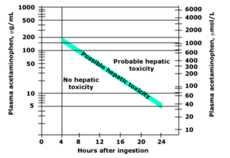
Acetaminophen Toxicity:

  • 4 grams is the daily maximum recommended dose
  • N-acetylcysteine is the treatment of choice in suspected acetaminophen toxicity -nmay be given IV, using a 20 hour protocol, or orally, using a 72 hour protocol
  • Use the Rumack-Matthew nomogram to elevate for poisoning severity

picture14

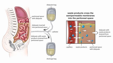
Options for renal replacement therapy (RRT):

  • Hemodialysis
  • Peritoneal dialysis
  • Transplantation
  • Non-dialytic management – manage symptoms and focus on QOL

Peritoneal dialysis – a dialysate solution is intermittently instilled into the peritoneal cavity via an indwelling catheter, and excess water/solutes are removed by osmosis and diffusion across the peritoneal membrane into the dialysis solution.

picture15picture16

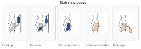
Two different options for PD exist:

  1. Continuous ambulatory peritoneal dialysis (CAPD)
  2. Automated peritoneal dialysis (APD)

    *CAPD does not require a machine, while APD relies on a “cycler” to fill and empty your belly 3-5 times during the night

    *There is no difference in clinical outcome between PD and HD – although there is a trend toward superior outcomes for PD patients with new-onset ESRD who have residual kidney function

Potential complications with PD:

  • Peritonitis – from frequent access via the catheter or from perforation or microperforation of bowel
  • Hernias at catheter site
  • Subcutaneous edema
  • Difficulty draining catheter (often due to constipation)
  • Pleural effusions
  • Sclerosing peritonitis: scarring/fibrosis from recurrent peritonitis – can lead to bowel constriction.  Prognosis is extremely poor (often fatal) and treatment requires surgical stripping.

Diagnosis of peritonitis requires 2 out of 3 of the following:

  • Clinical signs of peritonitis
  • Peritoneal fluid WBC > 100 cells/mm3 with >50% PMNs
  • Positive peritoneal fluid culture

Common clinical manifestations of PD related peritonitis:

  • Abdominal pain (79-88%)
  • Cloudy peritoneal effluent (84%)
  • Fever (>37.5 C) (29-53%)
  • Nausea/Vomiting (31-51%)
  • Hypotension (~18%)

Most common organisms of peritonitis in PD patients:

  • Gram-positive organisms ~ 50% -Coagulase-negative staph (S. epidermidis) and S. aureus
  • Gram-negative organisms ~ 15%
  • Fungal ~2%
  • Polymicrobial ~ 4%
  • Culture negative ~ 20%

Empiric Antibiotic Regimen:

  • Gram positive coverage – typically Vancomycin (or 1st generation cephalosporin)
  • Gram negative coverage – typically 3rd generation cephalosporin
  • Fungal prophylaxis while on ABX – typically nystatin

Indications for PD catheter removal:

  • Relapsing peritonitis – episode of peritonitis with the same organism that caused the preceding episode within 4 weeks of completing antibiotics
  • Refractory peritonitis – peritonitis that does not respond to appropriate antibiotics within 5 days
  • Refractory catheter infection – exit-site/tunnel infections
  • Fungal or mycobacterial peritonitis

AM Report 1/25/17: Secondary Adrenal Deficiency secondary to Megace

Adrenal Anatomy:

1818_the_adrenal_glands

Primary Adrenal Failure (Addison’s Disease):

  • Failure of the adrenal cortex
  • Prevalence 10-15/100,000 persons
  • Etiology – Autoimmune adenitis ~70-80%; 2/3 will have at least one other autoimmune endocrine disorder; TB infiltration ~ 7-20%

capture1

Diagnostic Algorithm:

capture2

Adrenocorticotropic Hormone Deficiency (secondary cortisol deficiency):

Etiology:

  1. Exogenous glucocorticoid use
  2. Damage to the pituitary gland/stock
  • Reminder of intact adrenal cortex will be a normal renin/aldosterone ratio
  • Less risk for hypotension, hyponatremi, adrenal crisis than primary failure
  • No hyperpigementation due to lack of hypersecrtion of ACTH

Multiple case reports of megase induced secondary adrenal insufficency

picture11picture12picture13

AM Report 1/18/17: Adult Onset Still’s Disease

Fever of Unknown Origin Criteria:

  • Fever > 38.3 on several occasions
  • Duration of fever > 3 weeks
  • Uncertain diagnosis after 1 week of inpatient investigation

5 Main Etiologies for FUO:

  • Infection
  • Malignancy
  • Connective tissue disease
  • Other
  • No diagnosis

picture1

1

picture2

Adult Onset Still’s Disease (AOSD):

  • Very uncommon – estimated annual incidence of 0.16 cases per 100,000 people
  • Equal distribution between the sexes
  • Bimodal age distribution: one peak between 15-25 and the other between 36-46 years old
  • Diagnosis of exclusion, must exclude infection, malignancy, or other rheumatologic disease.

AOSD is multisystem inflammatory disease characterized by 4 major findings:

  1. High spiking fevers
  2. Salmon-color rash
  3. Arthritis
  4. High neutrophil counts

AOSD Laboratory Findings:

  • Elevated ESR / CRP (99%)
  • Elevated Ferritin (~75%)
  • Leukocytosis > 10,000/mm3 (92%), >15,000/mm3 (81%) with neutrophil predominance (>80%)
  • Abnormal LFTs (~ 75%)
  • Negative ANA (92%)
  • Negative RF (93%)

Yamaguchi Criteria for AOSD:

  • Diagnosis requires 5 criteria total – with at least 2 major criteria

picture5picture6

AOSD Treatment:

Initial therapeutic decisions should be based on the degree of disease activity:

  • Mild disease: fevers, rash, and/or mild arthritis
  • Moderate disease: high fevers, debilitating joint symptoms, evidence of internal organ involvement that is not life-threating or severe
  • Severe disease: presence of life-threating organ involvement

Treatment options:

  • NSAIDS – effective alone for 20% of mild patients
  • Glucocorticoids – should be started in all patients meeting moderate disease criteria
  • Methotrexate – used in patient’s who fail to improve with steroids
  • Can also consider other DMARDs – will not be discussed here!

AM Report 1/30/17: Neurosyphilis

Which Asymptomatic patients should you screen for syphilis?

–HIV
–MSM
–Partner with syphilis
–High risk sexual behavior/history of incarceration or commercial sex work
–Pregnancy (r/o congenital syphilis)

Testing for Syphilis (Treponemal vs. Non-Treponemal)

BOTH type of tests required due to risk of false positives and false negatives (eg: immunosuppression/early disease)
-Can use direct visualization tests like DFA or Darkfield Microscopy but SEROLOGY is the most common method of testing.

Non-Treponemal testing

-Based on reactivity of serum of infected patients to a cardiolipin-cholesterol-lecithin antigen but non-specific
-Positive results are reported as a TITER (eg: 1:32) and wane over time with treatment 

RPR (Rapid Plasma Reagin)
–VDRL (Veneral Disease Research Laboratory)
–TRUST (Toluidine Red Unheated Serum Test)

Treponemal testing

-Directly evaluating for antibodies against Treponemal antigens(higher specificity)!
-Qualitative only (no titers!)  and remain positive for life

–FTA-ABS (Fluorescent Treponemal antibody absorption)
–TPPA (T.Pallidum particle agglutination assay)
EIA (Enzyme immunoassay)
–CIA (Chemiluminsence immunoassay)

At VMC, our policy is to do a Treponemal test (EIA) and confirm with a Non-Treponemal test (RPR with titer)

What if you suspect Neurosyphilis

-Must do LP and classically see elevated lymphocytes and protein on CSF
(+) VDRL in CSF is highly specific for neurosyphilis but poor sensitivity (can be negative in up to 70 % of cases!)
–If negative VDRL, can check FTA-ABS (higher sensitivity/poor specificity)

Clinical manifestations of Neurosyphilis

EARLY neurosyphilis

  • Asymptomatic! (+) CSF VDRL
  • Meningitis, cranial neuropathies
  • Syphilitic gummas (can occur any
  • Ocular syphilis/Oto-syphilis

LATE neurosyphilis

  • General paresis (General paralysis of the insane)-10 % of psych admissions prior to 1928
  • Tabes Dorsalis (posterior columns/dorsal roots)- + Argyll-Robertson pupil

Treatment of Neurosyphilis

  • Aqueous IV Pencillin G 4 million units q4h x 10-14d

AM Report 1/24/17: Pure Red Cell Aplasia (PRCA) due to Parvovirus B19

Etiology of Acquired PRCA (can also be congenital

IDIOPATHIC (most common)
-Drugs (Phenytoin, Bactrim, Zidovudine, Mycophenolate, INH etc.)
-Infections (Parvovirus B19, HIV, Viral Hepatitis)
Auto-Immune (SLE, RA, auto-immune hemolytic anemia, ABO incompatible HCT, Epo treatment)
Lymphoid malignancies (CLL, HL, NHL, Myeloma)
Myeloid malignancies (CML, myelodysplastic syndrome, other cancers)
-Thymoma
Pregnancy

Making the Diagnosis of PRCA (all 4 criteria must be met)

  • Normocytic Normochromic anemia
  • Very low or zero reticulocyte count with ARC <10,000/microL
  • NORMAL WBC count and platelet count


Bone Marrow Biopsy: Normocellular bone marrow with normal myeloid, lymphoid, and megakaryocytopoiesis but minimal erythroid precursors

LOW reticulocyte count=reduced bone marrow response

  • AOCD, mild/moderate IDA, renal failure, PRCA

HIGH reticulocyte count=preserved bone marrow response

  • Hemolysis, bleeding, Sickle cell, RBC membrane disorders

General treatment of PRCA 

-RBC transfusion for symptomatic anemia
-STOP any possible offending drugs (see above)
-Search and treat any associated conditions
Immunosuppression (Glucocorticoids/Cyclosporine/Cyclophosphamide etc)

Specific Treatment for Parvovirus B19 related PRCA

Immunocompetent (usually spontaneous resolution within 2-3 weeks)
Immunocompromised (Usually will not spontaneously resolve and can develop chronic infection) Treatment of choice is IVIG

AM Report 1/17/17: APML

APML (Acute Promyelocytic Leukemia)M3 variant of AML

Epidemiology

ANY age but uncommon before age 10 or after age 60
-RF include exposure to cytotoxic chemotherapy or radiation (esp. Topoisomerase inhibitors)

Timing

-Sub-acute

S&S

-Related to pancytopenia (eg: pallor,fatigability,  infections, gingival bleeding, ecchymoses etc.)
-APML in particular often presents with COAGULOPATHY due to DIC with Auer Rods seen on peripheral smear (see below)

auer rods.png

How do you make the diagnosis?

Molecular genetics via FISH analysis most common method

-Most common translocation is T(15:17) creating the fusion gene PML-RARA (see image below)
-Can also use Karyotype, RT-PCR, or anti-PML antibodies.
Bone Marrow Biopsyif high suspicion for APML based on cytologic and clinical criteria, it is recommended to START treatment prior to BMB due to high rate of early mortality. 

pml-rara
Treatment

-First line treatment is ATRA (All-Trans-Retinoic Acid) 

  • Combine with either Anthracycline based chemotherapy (Idarubicin or Daunorubicin and Cytarabine) or Arsenic Trioxide (ATO)

Which one do you choose?

  • Low/Intermediate risk: ATRA + ATO Lower risk of myelosuppression, cardiac toxicity, reduced risk of secondary leukemia
  • High risk APL (Initial WBC count >10^9 and platelet<40): ATRA + Anthracycline*

90 % of patients will achieve hematologic complete remission (CR) with induction chemotherapy

Supportive Care

High risk of DIC and coagulopathy-Monitor DIC labs and transfuse to keep platelet count >20k-30k, and fibrinogen>150

Complications-ATRA Differentiation Syndrome 

25 % of patients on ATRA may develop it within 2-25 days of treatment. However, can occur in those with APL who are NOT even treated with ATRA 

Signs and Symptoms

-Fever, pulmonary infiltrates, peripheral edema, hypoxemia, AKI and pleural/pericardial effusion
-Looks like ARDS or sepsis

Treatment is with Dexamethasone (10 mg IV q12h x 3d) and often cessation of ATRA treatment.

References

Images and information from UpToDate

AM Report 1/11/17: Myxedema Coma

Osborn Waves (J Waves):

  • The Osborn wave (J wave) is a positive deflection at the J point
  • It is usually most prominent in the precordial leads
  • Typically associated with hypothermia (typically < 30 C), but they are not pathognomonic; Also seen in: hypercalcemia, neurological insults, medication side effect
  • Height of the Osborn wave is roughly proportional to the degree of hypothermia


32.5 C

picture1

30 C

picture2

<27 C

picture3

Thyroid Regulation:

  • TRH (a tripeptide amide) formed in the hypothalamus and travels to the anterior pituitary where it stimulates the release of TSH.

TSH has 3 main effects:

  • ↑ release of preformed thyroid hormone
  • ↑ formation of thyroid hormone
  • ↑ size/number of thyroid cells

Synthesis of T3 and T4 (very complicated) requires two main components thyroglobulin and iodine.

4 physiologic effects of T3 and T4:

  • ↑ basal metabolic rate (↑ heat generation, ↑ O2 consumption)
  • ↑ metabolism (↑ gluconeogenesis, ↑ glycolysis, ↑ glucose absorption, ↑ lipolysis, ↑ protein turnover)
  • Stimulates bone maturation and growth
  • ↑ cardiac output (↑ HR, ↑ contractility)

picture5

Wolff Chaikoff effect: reduced thyroid hormone following large ingestion of iodine; explains potential hypothyroidism cased by amiodarone.

Jod-Basedow effect: iodine-induced hyperthyroidism in a patient with an endemic goiter

capture

Myxedema Coma:

3 key features:

  • Altered mental status: despite the name, most patients do not present in coma, but usually with confusion/lethargy
  • Hypothermia: due to decrease in thermogenesis that accompanies the decrease in metabolism
  • Precipitating event: look for cold exposure, infection, drugs (diuretics, sedatives, analgesics), trauma, stroke, heart failure, GI bleed, etc.

Pathogenesis:

capture

Typical presenting patient: older female presenting in the wintertime (often with a history of hypothyroidism)

  • Female > Male 4:1
  • Almost exclusively > 60 years old
  • 90% of cases during the winter months

If diagnosis is suspect, labs to get include:

  • TSH
  • FT4
  • Cortisol

Pearl: without a frankly low T4 level, myxedema coma is unlikely, regardless of the TSH elevation

Myxedema coma is an endocrine emergency and should be treated aggressively – mortality rate 20-40%

Treatment:

  • Thyroid replacement – controversial about type of replacement: Levothyroxine (T4) versus Liothyronine (T3), but initially route of administration should be IV given potential for impaired GI absorption
  • Stress dose steroids – patients with secondary hypothyroidism may have associated hypopituitarism and secondary adrenal insufficiency
  • Supportive measures – ICU management, mechanical ventilation (if necessary), IVF, vasopressors, rewarming, etc.

AM Report 1/3/17: Thyrotoxicosis

EKG findings with Atrial Fibrillation:

  • Irregularly irregular rhythm (no RR repetitive pattern)
  • No distinct P waves
  • f waves (small, irregular waves indicating rapid atrial activity ~150-300 bpm)

Common etiologies of Atrial Fibrillation:

  • Ischemic heart disease
  • Hypertension
  • Thyrotoxicosis
  • Drugs (sympathomimetics)
  • PE
  • Valvular heart disease (esp. mitral stenosis / regurgitation)
  • Electrolyte abnormalities (hypokalemia, hypomagnesaemia)
  • Cardiomyopathies (dilated, hypertrophic)

Different Classifications of Atrial Fibrillation:

  • Paroxysmal: AF that terminates spontaneously or with intervention within 7 days of onset
  • Persistent: sustained AF that lasts > 7 days
  • Long-standing persistent: sustained AF that lasts > 12 months
  • Permanent: persistent AF with either treatment failure or a decision to not pursue treatment

CHADS2 versus CHA2DS2VASc:

CHADS2 = CHF (1), HTN (1), Age ≥ 75 years (1), DM (1), Stroke/TIA (2)

CHA2DS2VASc = CHF (1), HTN (1), Age ≥ 75 years (2), DM (1), Stroke/TIA (2), Vascular disease (1), Age 65-74 (1),  Female sex (1)

  • Treat with anti-coagulation is score >2
  • Treatment options include: warfarin (goal INR 2-3), dabigatran, rivaroxaban, apixaban

Rate versus Rhythm Control:

AFFIRM trial

  • In patient’s with non-valvular AF, there is no survival benefit between rate and rhythm control
  • A non-significant trend towards decreased mortaility was associated rate control

Strict versus Lenient rate control:

RACE II trail

  • Among patients with permanent atrial fibrillation, lenient rate control (HR <110 bpm) is as effective as strict rate control (HR < 80 bpm) in preventing cardiovascular events
  • Of note, the strict group met resting targets in 78% of cases (compared with 98% in lenient) and required 9 times as many visits (684 vs. 75)

Physical Exam of Thryotoxicosis:

Skin: warm/moist skin, thin/fine hair, pretibial myxedema (Grave’s disease), thyroid acropachy (digital clubbing, swelling of digits)

Eyes: stare (lid retraction), lid lag, exophthalmos, periorbital/conjunctival edema, limitation of eye movement

CV: tachycardia, atrial fibrillation, systolic hypertension, high output CHF

GI: increased appetite, hyperdefecation

Neuro: fine tremor, hyperreflexia, proximal muscle weakness

Psych: anxiety, agitation, psychosis, depression, insomnia, mania

Thyroid: diffuse thyromegaly, thyroid bruit (specific for Graves)

Graves’ Disease Toxic adenoma / TMNG Subactue Thyroiditis
Epidemiology Females between 20-50 yo TA – 40-50 yo; TMNG > 50 yo Female > Male; young > old
Pathophysiology Antibodies against TSH receptors (TSI or TRAb) stimulate autonomous production of T4/T3 Autonomous production from activating mutations in TSH receptor Leakage of hormone from gland
Clinical Course / Physical Exam Exophthalmos,

Pre-tibial myexedema, Thyroid bruit

Hyperthyroid symptoms;

Palpable nodule(s)

Often preceded by URI, painful thyroid palpation, initially hyperthyroid => hypothyroid => normalizes
Diagnosis / Tests Low TSH, High FT4/T3, TSI, TBII; RAIU with markedly increased activity TSH, FT4, RAIU, thyroid US TSH, FT4

RAI – low uptake

Thyroid US – diffuse enlargement

Treatment Thionamides (PTU, methimazole); RAIA (avoid with ocular findings), surgery Thionamides (toxic patients), surgery for compression, RAIA NSAIDs, supportive care; prednisone if poor response, BB for symptom management

AM report 1/12/17: Accelerated Hypertension

Accelerated Hypertension ( Hypertensive Emergency /Malignant Hypertension)

 BP>180/120 + End Organ Damage

What is considered end-organ damage?  *Not an exhaustive list!

Cardiac-ACS/Aortic Dissection/CHF

Neuro-Stroke/Encephalopathy/Bleeding/Retinopathy/Papilledema

Renal-AKI/hematuria/proteinuria/MAHA/preclampsia/scleroderma renal crisis

Goal of Treatment Lower MAP with IV agents (eg: Labetalol/Esmolol/NTG/Nitroprusside/Nicardipine) by 10-20 % in minutes-2 hours, and then lower additional 5-15 % in next 23 hours.

Recommendations vary including lowering DBP<110 within 2-6 hours as tolerated

Compare to hypertensive urgency (no end-organ damage) where BP is lowered with oral medications and goal is to decrease BP in hours using PO agents with goal normal BP in 1-2 days

SPECIAL INSTANCE:

1)CHF and Accelerated HTN

-Treat with Diuretics and Nitroglycerin or Nitroprusside (watch for cyanide toxicity) –Avoid drugs that decrease contractility (eg: beta blockers), or increase cardiac work (Hydralazine)

Avoid Labetalol in cases of unopposed alpha states (eg: Pheochromocytoma/cocaine/methamphetamine)

EXCEPTIONS

1)Acute ischemic stroke-generally allow permissive hypertension unless BP>220/110 and not tPA candidate or >185/110 and tPa candidate

2)Aortic dissection-goal of 100-120 SBP to decrease wall stress (especially Type B aortic dissections which are medically managed)

Secondary Hypertension Etiologies and Workup

1)Renovascular HTN-90 % of cases due to atherosclerosis, but 10 % due to FBD, MRA superior to ultrasound in diagnosis

2)CKD-Check GFR, normocytic anemia, hx of DMII, PCKD, renal ultrasound

3)Primary Hyperaldosteronism-Check Renin/Aldo levels, only 50 % have hypokalemia and metabolic alkalosis-need to confirm that patient is off anti-hypertensives and aldosterone is suppressed with saline infusion challenge

4)OSA-based on history and sleep study

5)Drugs (eg: OCP/sympathomimetics/cocaine/METH)

6)Pheochromocytoma (24 hour urine metanephrines and catecholamines-sensitive and specific, serum metanephrines easier to test but lower specificity)

7)Cushing syndrome-Dexamethasone supression test

8)Coarctation of Aorta– Associated condition (eg: Turners), Radiofemoral delay

9)Primary Hyperparathyroidism-check PTH

10)Hypo or Hyperthyroidism-Check TFT

Causes of PAINLESS loss of vision is related to either RETINA or OPTIC nerve involvement

RETINA

1)Central Retinal Artery Occlusion (CRAO)-ischemic retina, see cherry red fovea (has its own blood supply)
2)Central Retinal Vein Occlusion (CRVO)-“blood and thunder” appearance
3)Retinal detachment-new onset floaters/black dots, can be related to trauma

OPTIC NERVE involvement

1)Ischemic optic neuropathy (rule out GCA!)
2)Optic neuritis (Multiple Sclerosis commonly)-(+) RAPD but not specific
3)Papilledema (any increase in ICP)
4)Compression of optic chiasm (eg: pituitary adenoma)

See examples below (courtesy of UpToDate)


AM Report 01/10/17: Dermatomyositis

Dermatomyositis and Polymyositis- Idiopathic Inflammatory Myopathies

-Present with PROXIMAL muscle weakness (eg: difficulty climbing stairs, getting up from chair)
-Evidence of muscle inflammation (Elevated CK, Aldolase but also includes elevated LDH, AST, ALT)
Skin manifestations (Dermatomyositis)

Problem Representation for Dermatomyositis (see exception below)

-Usually female (2:1 Female:Male involvement) at any stage of life but peak incidence age 40-50, who presents with gradual onset (weeks-months) of symptoms

Making the diagnosis of Dermatomyositis

-Ask about proximal weakness
-Ask about cutaneous eruptions (see below)
-Ask about systemic involvement (dysphagia from oropharyngeal or esophageal muscle involvement or dyspnea from ILD)
-Rule out drug myopathy (Glucocorticoids, Statins, Fibrates, Alcohol, Anti-Malarial drugs, Colchicine but LONG list of drugs that can cause myopathy

(+) autoantibodies in 80 % of patients with DM and PM (ANA/Anti-Jo-1/Anti-SRP/Anti Mi-2 among others)


Clinical presentation and physical exam

-90 % of patients will have muscle weakness (proximal and symmetric)
-Pain and stiffness are NOT prominent and only about 50 % of patients will have myalgias and muscle tenderness
Crackles on exam (if ILD is present)

Skin manifestations (pictures from UptoDate)

Anti-Synthetase Syndrome (~30 % of patients with DM/PM

ACUTE onset (different than usual sub-acute presentation) + constitutional symptoms + myositis + Raynaud + non-erosive arthritis + mechanic’s hands (see picture above) and positive anti-synthetase antibody (Anti-Jo)
-Important to recognize due to higher risk of developing Interstitial Lung Disease (ILD)

Testing

EMG

-Can support diagnosis of inflammatory myopathy but not diagnostic for PM or DM and can see similar findings in viral, infectious, toxic, or metabolic myopathies

Muscle Biopsy

Can distinguish DM, PM, and inclusion body myositis

Association with Malignancy

DM and PM (DM>PM) puts you at HIGHER risk for adenocarcinoma malignancy, most commonly OVARIAN cancer but includes cancer of cervix, lung, pancreas, bladder, and stomach among others.

-Can occur before, simultaneously, OR after diagnosis of inflammatory myopathy
-All patients with newly diagnosed PM or DM should be evaluated for possible malignancy, including pelvic ultrasound and age-appropriate screening

Treatment

                    First line therapy is GLUCOCORTICOIDS 

Steroid sparing agents like Azathiprine (check that TPMT level!), Methotrexate, and Hydroxychloroquine (good for skin manifestations) are often added to primary therapy
-IVIG and other therapies used for refractory cases
-Physical therapy
-Aspiration precautions (risk of dysphagia)
-Treatment/Prevention of Osteoporosis as on high dose steroids
-PJP prophylaxis if on high dose steroids (~>20 mg Prednisone/1 month)