
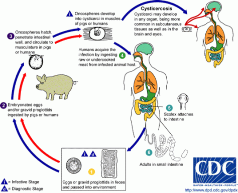
Cysticercosis
-Caused by the larval stage of the pork tapeworm (Taenia Solium)
-Humans get cysticercosis by drinking/eating water/food contaminated by tapeworm eggs (eg: infected pork)
-See life cycle below
Clinical syndromes
–Neurocysticercosis
Parenchymal
Extra-Parenchymal (Intraventricular/sub-arachnoid/intraocular/spinal)
-Extraneural cysticercosis
Clinical presentation
Parenchymal cysts
–Seizures/headache-Most common cause of adult onset seizures in many countries (70 % of patients)-esp. Latin America, India, Africa, and China
-Can be YEARS after infection. Most never cause symptoms and identified incidentally
Extraparenchymal cysts:
-Increased ICP- HYDROCEPHALUS, headache, nausea, vomiting, AMS
-Intraventricular cysts can cause obstructive hydrocephalus (nausea/vomiting/headache), subarachnoid cysts, spinal (<1%), ocular, extra-neural (subQ/intramuscular)
How do you make the diagnosis?
-Stool O&P usually negative as chronic infection
-Peripheral eosinophilia is NOT commonly seen
-If you have a patient from an endemic area with seizure and enhancing lesion on MRI-very likely to be Taenia Solium.
-See criteria below as definitive diagnosis requires at least one absolute criterion or two major plus one minor and one epidemiologic criterion

- Reference: UpToDate
–Note that identification of the Scolex (anterior end with hooks) in cystic lesion is pathognomonic.
-Serology with EITB (enzyme linked immunoelectrotransfer blot)antibody to T.solium, 83-100 % sensitive, 100 % specific but lab dependent)-takes a while to come back
-A detailed eye exam should be done to rule out ocular cysticercosis
-Brain biopsy rarely done as can be diagnosed by above


Differential Diagnosis (not complete) but do not miss other infectious causes!
–Toxoplasmosis
-Cryptococcus
-Brain abscess
-Nocardiosis
-Septic emboli
-TB/fungal
-Meningeal carcinomatosis
-Glioblastoma
Treatment
–Seizure control (controversial but esp. if multiple lesions, parenchymal involvement, or presenting with seizure)
-Treatment of increased ICP
–Antiparasitic therapy (Albendazole + Praziquantel with better efficacy, always given with or after anti-inflammatory therapy (steroids) due to inflammation with dying cysts. Can RECUR after treatment so needs to be tailored to imaging and symptoms.
-Surgical management if ocular or spinal lesions
