All posts by vmcimchiefs

AM Report 12/14/2016: Nephrotic Syndrome

Define:
Nephrotic Range Proteinuria:  >3g/24 hours without other findings
Nephrotic Syndrome:
– Proteinuria > 3.5 g/24 hours (protein/creatinine > 3.5 mg/mg)
– Hypoalbuminemia
– Clinical evidence of edema

Secondary Causes of Nephrotic Syndrome:
picture1

Primary Nephrotic Syndrome:
Minimal Change Disease:

Epidemiology:
– Children < 10 years old; can be primary or secondary
– 10% of cases in adults

Clinical Features:
– Sudden onset of edema
– Thrombotic episodes more common in adults
– AKI may be seen in 20% at presentation

Diagnosis:
– Renal biopsy

MCD on light microscopy:
– Appears essentially normal (hence the name minimal change!); tubules may show lipid accumulation
picture2
MCD on electron microscopy:
– Characteristic fusion and effacement of podocyte foot processes
picture3

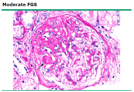
Focal Segmental Glomeruloscerlosis (FSGS):

Epidemiology:
– most common cause of primary nephrotic syndrome in the US
– can be primary, familial, or secondary
– African-Americans more common, but increasing incidence in all races

Clinical Manifestations:
– Asymptomatic proteinuria up to nephrotic syndrome ~ 2/3 at presentation!
– Hypertension usually seen in 30-50%
– Decreased GFR at presentation 20-30%

Diagnosis:
– Renal biopsy

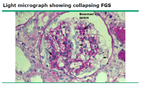
FSGS on light microscopy: scarring or sclerosis involving some (focal) glomeruli, which are affected only in a portion of the glomerular capillary bundle (segmental)
picture4
Collapsing FSGS – variant associated with HIV
picture5

Membranous Nephropathy (MN):

Epidemiology:
– most common in adults (>60 years old) and Caucasian
– can be primary (immune complex disease) or secondary (infection, autoimmune, cancer, drugs)

Potential complications:
– thrombotic disease – especially renal vein thrombosis

Diagnosis:
– renal biopsy
– PLA2R antibodies found in 75% of cases

Light microscopy for MN: diffuse thickening of the glomerular capillary wall
picture6

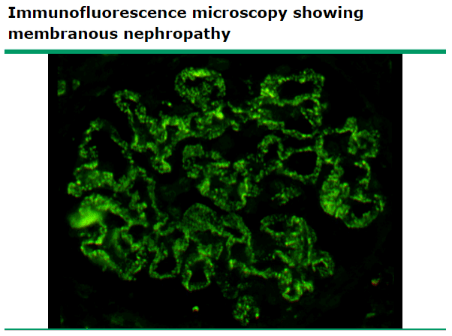
Immunofluorescence microscopy: diffuse, granular IgG deposition along capillary walls
picture7

Treatment:
– management is limited by a lack of clear evidence-based guidelines

General treatment:
– restrict dietary sodium to < 2 g/day
– restrict fluid intake to < 1.5 mL/day

Loop diuretics can be ineffective given that they are protein-bound and serum protein levels are reduced
Can add a thiazide diuretic and/or administer IV albumin bolus to improve diuresis

ACEi/ARBs – typicially used to reduce proteinuria although degree of benefit is unproven and evidence supporting routine use is conflicting
BP goal 130/80
Recent Cochrane review found no evidence to support the use of lipid-lowering agents in NS patients
Typically improves with resolution of disease
Corticosteroids are often used despite an absence of supporting evidence
Recent Cochrane review showed that combining alkylating agent (cyclophosphamide) with a corticosteroid has some short and long term benefits for MN
One exception – NS due to SLE – highly effective and supported by multiple studies


AM Report 12/12/16: Diverticulitis

Define:
Diverticulosis:
presence of diverticula ~ sac-like protrusion of colonic wall.
Diverticulitis:
inflammation of the diverticula

Epidemiology:
5-10% of patients > 45 years old
80% of patients > 85 years old

Risk Factors (formation of diverticulosis):
Lack of fiber / Western Diet (red meats/fats)
Decreased colonic motility
Decreased colonic wall resistance
Genetic susceptibility
NSAIDs/Aspirin
Age
Obesity
Smoking
Lack of exercise

DDx – Features:
Bowel Obstruction – abdominal pain, nausea/vomiting, abnormal imaging
Colorectal CA – weight loss, anemia, GI bleeding
Gastroenteritis – abdominal pain, nausea/vomiting
IBD – diarrhea, weight loss, rectal bleeding, mucus in stool
IBS – abdominal spasms relieved with defication
Ischemic colitis – abdominal pain out of portion to exam, arterial disease
Nephrolithasis – flank pain, hematuria, stone on imaging
Pancreatits – epigastric pain, nausea/vomiting

Likelihood Ratios for Acute Diverticulitis:
1

LLQ Imaging:

2

Accuracy of CT Finding for Diagnosing Acute Diverticulitis:

3

Treatment:
Inpatient ~ 6%
Any complicated case (abscess, obstruction, perforation, fistula)
Any uncomplicated with 1 (or more)
– Immunosuppression – Comorbidities
– Fever > 39 C – No PO intake
– Significant leukocytosis – Non-compliance
– Severe pain – Failed outpatient
– Old age

Outpatient ~ antibiotics 7-10 days
1) Cipro + Flagyl
2) Bactrim + Flagyl
3) Augmentin
4) Moxifloxacin

AM Report 12/07/16 TINU

Remember that red eye with WARNING symptoms like loss of visual acuity, ophthalmoplegia, pupillary involvement, photophobia, diplopia,  associated systemic symptoms 

Signs and Symptoms of Uveitis 

Exam: can see ciliary flush (circumcorneal injection), hypopyon (not specific, can be seen in many other eye conditions), redness at the limbus
Discharge: MINIMAL
Pupil: often constricted
Ocular pain: moderate to severe, deep aching pain
Vision: mild-moderately reduced
Cornea: often hazy in appearance
Treatment: Steroids

Etiology of Uveitis 

IDIOPATHIC (most common)
-Trauma
-Infectious (HSV, Zoster, Toxo, Syphillis, TUBERCULOSIS, West-Nile, cat-scratch disease)
-Sarcoidosis
-Vasculitis
-Spondyloarthropathies
-SLE/Sjogrens

AIN (Acute interstitial nephritis) 
Keep AIN in your differential for new onset AKI, especially if on antibiotics or NSAIDS!

Etiologies of AIN

1)Drugs (LOTS of them, commonly cephalosporins, NSAIDS, but includes famotidine, omeprazole)
2)Infections (Leptospira, legionella, staph, strep, etc.)
3)Sarcoidosis
4)Sjogrens syndrome
5)TINU
Many other immune and neoplastic disorders!

TINU (Tubulointerstitial nephritis and uveitis) 

RARE disease, ~250 cases since 1976
-Pathogenesis thought to be due to auto-immune T cell mediated with common modified C-reactive protein in both the uvea and renal tubular cells
Manifests as Uveitis and Interstitial Nephritis, however must RULE out other etiologies of uveitis and AIN, especially Sarcoidosis and Sjogren’s as can look very similar.
Treatment: Steroids, usually 1 mg/kg/day  (40-60 kg) for 3-6 months


AM Report 12/01/2016: Pulmonary HTN

Pulmonary HTN: Defined as mPAP>25 mmHg on right heart cath

WHO class Group 1 to Group 5

Group 1 (Pulmonary arterial hypertension)
-Idiopathic
-Heritable
-Drug and Toxin induced (toxic rapeseed oil, fenfluramine, likely methamphetamine, cocaine)
-Connective tissue Disease
HIV
Portopulmonary HTN
-Congenital heart disease
-Schistosomiasis
-Chronic hemolytic anemia (eg: SCD)
Group 2 (due to left heart disease)

-Systolic/diastolic dysfunction
-Valvular disease (aortic/mitral)

Group 3 (Due to lung disease or hypoxia)
-COPD -ILD, Pulmonary fibrosis
-Chronic exposure to high altitude

Group 4 (Chronic thromboembolic pulmonary  hypertension) 
-Recurrent PE

Group 5 (multifactorial)

-Hematologic disorders (myeloproliferative disorders)
-Systemic disorders (sarcoidosis, LAM, vasculitis)
-Metabolic disorders (glycogen storage diseases)

Vasoreactivity test

-Done with right heart cath to see if patient will respond to CCB.
-Most commonly iNO given during right heart cath and positive test is if mean pulmonary artery pressure decreases by at least 10 to a value less than 40.

Treatment

Group 1 PAH- No effective primary therapy, usually need advanced therapy (must confirm diagnosis with right heart cath)

a)Prostacyclins
b)Endothelin receptor antagonists
c)PDE5 inhibitors (eg: sildenafil)
d)iNO
e)CCB (if positive vasoreactivity test)

Group 2 PH- treat left heart disease
Group 3 PH: OXYGEN + CPAP (if due to OSA)
Group 4: Anticoagulation and Surgical Thromboendarterectomy
Group 5: varies based on etiology.


AM Report 12/6/2016: Toxic Megacolon

Risk factors for developing C. Diff associated diarrhea:

1

The pathogenesis of C. diff infection:

2

Treatment for C. diff associated diarrhea:

3

Two criteria necessary to distinguish toxic megacolon from other types of colonic distention:

  • Total or segmental non-obstructive colonic dilation
  • Systemic toxicity (not present in other forms of megacolon)

Causes of toxic megacolon:

  • Inflammatory
  • Infectious
  • Other (volvulus, obstructive colon cancer, etc.)

picture4

Diagnosis of toxic megacolon:

1) Radiographic evidence

2) Main Criteria (need 3):

  • Fever
  • Tachycardia
  • Leukocytosis
  • Anemia

3) Minor Criteria (need 1):

  • Dehydration
  • Altered level of consiousness
  • Electrolyte imbalances
  • Hypotension

* The overall clinical condition is more important than the absolute width of the dilated segment

Treatment of toxic megacolon:

  • Initial therapy is medical, which is successful in preventing surgery in up to 50% of patients
  • Specific medical therapies are aimed at the underlying etiology (i.e. steroids in IBD)

In patient’s with toxic megacolon due to C. diff colitis:

  • Stop any offending antibiotics
  • Start C. diff treatment: PO vancomycin 500 mg QID and IV metronidazole 500 mg Q8H
  • Colectomy in patient’s who fail to improve within 48 – 72 hours or show evidence localized perforation

AM Report 11/30/2016: Hepatorenal Syndrome

Some common physical exam findings of liver disease:

  • Fetor hepaticus (breath smells like a freshly opened corpse)
  • Spider nevi
  • Gynecomastia
  • Jaundice / Scleral icterus
  • Ascites
  • Caput medusae (dilated abdominal veins)
  • Rectal varices
  • Testicular atrophy
  • Palmar erythema
  • Dupuytren’s contracture

Picture7.png

MELD Score:

  • Predicts 3-month survival in patients with cirrhosis.  In cirrhotic patients, an increasing MELD is associated with increasing severity of hepatic dysfunction and increased 3-month mortality.

picture8

Factors that go into MELD: serum bilirubin, serum creatinine, INR, sodium (added 1/2016), +/- hemodialysis > 2 in the past week

MELD Interpretation:

picture9

Maddrey Discriminant Function:

DF = (4.6 x [prothrombin time] – [control prothrombin time]) + (serum bilirubin)

  • DF > 32 indicates SEVERE alcoholic hepatitis
  • High short term mortality (20-30% within 1 month; 30-40% within 6 months)
  • Patient may benefit from glucocorticoids
  • Prednisone requires hepatic conversion to the active form (prednisolone)
  • Multiple trials have showed a reduced short term mortality in patients with a DF >32 (NNT 3-68); no effect in patients <32

STOPAH Trial:

Picture10.png

  • Multicenter, double blinded, RTC
  • 1103 patients with DF >32 and TBili > 4.7
  • 4 treatment groups: (placebo, pentoxifylline + placebo, prednisolone + placebo, pentoxifylline + prednisolone)
  • Prednisolone had a significant 28 day mortality benefit (after adjusting factors), but the benefit was lost at 90 days and 1 year
  • Pentoxifylline did not improve survival compared to placebo

    Picture11.png

Hepatorenal Syndrome:

Characteristics of HRS:

  • Clinically evidence acute or chronic liver disease
  • Progressive rise in serum creatinine
  • Normal urine sediment
  • Absent/Minimal proteinuria
  • Low urine sodium (often < 10 mEq/L)
  • Oliguria

Types of HRS:

Type 1: more serious; at least a 2 fold increase in serum creatinine to >2.5 mg/dL in less than 2 weeks

Type 2: less severe; major clinical feature is ascites resistant to diuretics

Etiology:

  • Splanchnic vasodilation ⇒ reduced effective circulating volume ⇒ renal failure
  • MAP = CO x SVR; In patients with HRS, SVR is decreased (from splanchnic vasodilation); hence treatment is aimed at improving SVR and MAP.

Diagnosis of HRS is one of exclusion:

  • Normal urinary sediment
  • Absence of nephrotoxic meds
  • No hypotension
  • Urine studies similar to pre-renal AKI

Fluid challenge with albumin: 1 g/kg albumin (max 100 g) daily for 2 days

  • If renal function IMPROVES, suggestive of pre-renal AKI
  • If renal function continues to DECLINES, suggestive of HRS

Treatment of HRS:

  • Liver transplantation – TREATMENT OF CHOICE
  • Medical Management:
    • ICU: treat with norepinephrine and albumin
    • Non-ICU: treat with midodrine, octreotide, and albumin

AM Report 11/28/2016: Gastric Adenocarcinoma

Peritoneal Fluid Analysis:

  • Increased accumulation:
    • Increased capillary permeability
    • Increased venous pressure
    • Decreased protein (oncotic pressure)
  • Decreased clearance:
    • Increased lymphatic obstruction

Causes of ascities:

Transudative (<30 g/L ~ systemic disease)

  • Liver (cirrhosis)
  • Cardiac (RHF, constrictive pericarditis, etc.)
  • Renal failure
  • Hypoalbuminemia (nephrotic syndrome)

Exudative (>30 g/L ~ local disease)

  • Malignancy
  • Venous obstruction (Budd-Chiari)
  • Pancreatitis
  • Lymph obstruction
  • Infection (especially TB)

SBP:

  1. PMNs > 250 cells/mm3
  2. + bacterial culture
  3. Absence of secondary causes (i.e. bowel perforation, abscess, etc.)

SAAG:

  • >1.1 g/dL ~ portal hypertension (97% accurate) => Transudative effusion
  • <1.1 g/dL ~ absence of portal hypertension => Exudative effusion

Lymphocytic Ascities:

  • Peritoneal carcinomatosis (>50% lymphocytes)
  • Peritoneal TB (>70% lymphocytes)

Sister Mary Joseph’s Sign: hard periumbilica nodule ~ indicative of metastatic disease to abdomen – usually pelvic/GI primary.

picture5

Virchow’s Node: enlarged lymph node in the left supraclavicular fossa ~ indicates cancer in the abdomen.

picture6

AM Report 11/22/2016: Tuberous Sclerosis

Remember the 3 large distinctions of anemia types and potential etiologies:
Microcytic (MCV < 80)
Normocytic (MCV 80-100)
Macrocytic (MCV >100)

picture1

Know how to interpret an iron panel in a medically ill patient:

1) Ignore the transferrin saturation

  • Reason: does not discriminate between anemia of inflammation and iron deficiency

2) Assess ferritin

  • <20: Iron deficiency
  • 20-100: Iron deficiency likely if inflammation, chronic viral infection, etc.
  • 100-800: Unlikely iron deficiency (unless hepatitis, chronic HD)
  • >800: Extremely unlikely iron deficient

3)Assess the TIBC (preferred over transferrin level)

  • >400: Iron deficient
  • 300-400: Likely a component of iron deficiency
  • 200-300: Unlikely iron deficient
  • <200: Very unlikely iron deficient, usually inflammation, liver failure

Tuberous Sclerosis:

  • Autosomal dominant genetic disorder due to a mutation in either TSC1 or TSC2 gene.
  • Incidence of 1 in 5000-10000 live births
  • De novo mutations account for ~ 80% of TSC cases
  • TSC is highly variable in expression – thus the severity of disease can vary substantially among affected individuals within the same family
  • TSC is characterized by the development of a variety of benign tumors in multiple organs: brain, heart, skin, kidney, lung, and liver

81-95% of TSC patients have on the characteristic skin lesions:

  • Angiofibromas ~ typically on the malar region of the face
  • Hypopigmented macules AKA ash-leaf spots ~ elliptic in shape
  • Shagreen patches ~ usually over the lower trunk
  • A distinctive brown fibrous plaque on the forehead ~ usually the first and most readily recognized feature of TSC
  • Periungual/subungual fibromas develop during adolescence or adulthood; toenails > fingernails

CNS lesions characteristic of TSC include:

  • Glioneuronal hamartomas (corticol tubers)
  • Subependymal nodules
  • Subependymals giant cell tumors (SGCTs): the characteristic brain tumor of TSC with a prevalence of ~ 5-20%, with 6-9% symptomatic

Neurological complications include:

  • Seizures ~ 79-90% of patients, most often in the 1st year of life
  • Cognitive deficits ~ 44-65%, associated with a history of infantile spasms or refractory seizures
  • Behavioral problems ~ 40-90%; typically hyperactivity, inattention, and self-injury

Cardiovascular complications include:

  • Rhabdomyoma: a benign tumor that often presents as multiple lesions
  • No evidence for malignant transformation; no treatment is necessary for asymptomatic tumors
  • Unlike other lesions of TSC, cardiac rhabdomyomas often disappear in later life spontaneously

Renal complications include:

  • Angiomyolipomas ~ 55-75% (estimated incidence)
  • Benign tumors composed of abnormal vessels, immature smooth-muscle cells, and fat cells
  • Due to the abnormal vasculature and potential for aneurysms, spontaneously life-threatening bleeding is a potential complication

Pulmonary complication of TSC:

  • Lympangiomyomatosis (LAM); Women (almost exclusively) – Widespread pulmonary proliferation of abnormal smooth-muscle cells and cystic changes within the lung parenchyma
  • Two common intial manifestations of LAM: dyspnea and spontaneous pneumothroax

picture2

Diagnostic criteria for TSC:

Diagnosis is based on either genetic testing results and/or clinical findings

Genetic criteria:

  • Identification of either a TSC1 or TSC2 pathogenic mutation (i.e. a mutation that clearly inactivates the function of TSC1/2 proteins)

Clinical criteria:

  • 11 major and 6 minor features
  • Definite diagnosis requires 2 major or 1 major/>2 minor features

picture4

Management of TSC:

  • Requires a multidisciplinary approach of specialists including: neurologists, dermatologists, geneticists, and pulmonologists
  • Referral to a TSC clinic is recommended given the complexity of the disease (next slide)
  • Long-term follow-up including monitoring of lesion growth (angiomyolipomas and SGCTs); no conclusive guidelines exist
    • Standard practice: brain/abdominal imaging every 3 years; more frequently with known lesions
    • Genetic counseling for family planning (risk of affected child is 50%)

Therapeutic Options:

  • Sirolimus (mTOR inhibitor) has been identified as a potential option given tumor cells from patients with TSC active mTOR (studies ongoing)

AM Report 11/29/16: PJP Pneumonia

Risk Factors for PJP (Pneumocystis Jiroveci) Pneumonia

-Advanced immunosuppression (esp. HIV with CD4 count <200), hematologic malignancy, s/p BMT
-Previous episodes of PCP
-Oral thrush
-Recurrent bacterial PNA

Timing

SUBACUTE indolent symptoms (on average, ~3 weeks)-not as acute as bacterial PNA!

S&S

-Progressive exertional dyspnea (95 % of patients)
-Fever (90 % of patients)
-Non-productive cough (90 % of patients)
Pearl: make sure you walk your patients with suspected PCP as it will reveal their hypoxemia!


Labs to obtain to suggest diagnosis

-HIV (look for CD4 count <200, off Bactrim prophylaxis)
-ABG to evaluate for A-A gradient and hypoxemia
-LDH (elevated LDH has high specificity/low sensitivity)
-1,3 Beta D Glucan (>80 can support diagnosis, but not specific)

How do you make the diagnosis?

-Must visualize the cystic/trophic form directly, cannot be cultured
-Use a SILVER stain to make the diagnosis

Step 1: Sputum induction (sensitivity 55-90 %, specificity 100 %). If negative, go to step 2!
Step 2: Bronchoalveolar Lavage, 90-100 % sensitivity.

If still negative, lung biopsy has a sensitivity/specificity of 90-100 % but very invasive!

Treatment 

Bactrim 15-20 mg/kg x 21d

When do you add adjunctive steroids?

Only for moderate to severe hypoxemia! 
If PaO2<70, or A/A gradient >35, treat with Prednisone 40 PO BID x 5d, 40 mg PO daily x 5d, 20 mg PO daily x 11d.

See article here by NEJM and Cochrane study on the use of steroids in PCP.