All posts by vmcimchiefs

AM report 10/31/16 Pleural Tb

Most common etiologies for BLOODY pleural effusion

-Trauma
-Malignancy
-Pulmonary infarction
-Post-cardiac injury

Differential for Lymphocytic Exudative pleural effusion

TUBERCULOSIS
-Malignancy (Lung> BRCA, lymphoma, ovarian/gastric)
-Sarcoid
-Rheumatoid pleurisy
-Chylothorax

Workup of Suspected Pleural Tb

-Exudative lymphocytic pleural effusion, <10 % eosinophils
-High LDH (usually >500)
-AFB stain and culture (only positive 20-30 % of the time)
-Adenosine deaminase- HIGH sensitivity (if <40) and HIGH specificity (if >60), however depends on laboratory validity
-Pleural biopsy (positive 60-90 %)- can be done either via thoracoscopy or percutaneous needle biopsy)
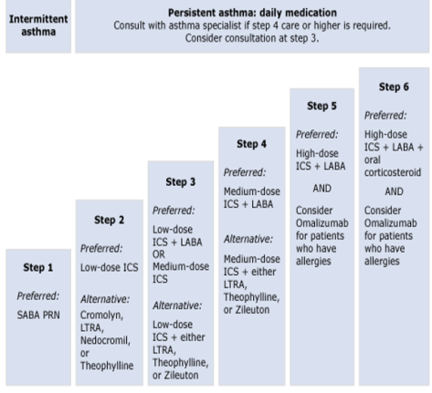
Treatment
-Airborne isolation (~50 % of patients with concomitant Pulmonary Tb)
-RIPE therapy (similar to pulmonary Tb)

AM Report 10/25/16: Bacterial Meningitis

Indications for CT before LP:

  • >60 years old
  • Immunocompromised state
  • History of CNS disease (mass lesion, stroke, focal infection)
  • Seizure within 1 week of presentation
  • Altered consciousness / focal neurological deficit

* Patients with suspected meningitis without the above findings are good candidates for LP without CT given their low risk for herniation.

CSF Studies: Normal Bacterial Meningitis Viral Meningitis Fungal/TB Meningitis
Opening Pressure (cm H20) <15 Normal/↑ Normal/↑
Protein (mg/dL) 15-45 Normal/↑
Glucose (mg/dL) 40-70 Normal
WBC Count (uL) 0-8 ↑↑ (>1000) ↑ (50-300)
WBC Differential Normal >80% PMNs >50% Lymphs, <20% PMNs 50-80% Lymphs

Common Signs/Symptoms:

  • Patient’s often present soon after onset of symptoms ~ 24 hours (range 1 hour – 14 days)

Classic Triad:

  • Fever (95-77% at presentation)
  • Nuchal Rigidity (94-83% at presentation)
  • Altered Mental Status (83-78% at presentation)
  • *Headache (94-79% at presentation)

*One review of 696 cases of community-acquired bacterial; only 44% had clinical triad, although almost all (95%) had at least 2 of 4 symptoms.

Laboratory Findings (besides CSF):

  • Routine blood work is often unrevealing, but leukocytosis can be present
  • Serum chemistry may reveal an AG metabolic acidosis or hyponatremia (30% in one study)
  • Blood cultures are often positive (50-90% of patients) and can help guide therapy

Common Pathogens:

1.jpg

  • Streptococcus pneumoniae (71%)
  • Neisseria meningitidis (12%)
  • Group B Streptococcus (7%)
  • Haemophilus influenza (6%)
  • Listeria moncytogenes (4%)

* Percentages based on CDC data of 1083 cases of bacterial meningitis from 2003-2007

Treatment for Suspected Bacterial Meningitis:

  • Empiric therapy should be directed at the most likely bacteria and must be started without delay

Typical empiric regimen:

  • 3rd generation cephalosporin (Ceftriaxone 2 g IV Q12H or Cefotaxime 2 g IV Q6H)
  • Vancomycin 15-20 mg/kg IV Q12H ~ goal trough 15-20 mcg/mL
  • Ampicillin in patients > 50 years old

Indications for Steroids:

  • Used in attempt to diminish the rate of neurological complications (seizures, hearing loss, cranial nerve deficits, etc.)
  • European studies showed a decreased mortality (7% versus 15% with placebo)
  • Only indicated for patients with suspected/confirmed S. pneumoniae
  • 15 minutes before administration of antimicrobial agents and continued for full course (0.15 mg/kg Q6H for 4 days)  in suspected/confirmed pneumococcal meningitis

AM report 10/26/16: Acute lymphoblastic leukemia

Epidemiology
Peak incidence ages 2-5, most common leukemia in children/teens but can occur in adults! (like our patient)

Timing

Sub-Acute

Signs & Symptoms

-60 % Hepatosplenomegaly
-50 % lymphadenopathy
-33 % musculoskeletal pain (bone pain)
-Symptoms of low hemoglobin/platelet (usually WBC<10k, platelets <1000)- eg: bruising, bleeding.
-Headache
-Can also see unilateral testicular enlargement and mediastinal mass (mimicking NHL)

Workup

Bone marrow biopsy showing >30 % lymphoblasts
-Chem 10, uric acid-HIGH risk of tumor lysis syndrome with ALL
-Flow cytometry- check if it is B cell (eg: CD10/CD19/CD20) or T cell (Cd2, 4,5,7,8), usually Tdt+, MPO- in ALL
-Philadelphia Chromosome (T:9:22)-If positive, associated with worse prognosis

Treatment

Induction-CALBG + tyrosine kinase inhibitor if + Philadelphia Chromosome
-Consolidation
-Maintenance
-CNS prophylaxis

See image below that shows  progenitors for lymphocytes which are from a common lymphoid progenitor while granulocytes, platelets, RBC, and mast cells originate from a common myeloid progenitor.

1280px-Hematopoiesis_simple.svg.png

Image by Mikael Häggström, from original by A. Rad – Image:Hematopoiesis (human) diagram.png by A. Rad, CC BY-SA 3.0, https://commons.wikimedia.org/w/index.php?curid=7351905

AM report 10/24/2016: Acute liver transplant rejection

Early acute cellular rejection (<90 days) differential diagnosis

1)Recurrent HBV/HCV virus- can be difficult to distinguish from cellular rejection
2)Functional cholestasis
3)Cyclosporine toxicity
4)Massive hemorrhage necrosis
5)INFECTIONS– CMV, EBV, HSV, HIV
6)Drug induced liver injury

Clinical presentation (nonspecific!)

-Fever
-Malaise
-Abdominal Pain
-Hepatosplenomegaly

Abnormal labs

-Elevated aminotransferases, bilirubin, alkaline phosphatase (suspect acute cellular rejection especially if elevated AST/ALT with rising bilirubin)

Workup (if you are worried about acute cellular rejection)

-Ultrasound liver with doppler- look at hepatic artery and portal vein flow as well as other abnormalities
-Check CMV, EBV, HCV/HBV, HIV
-Check drug levels (eg: cyclosporine, tacrolimus) to look for under-dosing or toxicity
-MRCP/ERCP if suspicion for choledocholithasis or other biliary tract abnormalities
Must do URGENT Liver Biopsy (trans-jugular vs. percutaneous) to make the diagnosis-                              Prefer trans-jugular if bleeding diathesis or large amount of ascites

Triad of biopsy results consistent with cellular rejection

-Mixed inflammatory infiltrate in portal triad
-Destruction/non-destructive non suppurative cholangitis involving the interlobular bile duct epithelium
Endothelilitis (inflammation within the endothelium)

Treatment: high dose steroids, Solumedrol 500-1000 mg daily for 1-3d with steroid taper (HOWEVER increased mortality with steroids or T cell depletion if due to HCV infection so important to make right diagnosis)

AM Report 10/20/16 – Diabetes Insipidus

Hypernatremia (>145 mEq/L):

Non-Renal Losses:
1) Excessive sweating/burns
2) Insensible respiratory tract losses
3) Diarrhea, vomiting, NG suctioning

Renal Losses:
1) Diuretics
2) Osmotic diuresis – hyperglycemia, urea, mannitol
3) Post-obstructive diuresis
4) Diuretic phase of ATN

Hypertonic Sodium Gain:
1) Salt intoxication (VERY RARE)
2) Hypertonic IV fluids
3) Primary hyperaldosteronism
4) Central DI
5) Nephrogenic DI

Diabetes Insipidus: passage of large volumes of dilute urine (> 3 L/d); primarily a problem with ADH.
Anterior Pituitary (FLAT PEG): FSH, LH, ACTH, TSH, Prolactin, Endorphins, GH
Posterior Pituitary: Oxytocin, ADH

Mechanism of ADH:
1) Membrane binds ADH
2) Receptor activates cAMP => secondary messenger
3) Insert aquaporin channels
4) Water is absorbed by osmosis

figure_19_10_labeled
Central DI: problem with ADH production
Etiology – idiopathic, neurosurgery, malignancy, infiltration (sarcoid, wegners, IgG4 disease, etc.), trauma, anorexia nervosa, familial

Nephrogenic DI: problem with ADH response
Etiology – Lithium, other medications (demeclocycine, ampho B, etc.), electrolytes (HyperCa, HypoK), UTI, amyloidosis, Sjogren syndrome, hereditary

Diagnosing DI:
HyperNa (>145) and low urine osm (<200) = DI possible; H20 deprivation test
Normal Na and urine osm > 600 = excludes DI

AM Report 10/18/16 – Asthma

Lung Volume loops:

1

Interpreting PFTs:
FVC – volume of air that can forcibly be blown out after full inspiration (L)
FEV1 – maximum volume of air that can forcibly blow out in the first second during the FVC (L)
FEV1/FVC – in healthy adults this should be >~75-80%

Restrictive Lung Disease: primarily due to decreased lung compliance (volume)

  • Examples: ILD, sacroidosis, neuromuscular disease, pulmonary fibrosis, silicosis
  • FEV1 and FVC are BOTH reduced proportionally and the FEV1/FVC value may be normal (or increased)
  • Flow volume curve is narrowed because of diminished lung volumes, but the shape is generally the same as normal

Obstructive Lung Disease: primarily due to increased airway resistance (flow)

  • Examples: ILD, sacroidosis, neuromuscular disease (AML), pulmonary fibrosis, silicosis
  • FEV1 and FVC are BOTH reduced proportionally and the FEV1/FVC value may be normal (or increased)
  • Flow volume curve is narrowed because of diminished lung volumes, but the shape is generally the same as normal

2.png
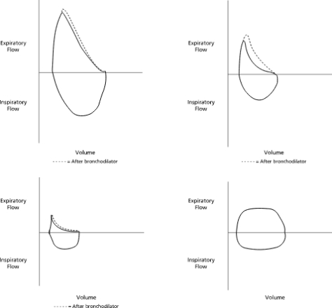
Top Right: Normal; Top Right: Asthma, Bottom Left: COPD, Bottom Right: Fixed Obstruction / Tracheal Stenosis

Asthma:

  • Common chronic respiratory condition characterized by reversible airway obstruction and bronchial hyper-responsiveness

Epidemiology: affects ~8% of US population; allergic asthma is strongly associated with personal/family history of allergies.  Higher prevalence and severity in people of lower income, children, and black populations

Pathogenesis: airway inflammation; chronic inflammation may result in epithelial damage, smooth muscle hypertrophy, airway fibrosis, and remodeling in some

Risk Factors: genetics, exposures to allergens, tobacco smoke, viruses

Symptoms/Clinical Evaluation: classically present with episodes of coughing, chest tightness, SOB, and wheezing

  • Consider other causes that may mimic asthma: COPD, vocal cord dysfunction, heart failure, bronchiectasis, ABPA, CF, mechanical obstruction, etc.
  • Obtain spirometry to assess for severity of airway obstruction and reversibility (FEV1 > 12% and 200 mL)
  • Chest radiographs are often normal

Asthma Syndromes:

  • Allergic Asthma – most common form in adults; family history is often positive
  • Cough-Variant Asthma – persistent/episodic cough without other symptoms
  • Exercise-Induced Bronchospasm
  • Occupational Asthma – related to workplace exposures (farmers, factory workers, hairdressers, etc.)
  • Aspirin-Sensitive Asthma – triad of severe persistent asthma, aspirin sensitivity, and hyperplastic eosinophilic sinusitis with nasal polyposis

Step Up/Down Treatment:

3

AM Report 10/19/16-Interstitial Lung Disease

If you see ground-glass opacities on CXR/CT scan and suspect patient has ILD, make sure you consider the differential diagnosis and you look for secondary etiologies!

Limited DDX: atypical PNA, fungal pneumonia (eg: Cocci, Blasto,Histo), CHF

Secondary etiologies (not an exhaustive list)

1)Environmental agents (Asbestos, Beryllium, Silicosis, Pneumoconiosis)
2)Drug induced (long list)-Include Amiodarone, Methotrexate, Bleomycin, Nitrofurantoin , NSAIDS
3)Connective Tissue Disease related-RA. SLE, Scleroderma, MCTD, Sjogrens
4)Radiation
5)Granulomatous diseases (eg: Sarcoid)
6)Vasculitis (eg: Churg Strauss)
7)LAM (lymphangioleiomyomatosis)-Usually young women of child-bearing age with recurrent PTX and chylous effusions, associated with Tuberous Sclerosis
8)Acute eosinophilic PNA-see >25 % eosinophils on BAL
9)Langerhans cell histiocytosis

If no etiology is known, then consider IDIOPATHIC INTERSTITIAL PNEUMONIA

This includes:

  1.  Idiopathic pulmonary fibrosis (IPF, also called Usual Interstitial PNA as the pathologic description)-WORST prognosis, does not respond to steroids, AND diagnosis can be made on HRCT if typical findings, especially if honey-combing (fibrosis)
  2. Desquamative interstitial PNA (DSIP)- strong association with smoking
  3. Respiratory bronchiolitis-ILD (RB-ILD)-strong association with smoking
  4. Non-Specific Interstitial PNA (NSIP)-can be due to HIV, CTD, hypersensitivity reactions or drug related
  5. Cryptogenic Organizing PNA (COP)-classically presents with patient with persistent symptoms despite multiple antibiotic treatments, responds to steroids
  6.  Acute interstitial PNA (AIP)-very rapid onset, produces ARDS like picture
  7. Lymphocytic interstitial PNA (LIP)-increased lymphocytes on BAL

S&S:

-Most commonly, progressive DOE and dry cough. Exam reveals crackles (“velcro-like”)

Workup:

-Look for secondary etiologies based on history and drug exposure.
-Check serology for auto-immune disease
-Bronchoscopy, BAL and trans-bronchial biopsy if necessary
-PFT to evaluate for restrictive disease
-VATS (video-associated thoracoscopic surgery) may be necessary if diagnosis is not clear (remember that IPF can sometimes be diagnosed via HRCT and other etiologies may be suggested on imaging)

Remember that IPF has the worst prognosis and does NOT respond to steroids so its important to make that distinction. 

AM Report 10/17/16-Vertigo

Step 1: Determine whether patient is having Vertigo (spinning sensation) or Pre-Syncopal sensation (about to faint/fall) as the differential and workup will be different

Vertigo: is it Central vs. Peripheral?
  Remember that ALL vertigo gets worse with movement! 

Peripheral Vertigo

-BPPV
-Vestibular neuritis (remember that labrynthitis refers to involvement of the inner ear while neuritis refers to involvement of the vestibular nerve)
-Meniere disease
-Ramsay Hunt Syndrome
-Perilymphatic fistula
-Otitis media

Central Vertigo

-Cerebellar infarction
-Cerebellopontine tumor
-Multiple Sclerosis
-Vestibular migraine
-Brainstem ischemia
-Vertebrobasilar insufficiency

How can you tell the difference between Central and Peripheral Vertigo? 

vertigo-worksheet

If someone has ongoing vertigo and nystagmus, The HiNTs exam can be used to differentiate  posterior circulation syndrome (brainstem or cerebellar stroke) vs. vestibular neuritis

1)Head impulse testing-“normal” test is POSITIVE meaning no saccade/correction on head rotation
2)Nystagmus-Nystagmus that changes direction or pure vertical/torsional nytagmus
3)Test of skew –Covering and uncovering each eye and uncovered eye demonstrates quick vertical gaze corrections

1/3 POSITIVE tests indicates patient may have a posterior circulation stroke. 

Check out this EMcrit video that has a blog and video on how to do the HiNTs exam!
See original study here in Stroke Journal-on this study, the maneuver was 100 % sensitive and 96 % specific for posterior circulation stroke

AM Report 10/12/2016 – Lympocytic Pleocytosis and IIH

Remember that there are different grades for Papilledema on the Frisen Scale – from 0 (none) to 5 (severe)

1

Remember the typical patient with IIH – female, obese, child-bearing age

Symptoms: headache (worse with valsalva / bending over), nausea (30%), visual loss (30-60%); diplopia (30%), neck stiffness, tinnitus, ataxia, dizziness

Signs: papilledema (100%), 6th nerve palsy (~10-20%) “false localizing sign”

Diagnosis of IIH:

  1. CSF opening pressure > 20 cm H20
  2. Normal CSF composition (possible exception of low protein)
  3. Signs / Symptoms of elevated ICP
  4. Normal radiographic imaging with exception of slit ventricles and/or empty sella

Be able to interpret CSF studies:

3

Remember that there is broad differential for lymphocytic pleocytosis of CSF fluid.

2

AM Report 10/4/2016 – UGIB / H. Pylori

Remember the use of likelihood ratios in your initial evaluation of a suspected UGIB!
Suspected UGIB:
– patient reported melena (LR 5.1 – 5.9)
– melena on exam (LR 25)
– blood/coffee ground on NG lavage (LR 9.6)
– BUN/Cr > 30 (LR 7.5)

Factors associated with severe bleeding:
– red blood on NG lavage (LR 3.1)
– tachycardia (LR 4.9)
– Hgb < 8.0 (LR 4.5 – 6.2)

Physical Examination:
Signs of hypovolemia – mild to moderate resting tachycardia
15% blood loss – orthostatic hypotension
40% blood loss – supine hypotension

Medications:
Acid Suppression: when PUD suspected use IV PPI
– decrease hospital stay, rebleeding rate, and need for transfusions
– also shown to promote hemostasis with lesions other than ulcers
Prokinetics: erythromycin/metoclopramide – used 30 minutes prior to EGD for improved visualization
Somatostatin Analogs: octreotide – use in suspected variceal bleeds to lower portal pressure through splenic vasoconstriction
Antibiotics: for cirrhotic patients; studies show prophylactic antibiotics reduce complications and mortality

Risk Scores:
Rockall Score: requires endoscopy for calculation

Blatchford Score: used at presentation, range from 0-23 (higher score worse). Utilizes BUN, SBP, pulse, presence of melena, syncope, hepatic disease, and/or cardiac failure to generate number.

Testing for H. Pylori:
Endoscopic biopsy: invasive and expensive; requires EGD for diagnosis
Urea Breath Test: non-invasive and inexpensive; sensitivity 88-95%, specificity 95-100%
Serology (IgG): non-invasive; sensitivity 90-100%; specificity 76-96% – not recommended in low prevalence populations (i.e. US resident without travel); conversion of positive serology to negative suggests cure.
Stool Antigen: non-invasive and inexpensive; sensitivity 94%, specificity 86% – good for documenting eradication; false negative if patient on PPI – MUST STOP 2 WEEKS PRIOR TO TESTING!

Treatment for H. Pylori:
Triple Therapy: PPI, amoxicillin (or metronidazole for penicillin allergy), clarithromycin
Quadruple Therapy: PPI/Rantidine, Bismuth Subsalicylate, Metronidazole, Tetracycline (or Doscycline)