Category Archives: Morning Report

AM report 01/04/17: Orbital Cellulitis, Angle Closure Glaucoma, Endophtalmitis, and Hypermucoid Klebsiella

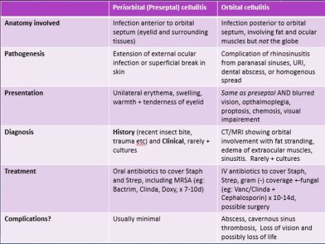
Distinguishing Pre-septal cellulitis (peri-orbital) and Orbital cellulitis  

preseptal vs orbital.JPG

Acute angle closure glaucoma

Clinical presentation 

Elevated IOP presenting as vision loss, acute SEVERE eye PAIN, frontal headache, nausea/vomiting with exam commonly showing corneal edema or cloudiness and mid-dilated pupil reacting poorly to light

                                             This is an eye emergency!

Treatment

You either want to DECREASE aqueous humor production or INCREASE OUTFLOW by constricting the eye. NEVER dilate the eye as it can decrease outflow and increase IOP

-Beta blockers, Acetazolamide, Mannitol (decrease production of aqueous humor)
-Alpha agonists, Pilocarpine (increase outflow)

Hypermucoid variant of KIebsiella Pneumoniae

Hypermucoviscosity virulence factor 
-Diagnosed in lab via STRING test and positive result is >5 mm viscous string from colony on agar plate
-Important to distinguish from non-virulent form as associated with metastatic involvement (endophthalmitis, meningitis, brain abscess but can go anywhere in the body). -Always check with the lab if not reported!

string test.jpg
KLA (Klebsiella Liver Abscess)-associated with Hypermucoid Klebsiella

-PRIMARY liver abscess in absence of hepatobiliary disease, diagnosed via ultrasound or CT scan
Risk factors include DMII, prior antibiotic use, and being of Asian descent (commonly from Taiwan) 
-Commonly presents with Fever (93 %), RUQ pain, N/V, leukocytosis, elevated alkaline phosphatase (78 %) and elevated AST/ALT (68 %)

Treatment of Hypermucoid Klebsiella

-Empiric treatment with Cephalosporin (commonly CTX) and Flagyl for empiric therapy, adjust based on sensitivities
Drainage of liver abscess if present (will not improve with antibiotics alone!)


AM report 12/27/16: ASA overdose

Management of ANY patient with suspected toxic ingestion:

-ABCs (Airway, Breathing, Circulation)
Call Poison Control! (1800 222-1222)
-Can patient get Activated Charcoal? (usually only within 1 hour of ingestion)
-Check Utox, Salicylate screen, acetaminophen screen, +- alcohol and volatile screen if suspected.
                                    You don’t want to miss a potential co-ingestion! 

ASA overdose

-Remember that ASA can be found in other compounds like topical salicyclic acid, herbal medications, bismuth subsalicyclate (part of Pepto-Bismol), and Oil of Wintergreen so don’t forget about those topical medications!
-Most sensitive vital sign abnormality in early ASA overdose is tachypnea with hyperventilation. 
-Classic acid/base abnormality is anion gap metabolic acidosis with respiratory alkalosis (see below)

How does ASA work? 

-Inhibits the COX-2 pathway, decreasing synthesis of prostaglandins, decreasing inflammation, and leading to platelet dysfunction
-Stimulates the chemoreceptor trigger zone to cause Nausea and Vomiting
-Activates the respiratory center in the medulla leading to hyperventilation and respiratory alkalosis
-Interferes with the Krebs cycle and decouples oxidative phosphorylation, leading to metabolic acidosis

Making the diagnosis

-Check Salicylate level and if elevated, check levels every two hours until two consecutive levels decrease from peak , value is less <40, and patient is asymptomatic.
-Check Serum CreatinineASA is renally excreted so significant renal failure will change management. 
-Check Potassium level-need to treat hypokalemia aggressively (see below)

Other labs that can support diagnosis but not required

-Coagulation studies (large overdose can cause hepatotoxicity and interfere with Vit K metabolism)
-Lactate (can be elevated due to uncoupling of oxidative phosphorylation)
-Anion Gap (Elevated due to ASA toxicity)
-ABG/VBG (Evaluate for resp.alkalosis/metabolic acidosis)
-CXR if concern for pulmonary edema (potential complication of ASA overdose)

TREATMENT of ASA overdose

-ABCs
-Activated Charcoal if <1 hour from ingestion
AVOID intubation if possible (remember that these patients have high minute ventilation (RR x TV) due to ASA effect on the medulla and this can be hard to reproduce on the ventilator without causing significant auto-peep)
-Volume resuscitation (be careful of pulmonary edema/cerebral edema)
-Alkalinize urine with sodium bicarbonate
Sodium Bicarbonate 1-2 meQ/kg IV bolus followed by 100-150 meQ/D5W and titrated to maintain urine pH of 7.5 to 8.0 and continued until salicyclate level <30. It is OK to continue sodium bicarbonate even with alkalemia as long as pH<7.60. Alkalinizing the urine keeps ASA in the non-acidic form (Sal-) , thus avoiding a lot of the complications of ASA overdose.  
-Treat hypokalemia aggressively to maintain alkalinization (see picture below). If hypokalemia is not corrected, the body will reabsorb potassium and acidify the urine, which is the opposite of what we want

-Consider giving glucose for neuro-glycopenic symptoms (controversial but patient can have neuro-glycopenic symptoms due to low CNS glucose even with a normal serum glucose)
-Call renal early if patient may need HEMODIALYSIS  (indications include AMS, cerebral edema/pulmonary edema, fluid overload, acute or chronic kidney injury, severe acidemia, or clinical deterioration despite aggressive care)

AM report 12/21/16: SJS/TEN

Warning symptoms for severe cutaneous reactions 

If any of these are present, consider life-threatening dermatological emergencies.

-Mucous membrane involvement!
-High fever (>38.5)
-Blisters
-Facial edema or erythema
-Lymphadenopathy

DRESS syndrome (Drug reaction with eosinophilia and systemic symptoms)

Commonly implicated drugs

-Antiepileptics
-Sulfa antibiotics (eg: Bactrim)
-Xanthine oxidase inhibitor (eg: Allopurinol)
-Sulfsalazine
-Abacavir (RTI)

-Remember that certain HLA haplotypes (eg: HLA B*58:01) are at higher risk for allopurinol related SJS or DRESS)
-Patients with HIV at >100x risk of developing DRESS compared to the general population

Timing

LONG latency period (2-8 weeks) compared to SJS/TEN

S&S 

Fever (85 %), Rash (75 %), Facial Erythema and Edema, Generalized lymphadenopathy, abnormal LFT.
Peripheral eosinophilia only seen in ~50 % and NOT required to make diagnosis

TEN/SJS 

-Extensive necrosis and detachment of the epidermis with >90 % having mucous membrane involvement, usually at two distinct sites (eg: oral, ocular, genital)
-<10 % BSA skin detachment is SJS while >30 % BSA skin detachment is TEN with SJS-TEN overlap in between

Most common implicated drugs

Same as DRESS syndrome but also includes NSAIDS!

Most common infectious trigger

Mycoplasma pneumoniae infection (more commonly in kids)

Timing

Usually 1-3 weeks after starting causative drug (note, more acute than DRESS)

S&S

-Dusky atypical targetoid skin lesions with at least two mucosal surfaces involved
-Rash is often painful, and diffusely involved.
(+) Nikolsky sign but also seen in SSSS and Pemphigus Vulgaris so not specific


Treatment

-WITHDRAWAL of culprit drug!
-Supportive care and management of complications
–Managing bacterial infections (MRSA, pseudomonas)
–Fluid and nutrition
–Wound care
–Pain control (remember that the rash can be very painful)
-Systemic steroids and IVIG controversial and NOT routinely recommmended

Management of Ocular symptoms

-Topical steroids, antibiotics
-Amniotic membrane transplanation (eg: Prokera ring) if extensive conjunctival involvement or pseudo-membrane formation.

prokera-ring
Prokera ring used as type of AMT for severe ocular inflammation in SJS/TEN

See article here by the NEJM for an excellent review of exanthematous drug eruptions 

AM Report 12/13/16: Cholangitis

Remember that Acute Cholangitis is a medical emergency that must be recognized and treated emergently!

CBD diameter

Remember that CBD diameter can be a clue to biliary obstruction
-95 % of normal patients have a CBD < 6 mm
-Can increase with age, usually upper limit corresponds to decade of life (70 year old upper limit of ~ 7 mm
-Can see CBD up to 10 mm if post-cholecystectomy!

common-bile-duct

If you suspect choledocholithiasis, the ASGE guidelines can help you decide whether you should do an MRCP or an ERCP

-Remember if you have any very strong predictors (see below), you should go directly to an ERCP!

Very strong predictors of choledocholithiasis

-CBD stone seen on trans abdominal ultrasound
-Bilirubin>4 mg/dl
-Clinical ascending cholangitis

acge-choledocholithiasis

riskfactorscholangitis.JPG
MRCP vs. ERCP

MRCP=diagnostic modality, NO contrast given, excellent sensitivity to evaluate for choledocholithasisis (90-100%)
ERCP=diagnostic and therapeutic modality, invasive (have to be in prone position!), risk of post-ERCP pancreatitis

Four main etiologies of biliary obstruction

Choledocholithasis (MCC)
-Biliary strictures
-Malignancies
-Biliary stent complication (eg: migration)

Etiology
GUT FLORA

-E.Coli (most common), Klebsiella, Enterobacter, Enterococcus, and Anaerobes (less common alone)

Key Clinical manifestations and lab findings for cholangitis and which ones are most common

-Fever (95 %)
-RUQ pain (90 %)
-Jaundice (80 %)
Charcot’s Triad=>Fever, RUQ, Jaundice
(+) Hypotension, Confusion, Leukocytosis, Cholestatic jaundice.

Treatment for empiric coverage for cholangitis

-Beta Lacam/Beta Lactamase inhibitor
-Flouroquinolone + Flagyl
-Carbapenem

Management for cholangitis in addition to antibiotics

-ERCP for source control, treatment of sepsis, and cholecystectomy

AM Report 12/20/2016: Hypercalcemia

Calcium correction:

  • Corrected Calcium = (0.8 (normal albumin – patient’s albumin)) + Ca2+
  • Check an ionized (free) calcium

Interpreting the degree of hypercalcemia:

  • Normal (8-10 mg/dL)
  • Mild hypercalcemia (10-12 mg/dL)
  • Moderate hypercalcemia (12-14 mg/dL)
  • Hypercalcemic crisis (>14 mg/dL)

1

Remember the clinical manifestations of hypercalcemia: “stones, bones, abdominal groans, thrones, and psychiatric overtones”

2

ECG in hypercalcemia:

  • the main ECG abnormality with hypercalcemia is shortening of the QT interval
  • in severe hypercalcemia, Osborn waves (J waves) may be seen

3 Main hormones involved in calcium homeostasis:

picture8

Etiologies of Hypercalcemia:

3

Pathophysiology/ Causes Calcium Level PTH Level 1, 25-vitamin D Phosphorous
Primary Hyperparathyroidism Overproduction of PTH, 85% due to single adenoma
Secondary Hyperparathyroidism Overproduction of PTH, commonly due to chronic renal failure ↔ or ↓ ↔ or ↑
Tertiary Hyperparathyroidism Overproduction of PTH, usually by autonomous hypersecretion of PTH ↑↑

4

Treatment of Hypercalcemia:

  • Any symptomatic patient with a calcium level > 12 mg/dL
  • Any patient with calcium level > 14 mg/dL

Treatment options:

  • IVF (NS) – enhances filtration/excretion of Ca2+; tailored towards urine output ~ 200 mL/hr
  • Loop Diuretics (Furosemide) – inhibits calcium reabsorption in the distal tubule; only use one volume status restored
  • Bisphosphonate – inhibits osteoclast action/bone reabsorption; indicated in hypercalcemia of malignancy; avoid in renal failure
  • Calcitonin – inhibits bone resorption and promotes Ca2+ excretion; recommended for severe cases after IV hydration
  • Glucocorticoids – inhibits vitamin D conversion to calcitriol; used for vitamin D intoxication, hematologic malignancies, and granulomatous disease
  • Dialysis – used for cases of resistant, life-threatening hypercalcemia

AM Report 12/14/2016: Nephrotic Syndrome

Define:
Nephrotic Range Proteinuria:  >3g/24 hours without other findings
Nephrotic Syndrome:
– Proteinuria > 3.5 g/24 hours (protein/creatinine > 3.5 mg/mg)
– Hypoalbuminemia
– Clinical evidence of edema

Secondary Causes of Nephrotic Syndrome:
picture1

Primary Nephrotic Syndrome:
Minimal Change Disease:

Epidemiology:
– Children < 10 years old; can be primary or secondary
– 10% of cases in adults

Clinical Features:
– Sudden onset of edema
– Thrombotic episodes more common in adults
– AKI may be seen in 20% at presentation

Diagnosis:
– Renal biopsy

MCD on light microscopy:
– Appears essentially normal (hence the name minimal change!); tubules may show lipid accumulation
picture2
MCD on electron microscopy:
– Characteristic fusion and effacement of podocyte foot processes
picture3

Focal Segmental Glomeruloscerlosis (FSGS):

Epidemiology:
– most common cause of primary nephrotic syndrome in the US
– can be primary, familial, or secondary
– African-Americans more common, but increasing incidence in all races

Clinical Manifestations:
– Asymptomatic proteinuria up to nephrotic syndrome ~ 2/3 at presentation!
– Hypertension usually seen in 30-50%
– Decreased GFR at presentation 20-30%

Diagnosis:
– Renal biopsy

FSGS on light microscopy: scarring or sclerosis involving some (focal) glomeruli, which are affected only in a portion of the glomerular capillary bundle (segmental)
picture4
Collapsing FSGS – variant associated with HIV
picture5

Membranous Nephropathy (MN):

Epidemiology:
– most common in adults (>60 years old) and Caucasian
– can be primary (immune complex disease) or secondary (infection, autoimmune, cancer, drugs)

Potential complications:
– thrombotic disease – especially renal vein thrombosis

Diagnosis:
– renal biopsy
– PLA2R antibodies found in 75% of cases

Light microscopy for MN: diffuse thickening of the glomerular capillary wall
picture6

Immunofluorescence microscopy: diffuse, granular IgG deposition along capillary walls
picture7

Treatment:
– management is limited by a lack of clear evidence-based guidelines

General treatment:
– restrict dietary sodium to < 2 g/day
– restrict fluid intake to < 1.5 mL/day

Loop diuretics can be ineffective given that they are protein-bound and serum protein levels are reduced
Can add a thiazide diuretic and/or administer IV albumin bolus to improve diuresis

ACEi/ARBs – typicially used to reduce proteinuria although degree of benefit is unproven and evidence supporting routine use is conflicting
BP goal 130/80
Recent Cochrane review found no evidence to support the use of lipid-lowering agents in NS patients
Typically improves with resolution of disease
Corticosteroids are often used despite an absence of supporting evidence
Recent Cochrane review showed that combining alkylating agent (cyclophosphamide) with a corticosteroid has some short and long term benefits for MN
One exception – NS due to SLE – highly effective and supported by multiple studies


AM Report 12/12/16: Diverticulitis

Define:
Diverticulosis:
presence of diverticula ~ sac-like protrusion of colonic wall.
Diverticulitis:
inflammation of the diverticula

Epidemiology:
5-10% of patients > 45 years old
80% of patients > 85 years old

Risk Factors (formation of diverticulosis):
Lack of fiber / Western Diet (red meats/fats)
Decreased colonic motility
Decreased colonic wall resistance
Genetic susceptibility
NSAIDs/Aspirin
Age
Obesity
Smoking
Lack of exercise

DDx – Features:
Bowel Obstruction – abdominal pain, nausea/vomiting, abnormal imaging
Colorectal CA – weight loss, anemia, GI bleeding
Gastroenteritis – abdominal pain, nausea/vomiting
IBD – diarrhea, weight loss, rectal bleeding, mucus in stool
IBS – abdominal spasms relieved with defication
Ischemic colitis – abdominal pain out of portion to exam, arterial disease
Nephrolithasis – flank pain, hematuria, stone on imaging
Pancreatits – epigastric pain, nausea/vomiting

Likelihood Ratios for Acute Diverticulitis:
1

LLQ Imaging:

2

Accuracy of CT Finding for Diagnosing Acute Diverticulitis:

3

Treatment:
Inpatient ~ 6%
Any complicated case (abscess, obstruction, perforation, fistula)
Any uncomplicated with 1 (or more)
– Immunosuppression – Comorbidities
– Fever > 39 C – No PO intake
– Significant leukocytosis – Non-compliance
– Severe pain – Failed outpatient
– Old age

Outpatient ~ antibiotics 7-10 days
1) Cipro + Flagyl
2) Bactrim + Flagyl
3) Augmentin
4) Moxifloxacin

AM Report 12/07/16 TINU

Remember that red eye with WARNING symptoms like loss of visual acuity, ophthalmoplegia, pupillary involvement, photophobia, diplopia,  associated systemic symptoms 

Signs and Symptoms of Uveitis 

Exam: can see ciliary flush (circumcorneal injection), hypopyon (not specific, can be seen in many other eye conditions), redness at the limbus
Discharge: MINIMAL
Pupil: often constricted
Ocular pain: moderate to severe, deep aching pain
Vision: mild-moderately reduced
Cornea: often hazy in appearance
Treatment: Steroids

Etiology of Uveitis 

IDIOPATHIC (most common)
-Trauma
-Infectious (HSV, Zoster, Toxo, Syphillis, TUBERCULOSIS, West-Nile, cat-scratch disease)
-Sarcoidosis
-Vasculitis
-Spondyloarthropathies
-SLE/Sjogrens

AIN (Acute interstitial nephritis) 
Keep AIN in your differential for new onset AKI, especially if on antibiotics or NSAIDS!

Etiologies of AIN

1)Drugs (LOTS of them, commonly cephalosporins, NSAIDS, but includes famotidine, omeprazole)
2)Infections (Leptospira, legionella, staph, strep, etc.)
3)Sarcoidosis
4)Sjogrens syndrome
5)TINU
Many other immune and neoplastic disorders!

TINU (Tubulointerstitial nephritis and uveitis) 

RARE disease, ~250 cases since 1976
-Pathogenesis thought to be due to auto-immune T cell mediated with common modified C-reactive protein in both the uvea and renal tubular cells
Manifests as Uveitis and Interstitial Nephritis, however must RULE out other etiologies of uveitis and AIN, especially Sarcoidosis and Sjogren’s as can look very similar.
Treatment: Steroids, usually 1 mg/kg/day  (40-60 kg) for 3-6 months


AM Report 12/01/2016: Pulmonary HTN

Pulmonary HTN: Defined as mPAP>25 mmHg on right heart cath

WHO class Group 1 to Group 5

Group 1 (Pulmonary arterial hypertension)
-Idiopathic
-Heritable
-Drug and Toxin induced (toxic rapeseed oil, fenfluramine, likely methamphetamine, cocaine)
-Connective tissue Disease
HIV
Portopulmonary HTN
-Congenital heart disease
-Schistosomiasis
-Chronic hemolytic anemia (eg: SCD)
Group 2 (due to left heart disease)

-Systolic/diastolic dysfunction
-Valvular disease (aortic/mitral)

Group 3 (Due to lung disease or hypoxia)
-COPD -ILD, Pulmonary fibrosis
-Chronic exposure to high altitude

Group 4 (Chronic thromboembolic pulmonary  hypertension) 
-Recurrent PE

Group 5 (multifactorial)

-Hematologic disorders (myeloproliferative disorders)
-Systemic disorders (sarcoidosis, LAM, vasculitis)
-Metabolic disorders (glycogen storage diseases)

Vasoreactivity test

-Done with right heart cath to see if patient will respond to CCB.
-Most commonly iNO given during right heart cath and positive test is if mean pulmonary artery pressure decreases by at least 10 to a value less than 40.

Treatment

Group 1 PAH- No effective primary therapy, usually need advanced therapy (must confirm diagnosis with right heart cath)

a)Prostacyclins
b)Endothelin receptor antagonists
c)PDE5 inhibitors (eg: sildenafil)
d)iNO
e)CCB (if positive vasoreactivity test)

Group 2 PH- treat left heart disease
Group 3 PH: OXYGEN + CPAP (if due to OSA)
Group 4: Anticoagulation and Surgical Thromboendarterectomy
Group 5: varies based on etiology.


AM Report 12/6/2016: Toxic Megacolon

Risk factors for developing C. Diff associated diarrhea:

1

The pathogenesis of C. diff infection:

2

Treatment for C. diff associated diarrhea:

3

Two criteria necessary to distinguish toxic megacolon from other types of colonic distention:

  • Total or segmental non-obstructive colonic dilation
  • Systemic toxicity (not present in other forms of megacolon)

Causes of toxic megacolon:

  • Inflammatory
  • Infectious
  • Other (volvulus, obstructive colon cancer, etc.)

picture4

Diagnosis of toxic megacolon:

1) Radiographic evidence

2) Main Criteria (need 3):

  • Fever
  • Tachycardia
  • Leukocytosis
  • Anemia

3) Minor Criteria (need 1):

  • Dehydration
  • Altered level of consiousness
  • Electrolyte imbalances
  • Hypotension

* The overall clinical condition is more important than the absolute width of the dilated segment

Treatment of toxic megacolon:

  • Initial therapy is medical, which is successful in preventing surgery in up to 50% of patients
  • Specific medical therapies are aimed at the underlying etiology (i.e. steroids in IBD)

In patient’s with toxic megacolon due to C. diff colitis:

  • Stop any offending antibiotics
  • Start C. diff treatment: PO vancomycin 500 mg QID and IV metronidazole 500 mg Q8H
  • Colectomy in patient’s who fail to improve within 48 – 72 hours or show evidence localized perforation