Category Archives: Morning Report

AM Report 11/7/16: Primary Polydipsia

Remember to check plasma osmolality first!

1

Next evaluate volume status!

2.png

If a patient is Euvolemic – evaluate urine osmolality and sodium

3.png

Practical approach to hyponatremia:

1.What is the normal/abnormal physiology?

2.Is it “true” hypo-osmolar hyponatremia?

  • Measure plasma osmolality

3.What is the volume and ADH status?

  • Urine Na and Urine Osmolality

4.Is it symptomatic?

5.What is my treatment?

  • Depends on volume status, ADH status, and +/- symptoms
  • Determine goal of correction
  • Avoid overcorrection

AM report 11/01/16 HOCM

Before considering HOCM, think about secondary causes of hypertrophic cardiomyopathy

-HTN
-Aortic Stenosis

Disease manifestations of Hypertrophic Cardiomyopathy

-LV outflow tract obstruction due to thickened septum (see picture below)
-Diastolic dysfunction
-Myocardial ischemia
-Mitral regurgitation

hocm.jpg


Clinical symptoms of HOCM

–Heart Failure (90 % present with DOE), due to LVOT, diastolic dysfunction, and MR
–Chest pain
–Arrhythmias-SVT and VT-risk of sudden cardiac death!
–Syncope

How to make the diagnosis? 

Start with an EKG!
-Look for severe LVH and biatrial enlargement, can suggest HOCM. Due to asymmetrical hypertrophy, can see deep Q waves in the lateral and inferior leads.

What do you see in this EKG of our patient who was found to have a new diagnosis of HOCM? See below for the answer

ekgprint
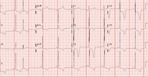
Severe LVH. Biatrial enlargement, deep T wave inversions throughout

Echocardiogram-see unexplained LV wall thickness >15 m (makes diagnosis), commonly asymmetric septal hypertrophy. Can also see SAM (systolic anterior motion of mitral valve, worsening obstruction), and LVOT.

Further workup

Ambulatory EKG monitoring:  24-48hr Holter to evaluate for arrhythmias
Exercise stress test-Risk stratification, GXT preferred. Evaluate degree of LVOT if not seen on echocardiogram
Cardiac MRI-risk stratification due to better visualization than TTE
-Note that genetic testing is NOT routinely done due to 11 genes, and >1500 mutations responsible, but can be considered as Autosomal Dominant inheritance.
Cardiac catheterization not routinely done unless high suspicion of ischemia

Medical treatment

-LOWER heart rate and DECREASE inotropy
-3 classes of medications commonly used
BETA BLOCKERS (first line)
Nondihydropyridine CCB (commonly Verapamil)
Disopyramide

-Classes of medications to AVOID

Diuretics-reduction in preload can worsen LVOT (don’t let them become hypovolemic!)
Vasodilators– decreased peripheral resistance can increase LVOT obstruction
–Positive Inotropes (eg: Dobutamine)

Non-pharmacologic therapies

1)Activity restriction-avoid high intensity exercise, need to be counseled on this as HOCM is the number one cause of sudden cardiac death in those under age 30!
2)Alcohol septal ablation
3)Surgical myomectomy

Who needs to get an AICD?

Only if you are HIGH risk based on risk stratification


-Highest risk if history of prior sudden cardiac arrest or sustained ventricular arrhythmias

Other high risk features include:
-Family History  of SCD
-Syncope
-NSVT
-Massive LVH>30 mm

Not everyone needs an AICD, eg: if asymptomatic and negative family history!

AM Report 11/3/16: TTP

Thrombocytopenia is due to three main problems: DECREASED PRODUCTION (bone marrow), INCREASED DESTRUCTION, SPLENIC SEQUESTRATION:

Decreased Production:

  • Aplastic anemia
  • Hematologic malignancies
  • Myelodysplastic syndrome
  • Chronic alcoholism
  • Infiltrative process (myelofibrosis, infection, etc.)

Increased Production:

  • Immune disorders
    • ITP
    • Drug induced (heparin, sulfonamides, thiazides, etc.)
  • DIC
  • TTP
  • HUS
  • Sepsis
  • AIDs/Viral infection
  • Liver Failure

Splenic Sequestration

Causes of MAHA:

  • DIC
  • TTP/HUS
  • Malignant hypertension
  • Mechanical valves
  • HELLP/Pre-eclampsia
  • Scleroderma renal crisis

36792tn

TTP Pentad: ONLY PRESENT IN 1/3 OF PATIENTS

  • Thrombocytopenia
  • MAHA (schistocytes on smear)
  • Acute renal insufficiency
  • Fevers
  • Encephalopathy

*Only need thrombocytopenia and MAHA to diagnose

Triad more common:

  • Elevated LDH (tissue hypoxia/injury)
  • Schistocytes
  • Thrombocytopenia

ADMTS 13 Testing

  • Protease that cleaves vWF multimers; deficiency leads to increase vWF leading to platelet aggregation (thrombocytopenia) and disruption/breaking of RBCs (MAHA)

Level of activity:

  • >50% ~ unlikely TTP
  • <10% ~ suggests TTP

*presence of ADAMTS 13 inhibitor suggests acquired TTP; negative suggests hereditary.

Treatment:

  • Plasma exchange
  • If unavailable – infuse FFP
  • Other options: steroids, splenectomy, rituximab, vincristine

* AVOID PLATELET TRANSFUSION AT ALL COSTS!

HUS:

  • Usually occurs in children ~ associated with E. coli 0157:H7
  • Most patients don’t have wide-spread symptoms; besides heme changes, renal involvement is the rule
  • In adults => progressive renal failure => ESRD
  • Supportive treatment in children, assume TTP in adults

AM Report 11/2/16: Vitamin B12 Deficiency

Pain and Temperature sensation are carried by the spinothalamic tract.  They decussate immediately in the spinal cord.

Vibration and Proprioceoption are carried by the posterior columns.  They ascend on the ipsilateral side of the spinal cord and decussate in the medulla (brain stem).

picture1

Interpretation of Vitamin B12 Levels:

  • > 300 pg/mL = NORMAL RESULT; vitamin B12 deficiency unlikely (sensitivity ~90%)
  • 200 – 300 pg/mL = BORDERLINE RESULT; vitamin deficiency possible
  • < 200 pg/mL = LOW RESULT; consistent with vitamin B12 deficiency (specificity 95-100%)

* If patient has BORDERLINE RESULT, but a high degree of suspicion for B12 or folate deficiency – check methylmalonic acid and/or homocysteine.

Interpretation of MMA and HC Levels:

  • Vitamin B12 deficiency – BOTH ARE ELEVATED
  • Folate deficiency – Homocystein is ELELVATED, while MMA is NORMAL

picture2

*Folate does not participate in MMA metabolism, but B12 is needed for both.

Clinical Manifestations of B12 Deficiency:

Hematologic

  • Macrocytic anemia (megaloblasic if folate also low)
  • Pancytopenia

Neurologic

  • Paresthesias
  • Peripheral neuropathy
  • Subactue combined degeneration (demyelination of dorsal columns and corticospinal tract)

Psychiatric

  • Irritability, personality changes
  • Mild memory impairment => dementia
  • Depression
  • Psychosis

Cardiovascular

  • Possible increased risk of MI (due to elevated homocysteine)

Vitamin B12 Absorption:

Step 1: acidic stomach breaks down B12 from food.
Step 2: intrinsic factor (IF), released from parietal cells, binds B12 in the duodenum.
Step 3: IF-B12 complex is absorbed by the terminal ileum.
Step 4: Once absorbed, B12 binds to transcobalamin II and is transported throughout the body.

Picture4.png

Pernicious Anemia:

  • Type of autoimmune disease that leads to destruction of gastric parietal cells.
  • Destruction of these cells leads to decreased production of intrinsic factor, and therefore, limits B12 absorption.

Testing for Pernicious Anemia:

  • Parietal cell antibodies (85-90% sensitive) – but non-specific (occurs in other autoimmune states)
  • Intrinsic factor antibody (50% sensitive) – but far more specific for diagnosis of pernicious anemia
  • Schilling Test: not routinely used given superior sensitivity of HC and MMA and difficulty with radiolabeled reagents
    • Stage 1: radiolabeled B12 is administered orally and 24-hour urine is measure for B12 excretion
    • Stage 2: (performed if stage 1 is abnormal) radiolabeled instrinsic factor and B12 are administered orally and another 24-hour urine is collected.

Treatment of B12 Deficiency:

  • Oral and parental B12 repletion are both potential options for treatment
  • Studies have shown that high dose oral B12 is as effective as parenteral therapy through a secondary pathway that does not require IF or a functioning terminal ileum
  • Treat B12 before folic acid to avoid precipitating subacute combined degeneration (giving folate first will turn the remaining B12 into methylcobalamin)

AM report 10/31/16 Pleural Tb

Most common etiologies for BLOODY pleural effusion

-Trauma
-Malignancy
-Pulmonary infarction
-Post-cardiac injury

Differential for Lymphocytic Exudative pleural effusion

TUBERCULOSIS
-Malignancy (Lung> BRCA, lymphoma, ovarian/gastric)
-Sarcoid
-Rheumatoid pleurisy
-Chylothorax

Workup of Suspected Pleural Tb

-Exudative lymphocytic pleural effusion, <10 % eosinophils
-High LDH (usually >500)
-AFB stain and culture (only positive 20-30 % of the time)
-Adenosine deaminase- HIGH sensitivity (if <40) and HIGH specificity (if >60), however depends on laboratory validity
-Pleural biopsy (positive 60-90 %)- can be done either via thoracoscopy or percutaneous needle biopsy)

Treatment
-Airborne isolation (~50 % of patients with concomitant Pulmonary Tb)
-RIPE therapy (similar to pulmonary Tb)

AM Report 10/25/16: Bacterial Meningitis

Indications for CT before LP:

  • >60 years old
  • Immunocompromised state
  • History of CNS disease (mass lesion, stroke, focal infection)
  • Seizure within 1 week of presentation
  • Altered consciousness / focal neurological deficit

* Patients with suspected meningitis without the above findings are good candidates for LP without CT given their low risk for herniation.

CSF Studies: Normal Bacterial Meningitis Viral Meningitis Fungal/TB Meningitis
Opening Pressure (cm H20) <15 Normal/↑ Normal/↑
Protein (mg/dL) 15-45 Normal/↑
Glucose (mg/dL) 40-70 Normal
WBC Count (uL) 0-8 ↑↑ (>1000) ↑ (50-300)
WBC Differential Normal >80% PMNs >50% Lymphs, <20% PMNs 50-80% Lymphs

Common Signs/Symptoms:

  • Patient’s often present soon after onset of symptoms ~ 24 hours (range 1 hour – 14 days)

Classic Triad:

  • Fever (95-77% at presentation)
  • Nuchal Rigidity (94-83% at presentation)
  • Altered Mental Status (83-78% at presentation)
  • *Headache (94-79% at presentation)

*One review of 696 cases of community-acquired bacterial; only 44% had clinical triad, although almost all (95%) had at least 2 of 4 symptoms.

Laboratory Findings (besides CSF):

  • Routine blood work is often unrevealing, but leukocytosis can be present
  • Serum chemistry may reveal an AG metabolic acidosis or hyponatremia (30% in one study)
  • Blood cultures are often positive (50-90% of patients) and can help guide therapy

Common Pathogens:

1.jpg

  • Streptococcus pneumoniae (71%)
  • Neisseria meningitidis (12%)
  • Group B Streptococcus (7%)
  • Haemophilus influenza (6%)
  • Listeria moncytogenes (4%)

* Percentages based on CDC data of 1083 cases of bacterial meningitis from 2003-2007

Treatment for Suspected Bacterial Meningitis:

  • Empiric therapy should be directed at the most likely bacteria and must be started without delay

Typical empiric regimen:

  • 3rd generation cephalosporin (Ceftriaxone 2 g IV Q12H or Cefotaxime 2 g IV Q6H)
  • Vancomycin 15-20 mg/kg IV Q12H ~ goal trough 15-20 mcg/mL
  • Ampicillin in patients > 50 years old

Indications for Steroids:

  • Used in attempt to diminish the rate of neurological complications (seizures, hearing loss, cranial nerve deficits, etc.)
  • European studies showed a decreased mortality (7% versus 15% with placebo)
  • Only indicated for patients with suspected/confirmed S. pneumoniae
  • 15 minutes before administration of antimicrobial agents and continued for full course (0.15 mg/kg Q6H for 4 days)  in suspected/confirmed pneumococcal meningitis

AM report 10/26/16: Acute lymphoblastic leukemia

Epidemiology
Peak incidence ages 2-5, most common leukemia in children/teens but can occur in adults! (like our patient)

Timing

Sub-Acute

Signs & Symptoms

-60 % Hepatosplenomegaly
-50 % lymphadenopathy
-33 % musculoskeletal pain (bone pain)
-Symptoms of low hemoglobin/platelet (usually WBC<10k, platelets <1000)- eg: bruising, bleeding.
-Headache
-Can also see unilateral testicular enlargement and mediastinal mass (mimicking NHL)

Workup

Bone marrow biopsy showing >30 % lymphoblasts
-Chem 10, uric acid-HIGH risk of tumor lysis syndrome with ALL
-Flow cytometry- check if it is B cell (eg: CD10/CD19/CD20) or T cell (Cd2, 4,5,7,8), usually Tdt+, MPO- in ALL
-Philadelphia Chromosome (T:9:22)-If positive, associated with worse prognosis

Treatment

Induction-CALBG + tyrosine kinase inhibitor if + Philadelphia Chromosome
-Consolidation
-Maintenance
-CNS prophylaxis

See image below that shows  progenitors for lymphocytes which are from a common lymphoid progenitor while granulocytes, platelets, RBC, and mast cells originate from a common myeloid progenitor.

1280px-Hematopoiesis_simple.svg.png

Image by Mikael Häggström, from original by A. Rad – Image:Hematopoiesis (human) diagram.png by A. Rad, CC BY-SA 3.0, https://commons.wikimedia.org/w/index.php?curid=7351905

AM report 10/24/2016: Acute liver transplant rejection

Early acute cellular rejection (<90 days) differential diagnosis

1)Recurrent HBV/HCV virus- can be difficult to distinguish from cellular rejection
2)Functional cholestasis
3)Cyclosporine toxicity
4)Massive hemorrhage necrosis
5)INFECTIONS– CMV, EBV, HSV, HIV
6)Drug induced liver injury

Clinical presentation (nonspecific!)

-Fever
-Malaise
-Abdominal Pain
-Hepatosplenomegaly

Abnormal labs

-Elevated aminotransferases, bilirubin, alkaline phosphatase (suspect acute cellular rejection especially if elevated AST/ALT with rising bilirubin)

Workup (if you are worried about acute cellular rejection)

-Ultrasound liver with doppler- look at hepatic artery and portal vein flow as well as other abnormalities
-Check CMV, EBV, HCV/HBV, HIV
-Check drug levels (eg: cyclosporine, tacrolimus) to look for under-dosing or toxicity
-MRCP/ERCP if suspicion for choledocholithasis or other biliary tract abnormalities
Must do URGENT Liver Biopsy (trans-jugular vs. percutaneous) to make the diagnosis-                              Prefer trans-jugular if bleeding diathesis or large amount of ascites

Triad of biopsy results consistent with cellular rejection

-Mixed inflammatory infiltrate in portal triad
-Destruction/non-destructive non suppurative cholangitis involving the interlobular bile duct epithelium
Endothelilitis (inflammation within the endothelium)

Treatment: high dose steroids, Solumedrol 500-1000 mg daily for 1-3d with steroid taper (HOWEVER increased mortality with steroids or T cell depletion if due to HCV infection so important to make right diagnosis)

AM Report 10/20/16 – Diabetes Insipidus

Hypernatremia (>145 mEq/L):

Non-Renal Losses:
1) Excessive sweating/burns
2) Insensible respiratory tract losses
3) Diarrhea, vomiting, NG suctioning

Renal Losses:
1) Diuretics
2) Osmotic diuresis – hyperglycemia, urea, mannitol
3) Post-obstructive diuresis
4) Diuretic phase of ATN

Hypertonic Sodium Gain:
1) Salt intoxication (VERY RARE)
2) Hypertonic IV fluids
3) Primary hyperaldosteronism
4) Central DI
5) Nephrogenic DI

Diabetes Insipidus: passage of large volumes of dilute urine (> 3 L/d); primarily a problem with ADH.
Anterior Pituitary (FLAT PEG): FSH, LH, ACTH, TSH, Prolactin, Endorphins, GH
Posterior Pituitary: Oxytocin, ADH

Mechanism of ADH:
1) Membrane binds ADH
2) Receptor activates cAMP => secondary messenger
3) Insert aquaporin channels
4) Water is absorbed by osmosis

figure_19_10_labeled
Central DI: problem with ADH production
Etiology – idiopathic, neurosurgery, malignancy, infiltration (sarcoid, wegners, IgG4 disease, etc.), trauma, anorexia nervosa, familial

Nephrogenic DI: problem with ADH response
Etiology – Lithium, other medications (demeclocycine, ampho B, etc.), electrolytes (HyperCa, HypoK), UTI, amyloidosis, Sjogren syndrome, hereditary

Diagnosing DI:
HyperNa (>145) and low urine osm (<200) = DI possible; H20 deprivation test
Normal Na and urine osm > 600 = excludes DI

AM Report 10/18/16 – Asthma

Lung Volume loops:

1

Interpreting PFTs:
FVC – volume of air that can forcibly be blown out after full inspiration (L)
FEV1 – maximum volume of air that can forcibly blow out in the first second during the FVC (L)
FEV1/FVC – in healthy adults this should be >~75-80%

Restrictive Lung Disease: primarily due to decreased lung compliance (volume)

  • Examples: ILD, sacroidosis, neuromuscular disease, pulmonary fibrosis, silicosis
  • FEV1 and FVC are BOTH reduced proportionally and the FEV1/FVC value may be normal (or increased)
  • Flow volume curve is narrowed because of diminished lung volumes, but the shape is generally the same as normal

Obstructive Lung Disease: primarily due to increased airway resistance (flow)

  • Examples: ILD, sacroidosis, neuromuscular disease (AML), pulmonary fibrosis, silicosis
  • FEV1 and FVC are BOTH reduced proportionally and the FEV1/FVC value may be normal (or increased)
  • Flow volume curve is narrowed because of diminished lung volumes, but the shape is generally the same as normal

2.png
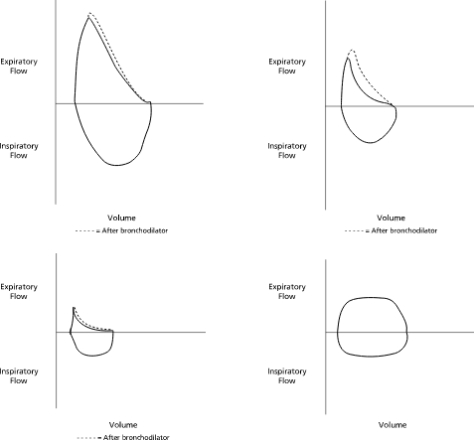
Top Right: Normal; Top Right: Asthma, Bottom Left: COPD, Bottom Right: Fixed Obstruction / Tracheal Stenosis

Asthma:

  • Common chronic respiratory condition characterized by reversible airway obstruction and bronchial hyper-responsiveness

Epidemiology: affects ~8% of US population; allergic asthma is strongly associated with personal/family history of allergies.  Higher prevalence and severity in people of lower income, children, and black populations

Pathogenesis: airway inflammation; chronic inflammation may result in epithelial damage, smooth muscle hypertrophy, airway fibrosis, and remodeling in some

Risk Factors: genetics, exposures to allergens, tobacco smoke, viruses

Symptoms/Clinical Evaluation: classically present with episodes of coughing, chest tightness, SOB, and wheezing

  • Consider other causes that may mimic asthma: COPD, vocal cord dysfunction, heart failure, bronchiectasis, ABPA, CF, mechanical obstruction, etc.
  • Obtain spirometry to assess for severity of airway obstruction and reversibility (FEV1 > 12% and 200 mL)
  • Chest radiographs are often normal

Asthma Syndromes:

  • Allergic Asthma – most common form in adults; family history is often positive
  • Cough-Variant Asthma – persistent/episodic cough without other symptoms
  • Exercise-Induced Bronchospasm
  • Occupational Asthma – related to workplace exposures (farmers, factory workers, hairdressers, etc.)
  • Aspirin-Sensitive Asthma – triad of severe persistent asthma, aspirin sensitivity, and hyperplastic eosinophilic sinusitis with nasal polyposis

Step Up/Down Treatment:

3