All posts by vmcimchiefs

Pneumothorax – 1/8/18

Primary spontaneous pneumothoax – that which happens without any underlying lung disease – in actuality, most of these patients just have undiagnosed lung disease

Secondary spontaneous pneumothorax – that which happens with known underlying lung disease

Risk factors – tall stature, male sex, Marfan’s, homocysteinuria, thoracic endometriosis, smoking

Clinical presentation – dyspnea, pleuritic chest pain, decreased breath sounds, hyperressonance to percussion, and decreased chest excursion

Patients are able to prevent hypercapnea because of their good lung but cannot prevent hypoxia because the areas of collapsed lung still receive perfusion.
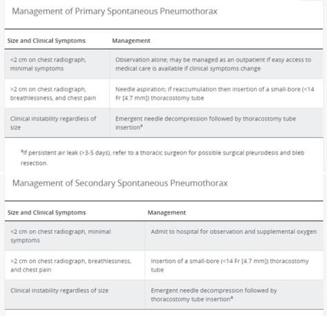
From MKSAP 17:

ptx

Patients with a primary spontaneous pneumothorax are safe to fly after re-expansion of the lung. Scuba diving is NOT safe even after re-expansion of the lung.

Recurrence rate for primary spontaneous is 23-50% over the first 5 years and 50% in secondary spontaneous pneumothorax.

Febrile neutropenia – 1/4/18

Who gets it? Cancer patients who are receiving cytotoxic medications that attack rapidly producing cells. The cells most affected are those in the bone marrow leading to immunosuppression and those in the GI tract leading to increased risk for translocation of gut bacteria into the bloodstream. Keep in mind that these patients may not show the normal inflammatory response and therefore, fever may be their only sign of infection!

What is neutropenia? ANC < 1500. Severe neutropenia is ANC < 500 or ANC expected to drop < 500 within 48 hours.

What is neutropenic feverAccording to the IDSA 2010 guidelines, neutropenic fever is a fever in a neutropenic patient of 38.3 once or 38 sustained over an hour.

How do you measure a fever? The best way to measure is oral (as compared to tympanic, axillary, or rectal) but the only time an oral temperature may be less accurate is if the patient has mucositis or oral ulcers.

What antibiotics should you start these patients on? The goal is to cover GPCs from the mouth and GNRs from the GI tract. The best 3 antibiotics are cefepime, meropenem, or zosyn. Add vanc if: evidence of mucositis, severe sepsis or patient unstable, pneumonia, patient with MRSA colonization, the patient has a central line, if the patient has been on prophylactic antibiotics previously, or if the patient has a soft tissue or skin infection. Add antifungals if: the patient has a persistent fever despite broad spectrum antibiotics or if the patient is hemodynamically unstable.

Low risk patients (those without comorbid conditions) should not be put on prophylactic antibiotics.

Acute coronary syndrome – 11/20/17

Unstable angina = symptoms with negative biomarkers and EKG
NSTEMI or STEMI = symptoms with positive biomarkers and EKG

Types of stress

  • Exercise
  • Dobutamine (works by increasing contractility)
  • Vasodilators – lexiscan or adenosine (not a true “stress” but causes vasodilation of the vessels and if there is an occlusion in one, then more blood will be shunted to the other ones leading to the “steal” phenomenon)

Types of imaging

  • EKG
  • Echo
  • Nuclear medicine study

When deciding what kind of stress and what kind of imaging to use you must take a few things into consideration:

  • If possible, always try to stress a patient with exercise as that gives you information about their exercise tolerance and functional capacity
    • Exercise can be paired with an EKG, echo, or nuclear medicine study
  • If a patient cannot exercise, consider dobutamine or a vasodilator
  • Dobutamine is best to use when a patient has a contraindication to a vasodilator e.g. a patient cannot use adenosine because they have bronchospastic airway disease (COPD or asthma)
    • Dobutamine can be paired with an echo or a nuclear medicine study
  • Vasodilators are ideal if a patient has a LBBB because it is not affected by the fact that in a LBBB you have a delayed contraction of the septum which can cause a false positive for obstruction if done with exercise or dobutamine
    • Vasodilators can only be paired with a nuclear study

Contraindications to using an EKG as your form of imaging are:

  • LBBB
  • Ventricular paced rhythm
  • ST changes > 1 mm

Anti-anginal medications:

  • BB
  • Nitrate (give a medication free period at night to avoid tachyphylaxis)
  • CCB
  • Ranolazine (4th line medication if the others have failed)

Myxedema Coma – 12/12/17

What is it?

  • Severe hypothyroidism leading to AMS and hypothermia
  • Can have other symptoms related to the slowing down of organs

Who gets it?

  • Usually older females with long standing hypothyroidism triggered by a precipitating event

How does it present?

  • Change in mental status (rarely presents as true overt coma)
  • Hypothermia
  • Hypotension
  • Bradycardia
  • Hyponatremia
  • Hypoglycemia
  • Hypoventilation

What labs should you check?

  • TSH, FT4
  • Cortisol (to rule out concurrent adrenal insufficiency)

What is the treatment?

  • IV T3 and/or T4 – data is mixed
    • Use IV because patient likely has gut edema so PO form may have decreased absorption
    • T3 has better bioavailability and is the active form
    • In acute illness, body’s normal conversion of T4 to T3 is impaired
    • Monitor patients on telemetry because biggest concern is arrhythmias
    • Use lower dosing in elderly patients or those with cardiac disease
    • Recheck TSH in one week – goal is drop by > 50%
  • Stress dose steroids (hydrocortisone 100 mg every 8 hours)
    • Until you rule out concomitant adrenal insufficiency
  • Supportive measures
    • Avoid dilute fluids which can worsen hyponatremia
    • Use passive rewarming
    • Pressors if needed