Tag Archives: Gastroenterology

Hepatitis B & HIV co-infection

We discussed a young male with past medical history of syphilis (incompletely treated) and recently diagnosed HIV (4 days prior to admission) who was admitted for elevated liver enzymes found incidentally. Liver enzymes consisted of mildly elevated alkaline phosphatase and bilirubin but extremely elevated AST/ALT in 4000s. Only a few entities cause elevation to the thousands. He was found to have acute co-infection of HIV and Hepatitis B. 

If AST/ALTs are in the thousands, there are only a few entities that can cause this:

  • Ischemia (shock liver)
  • Toxins (Tylenol is most common), Amanita aka magic mushrooms, herbal supplements (we don’t know what they put in these!)
  • Acute viral hepatitis (HAV, HBV, HCV, HEV, HSV, CMV, VZV, parvovirus)
  • Autoimmune hepatitis
  • Acute Budd Chiari
  • Reactivation HBV, HDV
  • HLH (we seem to see this a lot in this hospital for some reason?)
  • Malignant infiltration
  • HELLP
  • Wilsonian Crisis (severe hemolysis and impending acute liver failure in setting of Wilson’s)

According to the CDC, approx 10% of people with HIV in the US also have chronic or acute HBV. There is accelerated progression to liver disease and increased all cause mortality for HIV-HBV co-infection when compared to HIV mono infection. Monotherapy of HBV is not recommended in the HIV co-infected due to the evolution of HIV resistance. Recommended antiretroviral regimens for treating persons with HIV-HBV coinfection should include three medications that are active against HIV and two medications that are active against HBV.The preferred regimens include tenofovir alafenamide-emtricitabine, tenofovir DF-emtricitabine, or tenofovir DF plus lamivudine as part of a fully suppressive antiretroviral regimen.

Alcoholic hepatitis

We discussed a middle aged man with heavy alcohol use presenting with jaundice found to have profound liver failure. It is important to note acute liver failure warrants consideration for transplant candidacy urgently. However, previous records revealed this patient had a history of cirrhosis and prior alcoholic hepatitis, this patient likely suffered from acute on chronic liver failure from alcoholic hepatitis. We reviewed the broad differential for jaundice and specifically for conjugated hyperbilirubinemia (attached). Alcoholic hepatitis may manifest as fever, jaundice, hepatomegaly and leukocytosis. Maddrey’s DF score is used to prognosticate. A score >32 warrants consideration of corticosteroids. STOPAH trial shows nonsigifnicant survival advantage at day 28 in patients with prednisolone vs no prednisolone.  

Hypertriglyceridemia induced acute pancreatitis

We discussed a case of a man w/ HLD, IDDM2, Obesity, hx of pancreatitis, who presented to the hospital for acute epigastric pain, decreased PO intake, and non-bloody / non-bilious emesis. He was diagnosed with Hypertriglyceridemia-induced Acute Pancreatitis.
We discussed how to approach abdominal pain as well as pearls when treating hypertriglyceridemia-induced acute pancreatitis.
Framework for abdominal pain
  • R/o emergencies: obstruction, perforation, vascular, ectopic pregnancy.

Hypertriglyceridemia-induced acute pancreatitis

  • Triglycerides are broken down into free fatty acids that accumulate to toxic levels, which can cause inflammation of the pancreas.
  • Early (within ~48 hrs) enteral refeeding is encouraged.
    • Enteral (PO) feeding is recommended over parenteral (IV) to help maintain intestinal barrier and prevent bacterial translocation from the gut.
  • Insulin is used to treat hypertriglyceridemia-induced acute pancreatitis. Insulin promotes storage of triglycerides and inhibits breakdown of triglycerides into free fatty acids.

Hypertriglyceridemia induced acute pancreatitis Summary pdf

HTG-Panc – Don’t Pan(i)c!

Today we discussed a case of hypertriglyceridemia-induced pancreatitis (TG >4k). We first utilized this case as an opportunity to provide a comprehensive overview of acute and chronic pancreatitis:


We then discussed the management strategy:

General Pancreatitis Management

  1. Assess severity of pancreatitis with SIRS
  • Mild – absence of organ failure and local systemic complications
  • moderate – local complications and +/- transient organ failure (<48hrs)
  • Severe – persistent organ failure (>48hrs)
  1. Aggressive hydration at 5-10mls/kg/hr
  • Adjust based on vital signs, exam, urine output BUN, HCT
  1. Pain Control
  2. Nutrition
  • Mild
    • initiate PO intake within 48 hours (based on symptoms, NOT lipase reduction)
    • Go straight to low residue, low fat, soft diet
  • Moderate – Severe
    • If unable to tolerate PO, consider NJ vs. NG tube after 3-4 days
  • Enteral nutrition >>> parenteral nutrition
  1. Antibiotics?
  • not recommended regardless of pancreatitis severity or type (i.e interstitial or necrotitizing) unless extrapancreatic infection suspected (blood stream, PNA, UTI etc.) OR necrotizing fluid collection suspected to be secondarily infected (systemic infection OR radiographic evidence of infection)

HTG-Pancreatitis Management

  • Insulin Therapy
  • Lipid Lowering Therapy
  • Plasmapheresis (controversial)
    • invasive
    • expensive
    • requires AC
    • Despite that – UTD recommends apheresis in patients with any of the following (hypocalcemia, lactic acidosis, two or more signs of worsening inflammation, worsening organ dysfunction)
    • Two studies of note that discuss this
      • 1) Head-to-head RCT showed quicker time to TG reduction compared to insulin but worse clinical outcomes
      • 2) Addition of apheresis to patients already receiving insulin showed no clinically significant differences in outcome
    • Bottom Line: based on the limited data above, some experts favor an insulin only approach
    • For an in-depth analysis of this topic – please see this fantastic PulmCrit blog post: https://emcrit.org/pulmcrit/hypertriglyceridemic-pancreatitis/

Lower GI Bleeding

Today we discussed a case of a patient with chronic constipation who presented with abdominal pain and hematochezia. A CT scan revealed a large fecaloma, which led to stercoral ulceration of the rectum, colitis and eventually perforation.

We first reviewed the etiologies of hematochezia, remembering that in up to 15% of cases, a brisk UGI source is the etiology. When this occurs it is typically in patients who have hemodynamic instability. Some aspects to aid in the diagnosis were put together in an excellent Rational Clinical Examination article in JAMA that Dr. Jacobson referenced. Findings that have a high LR of a brisk UGI are melenic stool on examination, an elevated BUN/Cr ratio  30, an NG lavage with blood or CGE . Findings that have a high LR of the etiology being a LGIB is a history of LGIB and clots in stool.

IsItUGI.

https://jamanetwork.com/journals/jama/fullarticle/1105075

We then discussed a framework for LGIB:

LGIBDDx

We discussed the complications of constipation:

  • Decreased quality of life
  • Hemorrhoids
  • Anal Fissures/Tears
  • Diverticulosis
  • Rectal Prolpase
  • Stercoral Ulceration

Stercoral Ulceration/Colitis/Perforation 101

  • most cases have been reported in elderly, psychiatric, bedridden, or narcotic-dependent patients with a history of constipation
  • Fewer than 150 causes of perforation have been described in the literature
  • Severe chronic constipation thought to induce the formation of stone-hard fecalomas and maintain a continuous peressure over the bowel wall leading to pressure necrosis
  • This ulceration sometimes leads to a state of stercoral colitis (localized ischemia due to increased luminal pressure)
  • CT findings of perforation are fecal impaction, colonic dilatation, colonic wall thickening, discontinuity in the enhancement of the bowel wall in relation to focal fecal distension of the colonic lumen, and extraluminar free air
  • A mortality rate of ~30% has been mentioned
  • Treatment is generally surgical

Constrictive Pericarditis

Thanks to Phuong today for presenting the case of a young woman who presented with volume overload, found to have constrictive pericarditis!


Clinical Pearls

  • Constrictive pericarditis and restrictive cardiomyopathy can have similar clinical presentations.  Cardiac cath is generally needed to help distinguish between the two.
  • The most common cause of constrictive pericarditis is idiopathic!
  • Exam findings in constrictive pericarditis include volume overload, pulsus paradoxus, Kussmaul’s sign, pericardial knock, and occasionally (<20% of the time) pericardial friction rub.
  • ECG and CXR can be normal in constrictive pericarditis.
  • Treatment of early disease is supportive care.  Treatment of late stage constrictive pericarditis is pericardiectomy.

Etiologies of constrictive pericarditis:

  • Idiopathic (42-61%) ⇒ most common cause!
  • Post-cardiac surgery (11-37%)
  • Post-radiation therapy (2-31%) particularly after Hodgkin disease or breast cancer
  • Connective tissue disorder (3-7%)
  • Post-infectious – TB or purulent pericarditis (3-15%)
  • Miscellaneous causes (malignancy, trauma, drug-induced, asbestosis, sarcoidosis, uremic pericarditis) (1-10%)

Clinical Presentation

  • Symptoms related to fluid overload
  • Symptoms related to diminished cardiac output in response to exertion
  • Exam:
    • Elevated JVP
    • Pulsus paradoxus – drop in SBP >10 mmHg due to drop in stroke volume and cardiac output with inspiration (20%)
    • Kussmaul’s sign – lack of an inspiratory decline in JVP.  (Also present in people with severe tricuspid valve disease or R heart failure.
    • Pericardial knock – 47%
    • Pericardial friction rub – 16%
    • Stigmata of heart failure
  • ECG: can be normal
  • CXR: Majority of people do NOT have pericardial calcifications
    • Interestingly, calcifications are more common in people with idiopathic disease, a longer duration of symptoms, and those with TB!

Management of Constrictive Pericarditis

  • Early disease is usually managed with supportive care.  Diuretics can help mitigate symptoms of volume overload but must be used cautiously due to preload dependent physiology.
  • Late stage disease is treated with pericardiectomy.  Complication rates tend to be high and operative mortality can reach 12%!

Constrictive Pericarditis vs Restrictive Cardiomyopathy:

CP vs RCM

We also talked about a helpful way of breaking up new onset ascites to help generate a DDx:

Ascites fluid distribution

Strongyloidiasis

Thanks to the Human Dx Project for providing us with this fascinating case of a middle aged woman with history of asthma who presented with acute onset of fever and epigastric abdominal pain as well as a chronic progressive cough, found to be febrile, tachycardic, and ill appearing, with E coli bacteremia of unknown source.  Further history taking revealed a similar hospitalization several months prior with idiopathic E coli bacteremia.  Strongyloides titers were sent and markedly elevated.  She was treated with ceftriaxone and ivermectin and made a full recovery.


Clinical Pearls: 

  • Absence of eosinophilia does not rule out strongyloides.  Keep in mind that those presenting with severe illness and hemodynamic instability are commonly in a high cortisol state which can lead to eosinophil apoptosis.  Also, in those with history of steroid use (even for short periods of time), eosinophil count can be negative.
  • Think of strongy in anyone with the right travel history, older age, malnutrition, HIV, or steroid use.
  • Signs and symptoms can be quite non-specific so a high index of suspicion is required to make the diagnosis.
  • Think of strongyloides in a patient with history of recurrent GNR bacteremia of unknown etiology!

Strongyloidiasis 

Epidemiology

  • Higher incidence noted going from yellow to orange to red on the map above
  • Epidemiology
    • Typically in travelers to endemic areas, immigrants from endemic regions, or anyone with barefoot contact with infested soil.
    • Risk factors include older age, malnutrition, HIV, and steroid use
  • Signs and symptoms
    • Infected people can be asymptomatic or minimally symptomatic for years:
      • Could also have mild waxing and waning GI, skin, or pulmonary symptoms for years
      • Eosinophilia without symptoms
    • Skin: urticarial, larvae currens (see picture below), angioedema, erythrodermalarvae currens
    • Pulmonary: chronic cough, hemoptysis, recurrent pneumonia, astham that gets worse with steroids
    • GI: upper abdominal pain, duodenitis, diarrhea, anorexia, recurrent enteric GNR bacteremia
    • Disseminated disease/hyperinfection syndrome:
      • Increased parasite burden due to autoinfection (see picture below)
      • Massive dissemination of larvae to lungs, liver, heart, CNS, and endocrine glands
      • Can present with septic shock or multiorgan failure

        Strongy life cycle
        Greaves, D. BMJ 2013; 347:f4610
  • Diagnosis:
    • Stool O&P: <50% sensitive and requires multiple samples due to intermittent shedding
    • Serologies: 89% sensitive
  • Treatment:
    • Ivermectin or albendazole
    • Hyperinfection/disseminated disease: above PLUS broad-spectrum antibiotics

Quick review of endemic dimorphic fungi:

  • Southwest US ⇒ Cocci
  • Ohio & Mississippi River Valley ⇒ Histo
  • Southeast/South-central US ⇒ Blasto
  • Southeast Asia ⇒ Penicillium
  • South America ⇒ Paracocci, histo, blasto, cocci

Monthly M&M 5/10/2019

Thank you everyone for coming to our monthly M&M Conference!

This was case of a young man with undisclosed chronic HBV and cocaine use who presented with first onset hematemesis and melena. On presentation, he was tachycardic, anemic, but normotensive. His heart rate improved with more fluid resuscitation and at the time, he did not require ICU level of care.

While on the floor, his tachycardia gradually worsened, and his Hgb also downtrended from 9.6 from 7.8 in the span of 12 hours, which was initially attributed to dilutional error given he was given 3 L of fluids. He acutely decompensated in the evening, and despite our best efforts, we could not keep up with the amount of hemorrhage likely from variceal bleeding.


Cocaine

Mechanism in a nutshell: Inhibits catecholamines reuptake, leading to to increased levels or norepi, dopamine, and serotonin. This subsequently leads to elevated BP, tachycardia, fever, and inc risk of coronary artery vasospasm, arrhythmia, and QTc prolongation.

Onset of action: Peak cardiovascular effect seen 5-15 minutes after use.

Half life: 45-90 minutes

Metabolism: Metabolized in seurm and liver into ecgonine methyl ester and benzoylecgonine, which is what Utox picks up! Utox will be positive for cotaine 48-72 hours after use, and it can remain positive up to 10 days (even longer for chronic users).


Cocaine & Anesthesia

  • Compared to controls, pts who are cocaine positive on utox undergoing elective procedures requiring general anesthesia are no different in terms of:
    • Arrhythmia risk
    • Cardiovascular events (fluctuations in BP, HR)
    • Anesthesia duration
    • Medical dosage
    • PACU recovery time
  • This study in particular was insightful, but keep in mind that patients > 70, ESRD, or acutely intoxicated were excluded.

Hemorrhage Shock

Capture.JPG

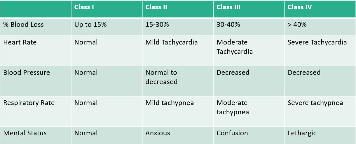
To put this into perspective, a patient (let’s say “Average” 70 kg adult) presenting with GIB can lose up to 1.5L of blood prior to seeing any chances in blood pressure. The first signs of clinical instability is HR, with mild tachycardia up to the lower 100s!

Also the Glawgow Blatchford Score is useful for risk stratification. If pt scores 0, there is a very low risk of complications. The higher the score is, the higher in terms of potential for complications and need for more urgent intervention.


When to Transfuse

See this study for more details. In general for UGIB, a restrictive transfusion strategy (transfuse when Hgb < 7 g/dL) has better outcomes (survival, rebleeding, mortality) vs a liberal approach (transfuse when Hgb < 9 g/dL).


In cirrhotic patients, is there such thing as over-resuscitation? YES.

In animal studies, severity of bleeding was directly related to portal venous pressure. Restrictive transfusion protocol also showed less portal pressure gradient vs the liberal arm, and less rebleeding and improved survival. Per the Baveno V Consensus, for variceal bleed, the target Hgb is between 7-8 g/dL.


What about when stuff hits the fan?

ABC comes first, don’t even worry about the numbers any more.

Remember when you order blood, it needs to be typed, screened, and cross-matched. It can take hours!

Know when to call the massive transfusion protocol (MTP). You can find the protocol on HHS-Connect on any desktop.

In a nutshell, activate MTP if you have a patient you anticipate requiring > 4 units PRBC within the first hour, or with high probability of requiring > 10 units within 12 hours. The blood bank will then start bringing in uncrossed blood, platelet, and FFP very quickly. Make sure to deactivate the protocol once your patient has been stabilized.

Also plan ahead for access! AT LEAST 2x large bore IV (18g or bigger) but know that in an acute situation, two PIVs can be easily overwhelmed. If you anticipate risk for decompensation, establish plan for access early on so you won’t get overwhelmed when the patient decompensates (i.e. IO kit or find an opportunity for a Cordis catheter).

Hepatocellular Carcinoma 5/6/2019

Katie presented an elderly man presenting with few weeks of unintentional 40lbs weight loss and abdominal pain, found to be jaundiced on exam with notable hepatomegaly. Labs notable for mild hepatitis and mix conjugated and unconjugated hyperbilirubinemia with mild coagulopathy. He was ultimately diagnosed with cirrhosis and extremely likely HCC with “numerous” masses of varying size (largest one was 10 cm) with portal vein invasion.


Please refer to this previous post on etiology of hyperbilirubinemia.

Please refer to this other post on hepatitis serologies made ridiculously simple.


Hepatomegaly and jaundice: Think infiltrative/malignant process!

For patients with cirrhosis and abdominal distension, palpating and percussing the liver can be challenging.

A strategy we went over during the last physical exam round was the scratch test, which relies on the principle of the different of sound transmission through materials of various densities.

To perform the scratch test, place your stethoscope over the RUQ just above the costal margin or just below the xiphoid, and from the RLQ, lightly scratch the patient horizontally and then slowly move superiorly toward the costal margins until the sound intensities. The location of sound intensification marks the inferior edge of the liver.

Small study re: accuracy of using the scratch test.


Hepatocellular Carcinoma

Epidemiology

  • Most of the time a complication from liver cirrhosis
  • Risk Factors
    • Cirrhosis, any etiology
    • Chronic HBV even without cirrhosis (oncogenic virus)
    • HCV with cirrhosis
    • Fungal aflatoxins
  • Higher prevalence in East and SE Asia + Sub-Saharan African nations
  • HBV inc risk by 100x

Presentation

  • Variable initial presentation
    • Asx
    • Decompensated cirrhosis
    • Jaundice, abd pain, B-sx, +/- palpable pass
    • Variceal hemorrhage
    • Tumor rupture leading to acute hemorrhagic shock
    • Obstructive jaundice due to biliary tree invasion
    • 10-15% with metastatic dz at time of dx.
      • Most common sites: Lung, lymph nodes, adrenal glands.
    • Paraneoplasic: Hypoglycemia (adv HCC), erythrocytosis, hypercalcemia, diarrhea

Diagnosis

  • Imaging: Sensitivity generally dec with small lesions, but imaging alone to establish dx of HCC without a biopsy in certain patient populations.
    • Contrast enhanced CT (triphasic)
      • Arterial phase hyperenhancement: Characteristic of HCC lesions but not specific, can be small hemangiomas, focal nodular hyperplasia, atypical focal fibrosis, non-HCC malignancy
      • Venous phase Washout: Again characteristic of HCC but not specific, cirrhosis nodules can be similar.
      • Capsular appearance: Pretty specific for HCC
      • All 3 of the above = diagnostic of HCC, very specific not but as sensitive.
      • Highest PPV for pts with cirrhosis with lesions > 2cm
    • MRI
      • Contraindicated in GFR < 30, Nephrogenic systemic fibrosis
      • More sensitive than CT, ~ specificity,
    • US: Can be use as diagnosis but cannot evaluate disease burden, transplant candidacy, operator dependent. 90% sensitive and 97% specifc
    • LIRADS
      • Should only be applied to pts with cirrhosis, chronic HBV, lesions identified on surveillance US for HCC, current or prior dx of HCC.
      • Should NOT be applied to: no risk factors for HCC, < 18, cirrhosis secondary to congenital hepatic fibrosis or vascular etiology.
    • LIRADS definitions for hepatocellular carcinoma based on ACR v2017:
      • LR-1: Definitely benign
      • LR-2: Probably benign
      • LR-3: Intermediate probability for HCC
      • LR-4: Probably HCC
      • LR-5 – Definitely HCC
      • LR-5V: Definitely HCC with tumor in vein
      • LR-M: Probably malignancy, not specific for HCC
      • LR-TR Viable: Treated, probably or definitely viable HCC
      • LT-TR Nonviable: Treated, probably or definitely not viable
      • LR-TR Equivocal: Treated, equivocally viable
      • LR-TR Nonevaluable: TReated, response not evaluable
  • Labs
    • Alpha-fetoprotein: Elevated in 40-65% of patients with HCC
      • Normally produced during gestation, not during adulthood.
      • Levels do not correlate well with degree of disease
      • Sensitivity: 60%, spec: 80%, not good as a screening tool.
      • Higher levels > 400 are very specific for HCC.
      • May be seen in chronic liver disease so not very sensitive.
      • Elevated levels are associated with advanced fibrosis, pregnancy
  • Biopsy: Reserved for indeterminate nodules that do not meet radiologic criteria for HCC.
    • Not recommended for LR1, LR2, LR3, or LR5 lesions
    • Risk: spread of tumor along needle track, sampling error (false negative), usual surgical risk (bleeding, infection, etc)
  • Staging:
    • TNM
    • Barcelona Clinic Liver Cancer staging system

Management

  • Surveillance for at risk patients:
    • Cirrhosis or HBV: Q6mo liver US
  • Resection
    • Preferred therapy for localized disease
    • Sufficient liver reserve, can’t be worse than Child Pugh A cirrhosis
    • 5 year survival rate as high as 90%
    • Stage IIIB, IVA, and IVB are incurable by resection (any invasion of a major portal or hepatic vein, other organs, visceral peritoneum, nodal mets)
  • Antiviral
    • Recommended for those with active viral infection and HBV related HCC.
  • Liver transplantation
    • Milan criteria widely accepted, to be considered a candidate for transplant, pt must meet all criteria
      • Solitary tumor < 5cm or up to 3 tumors all < 3cm
      • No evidence of regional nodal or distant mets
      • No evidence of vascular invasion
  • Ablation
    • Radiofrequency or microwave or localized ethanol/acetic acid/cryo localized ablation
    • Best outcomes for tumor size < 4 cm
    • Cirrhosis: Restricted to Child Class A or B
    • Can be used to bridge to liver transplant
  • TACE
    • Disruption of HCC supply, usually derived from the hepatic artery
    • Leads to tumor necrosis from ischemia.
    • Usually used for tx of large unresectable HCC not amenable to other tx i.e. resection or RFA.
    • Best candidates: No vascular invasion or extrahepatic spread, Child Puph A or B
    • Relative contraindication:
      • bili > 2
      • LDH > 425
      • AST > 100
      • Tumor > 50% of liver
      • Untreated EV
      • Significant medical comorbidities
  • Radiation
    • Localized external radiation vs radioembolization, HCC is radiation sensitive.
  • Systemic chemo
    • Sorafenib
      • SHARP trial, prolongs survival over supportive care in pts with adv HCC)
      • Might be more beneficial for HCC related to viral etiology
    • Other agents: Regorafenib, Lenvatinib
    • Complication during tx
      • Reactivation of viral hepatitis

Prognosis

  • 10-20% of cases are curable (resectable disease)
  • 5-yr survival rates of about 5% or less if beyond stage III (portal vein invasion)
  • Some evidence that sorafenib improves survival by around 3 months but it is a costly medication (60 tablets can cost up to $9000 after discount!)

IgG4 related disease!

Thanks to John for presenting the case of a middle aged man from Vietnam with history of smoking who presented to the hospital with painless jaundice with imaging concerning for malignancy, found to have IgG4 related autoimmune pancreatitis!


Clinical Pearls

  • For imaging of the biliary tree:
    • Ultrasound is best for stones
    • CT is better for parenchymal disease
    • ERCP/MRCP is best for intraductal masses or stones
  • IgG4 related disease is more common in men >50 years of age.  It can affect many organs, similar to sarcoid.
  • It is an inflammatory and fibrotic systemic conditions where organs form tumefactive lesions rich in IgG4 plasma cells.
  • Diagnosis requires biopsy.  Serum IgG levels may be normal even in active disease.  However, a significantly elevated level is highly sensitive/specific (>95%) for IgG4 related disease.
  • Treatment involves steroids + immunomodulating therapy.

Approach to high bilis!

conjugated hyperbili

unconjugate hyperbili

DDx for painless jaundice:

  • Cancer (pancreatic, cholangiocarcinoma)
  • Meds/toxins
  • Viral hepatitis
  • ESLD
  • CHF
  • Hemolysis
  • PBC/PSC

IgG4 related disease

  • First described in 2003
  • Majority men (62-83%) and > 50 years of age.
  • Inflammatory and Fibrotic systemic condition where organs have tumefactive (tumor forming) lesions with an infiltrate rich in IgG4 plasma cells and often elevated IgG4 serum levels (but not always!)
  • The pathophys is poorly understood but thought to be a combination of autoimmune and allergic mediated processes.

Clinical features:

  • Subacute onset, typically few systemic signs/symptoms
  • 30% of those with autoimmune pancreatitis also have tubulointerstitial nephritis at presentation.
  • Pancreatic involvement 
    • Manifestations include a uniformly enlarged pancreas (sausage pancreas on imaging), pancreatic mass which can mimic cancer, recurrent pancreatitis, or strictures.
  • Biliary involvement
    • Biliary strictures leading to obstructive jaundice as well as sclerosing cholangitis
  • ANY organ can be affected (eg: thyroiditis, interstitial nephritis, salivary involvement), much like sarcoid

Spectrum of IgG4-related disease

IgG4 spectrum
Source: UpToDate

Diagnosis:

  • Need tissue biopsy for diagnosis.  Serum serologies are only suggestive.

Treatment:

  • Steroids + immunomodulating therapy.  

Chronic pancreatitis

Etiologies of Chronic Pancreatitis (progressive inflammatory changes in the pancreas that result in permanent structural damage and histologic fibrosis)

  • Alcohol abuse(45%) as well as cigarette use
  • Recurrent acute pancreatitis
  • Genetic (eg: CFTR, SPINK mutations)
  • Chronic ductal obstruction
  • Systemic diseases (eg: SLE, hyperparathyroidism, hypertriglyceridemia)
  • Idiopathic
  • Autoimmune: Can be Type 1 (part of IgG4 related disease) or Type 2 (idiopathic) 

Clinical manifestations of chronic pancreatitis

  • Can be ASYMPTOMATIC
  • Epigastric abdominal pain most common symptom however
  • Pancreatic insufficiency (only after 90 %of pancreatic function lost) and manifests as steatorrhea and glucose intolerance/diabetes
  • Remember that chronic pancreatitis puts you at increased risk for PANCREATIC CANCER

Lab/Imaging

  • Amylase/Lipase usually NORMAL so not as helpful
  • 72 hour quantitative fecal fat (steatorrhea alone is non-specific!)
  • Fecal elastasehas high sensitivity and specificity for chronic pancreatitis
  • KUB can show calcificationshinting towards chronic pancreatitis and MRCP/ultrasound can show pancreatic duct obstructions, dilations, strictures or fluid collections

Treatment

  • Alcohol and smoking cessation!
  • Creon supplementation, may also need fat-soluble (A,D,K,E) supplementation
  • Analgesics for abdominal pain which is extremely hard to control. Minimize opioids but may be necessary for refractory pain.
  • Specialized approaches include celiac nerve blocks, endoscopic surgery and surgical resection