Tag Archives: Morning Report

Disseminated gonorrhea

We discussed a case about a young woman with hx of STIs (syphilis and chlamydia s/p treatment), subacute hx of migratory polyarthralgias, who presented with fevers and acute arthritis / tenosynovitis of her left index finger + thumb. She was found to have gram negative diplococci bacteremia and diagnosed with disseminated gonorrhea.

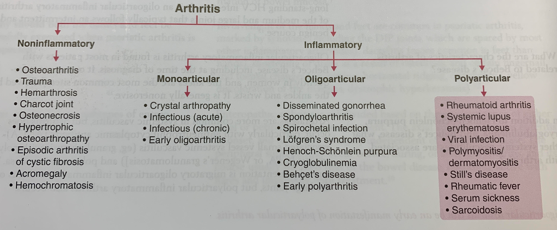
Framework for arthritis

  • Categorize differentials based on non-inflammatory vs inflammatory, as well as mono- / oligo- / polyarticular.

CDC screening recommendations

  • Men: those at high risk (MSM)
  • Women: can be asymptomatic -> complications of STIs (eg PID, infertility)
    • < 25 yrs old    AND
    • ≥ 25 yrs old + STI risk factors

Preferred screening / diagnostic testing

  • Uncomplicated: NAAT (urine, genitals / throat / rectal swab)
  • Disseminated: Blood, joint, abscess, and/or CSF cultures

Gonorrhea treatment

  • CDC reports increasing azithromycin resistance
  • Ceftriaxone is first line

!Bonus learning! Chlamydia treatment

  • Also increasing azithromycin resistance
  • Doxycycline 100mg BID x7 days is first line

Hepatitis B & HIV co-infection

We discussed a young male with past medical history of syphilis (incompletely treated) and recently diagnosed HIV (4 days prior to admission) who was admitted for elevated liver enzymes found incidentally. Liver enzymes consisted of mildly elevated alkaline phosphatase and bilirubin but extremely elevated AST/ALT in 4000s. Only a few entities cause elevation to the thousands. He was found to have acute co-infection of HIV and Hepatitis B. 

If AST/ALTs are in the thousands, there are only a few entities that can cause this:

  • Ischemia (shock liver)
  • Toxins (Tylenol is most common), Amanita aka magic mushrooms, herbal supplements (we don’t know what they put in these!)
  • Acute viral hepatitis (HAV, HBV, HCV, HEV, HSV, CMV, VZV, parvovirus)
  • Autoimmune hepatitis
  • Acute Budd Chiari
  • Reactivation HBV, HDV
  • HLH (we seem to see this a lot in this hospital for some reason?)
  • Malignant infiltration
  • HELLP
  • Wilsonian Crisis (severe hemolysis and impending acute liver failure in setting of Wilson’s)

According to the CDC, approx 10% of people with HIV in the US also have chronic or acute HBV. There is accelerated progression to liver disease and increased all cause mortality for HIV-HBV co-infection when compared to HIV mono infection. Monotherapy of HBV is not recommended in the HIV co-infected due to the evolution of HIV resistance. Recommended antiretroviral regimens for treating persons with HIV-HBV coinfection should include three medications that are active against HIV and two medications that are active against HBV.The preferred regimens include tenofovir alafenamide-emtricitabine, tenofovir DF-emtricitabine, or tenofovir DF plus lamivudine as part of a fully suppressive antiretroviral regimen.

Pulmonary emergencies featuring Dr. Halley Tsai

  1. Attached a good and recent NEJM review on supplemental O2. There’s an video too that has everything in the text: https://www.nejm.org/doi/full/10.1056/NEJMvcm2035240
  1. ABG interpretation, comprehensive chapter on CHEST: https://www.thoracic.org/professionals/clinical-resources/critical-care/clinical-education/abgs.php
  1. Attached the CHEST cheat-sheet quick ABG I had in my talk. If people want very easy one-look cheat-sheets like that, the “All Resources” section has great ones to click through if people have time: https://www.chestnet.org/topic-collections/covid-19/clinical-resources

DKA & acid-base analysis

We discussed a young male with no past medical history with 4 days of abdominal pain, nausea, vomiting, constipation found to be kussmaul breathing on physical exam. Patient presented with anion gap metabolic acidosis. 

Learn how to calculate acid base

  1. Anion gap = Na-Cl-bicarb
  2. Determine pH (7.35-7.45 is normal)
  3. Identify primary disorder (i.e. respiratory vs metabolic)
    • If metabolic: bicarb <22, think primary metabolic acidosis; if bicarb>28, think primary metabolic alkalosis
    • If respiratory: pCO2 <35, thik primary respiratory alkalosis; if pCO2>45, think primary respiratory acidosis
  4. To look for concomitant respiratory process on top of primary metabolic acidosis, calculate Winters Formula: expected pco2 = 1.5*(bicarb)+8
  5. To look for comitant metabolic process on top of primary metabolic process, calculate Delta gap:(patient’s AG – normal AG) – (patient’s bicarb-normal bicarb)
    • Delta gap>6 indicates metabolic alkalosis on top of AGMA 
    • Delta gap close to 0 is normal and indicates pure AGMA
    • Delta gap <-6 indicates NAGMA on top of AGMA  

Our patient had primary AGMA with metabolic alkalosis with superimposed respiratory acidosis. 

Learn differential for AGMA
GOLDMARK:

  1. Glycols – ethylene glycol/propylene glycol 
  2. Oxoproline (acetaminophen)
  3. L-lactate
  4. D-lactate
  5. Methanol
  6. Aspirin
  7. Renal Failure
  8. Ketoacidosis

Hyperkalemia

We discussed a case about a middle aged man with uncontrolled HTN and T2DM, presenting with subacute bilateral lower extremity edema and sacral edema, found to have Hyperkalemia likely due to CKD from uncontrolled HTN and T2DM. Teaching points focused on going over a framework to organize causes of hyperkalemia, EKG features of hyperkalemia, and treatment options for hyperkalemia.

EKG features of hyperkalemia

  • Tall peaked T waves, loss of P waves, widening QRS, sine wave / ventricular arrhythmia / asystole

Hyperkalemia tx (Check out the hyperkalemia order set!)

  • Calcium
    • Stabilize cardiac membrane action potential
    • Rapid onset (minutes); short duration (30-60 min)
    • Contraindicated in digoxin toxicity
  • Insulin + Dextrose
    • Intracellular shift of K via ↑ Na-K-ATPase
    • Rapid onset (10-20 min); lasts hours
  • NaHCO3-
    • Intracellular shifts of K
    • Rapid onset (minutes); short duration
  • Beta-2-adrenergic agonists
    • Intracellular shifts of K
    • Rapid onset (minutes)
    • Watch for tachycardia and angina
  • Loop diuretics
    • Excretion of K, prevention of K absorption
    • IV Lasix peak effect (30 min); IV Lasix duration (2hrs)
  • GI cation exchangers
    • Bind K in GI lumen -> excretion
    • Slow onset (many hrs); long duration (many hrs to days)
    • SPS (Kayexalate) associated w/ intestinal ischemia
  • Dialysis

Pulmonary embolism

We discussed a case of a middle aged woman who presented with subacute to acute shortness of breath on exertion. We discussed her EKG with S1Q3T3 findings which is not specific for PE but indicative of cor-pulmonale. The differential for cor-pulmonale is COPD, ARDS, PNA, Pneumothorax and PE. More specifically, her CTA showed bilateral pulmonary embolus and RV enlargement and her labs indicated elevation in BNP and troponin. Due to her hemodynamic stability but clear evidence of RV strain, troponin and BNP elevation – she was classified as submassive PE (these days known as intermediate high risk classification). In addition to starting heparin infusion, with all submassive PE, it is important to consider systemic thrombolysis (low dose vs high dose) AND catheter directed thrombolysis / thrombectomy. This will be a nuanced discussion but admission to ICU and early consults to IR, Cardiology and Pulmonology are required.

AML and leukostasis

We discussed a case of a man with weakness, found to have hyperleukocytosis. He was diagnosed with AML. There was a concern for leukostasis due to the tremendous amount of blasts on peripheral smear.We reviewed characteristics of AML vs ALL vs CML vs CLL and discussed four emergencies (TLS, DIC / bleeding, febrile neutropenia, and leukostasis), with a deeper discussion about leukostasis.

AML
– Adults (60s);  sxs: fatigue (anemia) + bleeding / bruising (thrombocytopenia) + infection (ineffective neutrophils);  dx by peripheral smear or bone marrow biopsy with >20% blasts;  Auer rods can be seen on smear

Leukostasis
– Hyperleukocytosis = total WBC > 50k to 100k
– Lots of blasts → ↑ blood viscosity → blockage/ischemia
– Respiratory distress and neurologic symptoms (eg stroke) are leading causes of early death
– Treatment: cytoreduction (chemotherapy, hydroxyurea, leukapheresis-controversial)

Alcoholic hepatitis

We discussed a middle aged man with heavy alcohol use presenting with jaundice found to have profound liver failure. It is important to note acute liver failure warrants consideration for transplant candidacy urgently. However, previous records revealed this patient had a history of cirrhosis and prior alcoholic hepatitis, this patient likely suffered from acute on chronic liver failure from alcoholic hepatitis. We reviewed the broad differential for jaundice and specifically for conjugated hyperbilirubinemia (attached). Alcoholic hepatitis may manifest as fever, jaundice, hepatomegaly and leukocytosis. Maddrey’s DF score is used to prognosticate. A score >32 warrants consideration of corticosteroids. STOPAH trial shows nonsigifnicant survival advantage at day 28 in patients with prednisolone vs no prednisolone.  

Seizures due to Neurocysticercosis

We discussed a case of a young man who presented to the hospital after having a seizure. He had a history of neurocysticercosis complicated by seizures many years ago. His most recent seizure was in the setting of restarting albendazole.
We discussed how to come up with a broad and organized ddx for the cause of seizures using the MIST mnemonic, practical knowledge for acute seizure management, and highlights regarding the management and treatment of neurocysticercosis.
Acute seizure management
  • Check the patient’s ABCs and IV access
  • Check for hypoglycemia
  • Ativan 2-4 mg IV pushes x3
  • Status epilepticus:
    • continuous seizure that lasts ≥5 min   OR
    • ≥2 discrete seizures w/out return to neuro baseline between seizures
Neurocysticercosis
  • Neuroimaging can definitely diagnose neurocysticercosis
  • Serologies can be helpful. The test of choice is enzyme-linked immunoelectrotransfer blot (EITB)
  • Antiparasitic therapy can cause degeneration of cysticerci -> inflammatory response.
    • In patients with ocular disease, the inflammatory response can cause edema and lead to blindness. Therefore, always consult ophthalmology to rule out ocular neurocysticercosis prior to starting antiparasitics.
    • In patients with CNS disease, the inflammatory response can cause edema and lead to seizures. Therefore, concomitant corticosteroids with antiparasitic medications (and AEDs) is recommended.

Neurocysticercosis Summary pdf