Tag Archives: Infectious Disease

Disseminated gonorrhea

We discussed a case about a young woman with hx of STIs (syphilis and chlamydia s/p treatment), subacute hx of migratory polyarthralgias, who presented with fevers and acute arthritis / tenosynovitis of her left index finger + thumb. She was found to have gram negative diplococci bacteremia and diagnosed with disseminated gonorrhea.

Framework for arthritis

  • Categorize differentials based on non-inflammatory vs inflammatory, as well as mono- / oligo- / polyarticular.

CDC screening recommendations

  • Men: those at high risk (MSM)
  • Women: can be asymptomatic -> complications of STIs (eg PID, infertility)
    • < 25 yrs old    AND
    • ≥ 25 yrs old + STI risk factors

Preferred screening / diagnostic testing

  • Uncomplicated: NAAT (urine, genitals / throat / rectal swab)
  • Disseminated: Blood, joint, abscess, and/or CSF cultures

Gonorrhea treatment

  • CDC reports increasing azithromycin resistance
  • Ceftriaxone is first line

!Bonus learning! Chlamydia treatment

  • Also increasing azithromycin resistance
  • Doxycycline 100mg BID x7 days is first line

Hepatitis B & HIV co-infection

We discussed a young male with past medical history of syphilis (incompletely treated) and recently diagnosed HIV (4 days prior to admission) who was admitted for elevated liver enzymes found incidentally. Liver enzymes consisted of mildly elevated alkaline phosphatase and bilirubin but extremely elevated AST/ALT in 4000s. Only a few entities cause elevation to the thousands. He was found to have acute co-infection of HIV and Hepatitis B. 

If AST/ALTs are in the thousands, there are only a few entities that can cause this:

  • Ischemia (shock liver)
  • Toxins (Tylenol is most common), Amanita aka magic mushrooms, herbal supplements (we don’t know what they put in these!)
  • Acute viral hepatitis (HAV, HBV, HCV, HEV, HSV, CMV, VZV, parvovirus)
  • Autoimmune hepatitis
  • Acute Budd Chiari
  • Reactivation HBV, HDV
  • HLH (we seem to see this a lot in this hospital for some reason?)
  • Malignant infiltration
  • HELLP
  • Wilsonian Crisis (severe hemolysis and impending acute liver failure in setting of Wilson’s)

According to the CDC, approx 10% of people with HIV in the US also have chronic or acute HBV. There is accelerated progression to liver disease and increased all cause mortality for HIV-HBV co-infection when compared to HIV mono infection. Monotherapy of HBV is not recommended in the HIV co-infected due to the evolution of HIV resistance. Recommended antiretroviral regimens for treating persons with HIV-HBV coinfection should include three medications that are active against HIV and two medications that are active against HBV.The preferred regimens include tenofovir alafenamide-emtricitabine, tenofovir DF-emtricitabine, or tenofovir DF plus lamivudine as part of a fully suppressive antiretroviral regimen.

Seizures due to Neurocysticercosis

We discussed a case of a young man who presented to the hospital after having a seizure. He had a history of neurocysticercosis complicated by seizures many years ago. His most recent seizure was in the setting of restarting albendazole.
We discussed how to come up with a broad and organized ddx for the cause of seizures using the MIST mnemonic, practical knowledge for acute seizure management, and highlights regarding the management and treatment of neurocysticercosis.
Acute seizure management
  • Check the patient’s ABCs and IV access
  • Check for hypoglycemia
  • Ativan 2-4 mg IV pushes x3
  • Status epilepticus:
    • continuous seizure that lasts ≥5 min   OR
    • ≥2 discrete seizures w/out return to neuro baseline between seizures
Neurocysticercosis
  • Neuroimaging can definitely diagnose neurocysticercosis
  • Serologies can be helpful. The test of choice is enzyme-linked immunoelectrotransfer blot (EITB)
  • Antiparasitic therapy can cause degeneration of cysticerci -> inflammatory response.
    • In patients with ocular disease, the inflammatory response can cause edema and lead to blindness. Therefore, always consult ophthalmology to rule out ocular neurocysticercosis prior to starting antiparasitics.
    • In patients with CNS disease, the inflammatory response can cause edema and lead to seizures. Therefore, concomitant corticosteroids with antiparasitic medications (and AEDs) is recommended.

Neurocysticercosis Summary pdf

Hemoptysis & Bronchiectasis

Today we learned about a patient who presented with hemoptysis in the setting of latent TB that was diagnosed as TB Bronchiectasis. We discussed the framework for hemoptysis:

Hemoptysis

We then reviewed the CT and determined it was bronchiectasis:

  • Bronchioectasis is defined as “Irreversible dilation and destruction of larger bronchi caused by chronic infection and inflammation”
  • Development of bronchiectasis always requires two factors
    • An infectious insult
    • Impaired drainage, airway obstruction or a defect in host defense
  • Etiologies
    • Any pulmonary Infections
      • Childhood infections (bacterial, viral or mycoplasma PNA)
      • Mycobacterial infections
    • Cystic Fibrosis
      • up to 7 percent of patients with cystic fibrosis (CF) are diagnosed at age 18 years or older
      • Sinusitis and bronchiectasis are the major respiratory manifestations of CF in adults
    • Airway obstruction
      • FBA or any other intraluminar obstructing lesion (such as a carcinoid tumor) or extraluminal compression
    • Tracheobronchomalacia/tracheobronchomegaly
    • Defective host defenses
      • Local: Ciliary dyskinesia
      • Systemic: hypogammaglobulinemia/prolonged immunosuppression
    • Young Syndrome
      • Bronchiectasis, sinusitis and obstructive azoospermia who have no evidence of cystic fibrosis
    • Rheumatic/Systemic Disease
      • RA/Sjogrens
    • Primary Ciliary Dysfunction
    • Allergic bronchopulmonary aspergillosis
      • should be suspected in patients with a long history of asthma that is resistant to bronchodilator therapy and associated with a cough often productive of sputum that is mucopurulent or contains mucous plugs.
    • Alpha-1 antitrypsin deficiencies
  • Signs and Symptoms
    • cough, mucopurulent sputum production, dyspnea, rhinosinusitis, hemoptysis (27%) and recurrent pleurisy
    • on exam crackles and wheezing are common
  • Radiographic Findings
    • The internal diameter of the bronchus is larger than that of its accompanying vessel
    • the bronchus fails to taper in the periphery of the chest
  • Treatment of Acute Exacerbation
    • Deciding when a patient has an acute exacerbation requires clinical judgement as there is no laboratory features specific for an exacerbation
    • Antibiotics decrease the existing bacterial burden and can decrease systemic inflammatory mediators
    • 10-14 day treatment course is appropriate (though the ERS 2017 guidelines suggest 14)

Adult Onset Still’s Disease

Thanks to Dr. Szumowski for sending us the case of a middle aged man who presented with acute L knee swelling and pain one week after a viral URI syndrome, initially concerning for septic joint.  His clinical course was complicated by recurrent high daily fevers, a diffuse maculopapular rash, and knee arthrocenteses and joint washes that were clean leading to a diagnosis of Still’s disease!


Clinical Pearls

  • Still’s disease is a diagnosis of exclusion! Yamaguchi criteria can help with ruling in the diagnosis.
  • Still’s remains a multi-systemic disorder of unknown etiology because it’s difficult to diagnose and rare (0.16 cases per 100,000).
  • RF and ANA are generally negative but can be positive in <10% of patients with Still’s in low titers.
  • ~66% of patients present with sore throat secondary to cricothyroid perichondritis or aseptic nonexudative pharyngitis.
  • The disease is often recurrent. Predictors of poor outcome include erosive polyarthritis on presentation and shoulder/hip involvement.

Still’s disease:

  • Described in 1897 by George Still, it is a systemic inflammatory disorder of unknown etiology
  • Some clarifications on nomenclature:
    • Systemic juvenile idiopathic arthritis (sJIA): first presentation <17 years old, previously referred to as Still’s disease
    • Adult onset Still’s disease (AOSD): first presentation  > 17 years old
  • Epidemiology of AOSD:
    • 0.16 cases per 100,000
    • No sex predominance (F=M)
    • Bimodal age distribution with peak between 15-25 and another 36-46 years of age.  New diagnosis in patients >60 have been reported.
  • Etiology:
    • Poorly understood but likely a combination of genetic predisposition, environmental triggers (viruses such as echo, coxsackievirus B4, mycoplasma, yernisnia, lyme, etc), activated innate immunity leading aberrant production of pro-inflammatory cytokines
  • Diagnostics:
    • High ESR and CRP
    • Very high ferritin levels
    • Ultimately a clinical diagnosis so it’s important to exclude potential mimickers
      • Yamaguchi criteria are the most sensitivity (93.5%)
      • Fautrel’s Criteria are the most specific (98.5%)Diagnostic criteria
  • Treatment
    • Largely empirical since clinical trial data is lacking
    • High dose steroids are first line when systemic symptoms predominate
    • MTX is second line
    • NSAIDs are not good
    • Biologic agents for refractory cases (IL1 antagonist anakinra or canakinumab), IL6 antagonist tocilizumab, or TNF inhibitors.
  • Course:
    • Monocyclic pattern (systemic single episode)
    • Polycyclic pattern (multiple episodes, usually <1 /year)
    • Chronic pattern (persistently active disease with poly arthritis)

      AOSD patterns
      Giacomelli, R. et al. Journal of Autoimmunology. 2018
  • Prognosis:
    • Poor prognostic indicators:
      • Hip and shoulder involvement
      • Erosive polyarthritis at initial diagnosis

Strongyloidiasis

Thanks to the Human Dx Project for providing us with this fascinating case of a middle aged woman with history of asthma who presented with acute onset of fever and epigastric abdominal pain as well as a chronic progressive cough, found to be febrile, tachycardic, and ill appearing, with E coli bacteremia of unknown source.  Further history taking revealed a similar hospitalization several months prior with idiopathic E coli bacteremia.  Strongyloides titers were sent and markedly elevated.  She was treated with ceftriaxone and ivermectin and made a full recovery.


Clinical Pearls: 

  • Absence of eosinophilia does not rule out strongyloides.  Keep in mind that those presenting with severe illness and hemodynamic instability are commonly in a high cortisol state which can lead to eosinophil apoptosis.  Also, in those with history of steroid use (even for short periods of time), eosinophil count can be negative.
  • Think of strongy in anyone with the right travel history, older age, malnutrition, HIV, or steroid use.
  • Signs and symptoms can be quite non-specific so a high index of suspicion is required to make the diagnosis.
  • Think of strongyloides in a patient with history of recurrent GNR bacteremia of unknown etiology!

Strongyloidiasis 

Epidemiology

  • Higher incidence noted going from yellow to orange to red on the map above
  • Epidemiology
    • Typically in travelers to endemic areas, immigrants from endemic regions, or anyone with barefoot contact with infested soil.
    • Risk factors include older age, malnutrition, HIV, and steroid use
  • Signs and symptoms
    • Infected people can be asymptomatic or minimally symptomatic for years:
      • Could also have mild waxing and waning GI, skin, or pulmonary symptoms for years
      • Eosinophilia without symptoms
    • Skin: urticarial, larvae currens (see picture below), angioedema, erythrodermalarvae currens
    • Pulmonary: chronic cough, hemoptysis, recurrent pneumonia, astham that gets worse with steroids
    • GI: upper abdominal pain, duodenitis, diarrhea, anorexia, recurrent enteric GNR bacteremia
    • Disseminated disease/hyperinfection syndrome:
      • Increased parasite burden due to autoinfection (see picture below)
      • Massive dissemination of larvae to lungs, liver, heart, CNS, and endocrine glands
      • Can present with septic shock or multiorgan failure

        Strongy life cycle
        Greaves, D. BMJ 2013; 347:f4610
  • Diagnosis:
    • Stool O&P: <50% sensitive and requires multiple samples due to intermittent shedding
    • Serologies: 89% sensitive
  • Treatment:
    • Ivermectin or albendazole
    • Hyperinfection/disseminated disease: above PLUS broad-spectrum antibiotics

Quick review of endemic dimorphic fungi:

  • Southwest US ⇒ Cocci
  • Ohio & Mississippi River Valley ⇒ Histo
  • Southeast/South-central US ⇒ Blasto
  • Southeast Asia ⇒ Penicillium
  • South America ⇒ Paracocci, histo, blasto, cocci

Pneumocystis jiroveci pneumonia with a cavitary lesion 5/8/2019

Credit goes to Dr. Scott Burns from Roper Hospital in Charleston for his case on the Human Diagnosis Project.

Today we discussed a case of a young man with otherwise no medical history presenting with subacute dry cough, malaise, and weight loss. On exam he was septic on presentation with notable oral thrush. CXR revealed bilateral interstitial infiltrates and a RUL cavity with air fluid level which was confirmed on CT. LDH was elevated. He was confirmed HIV positive with a low CD4 count in the single digits, and BAL confirmed the diagnosis of PCP/PJP pneumonia!


Oral Thrush: NEVER assume normal, extremely rare in immunocompetent patients, so if you see this, consider whether the patient could be immunocompromised.


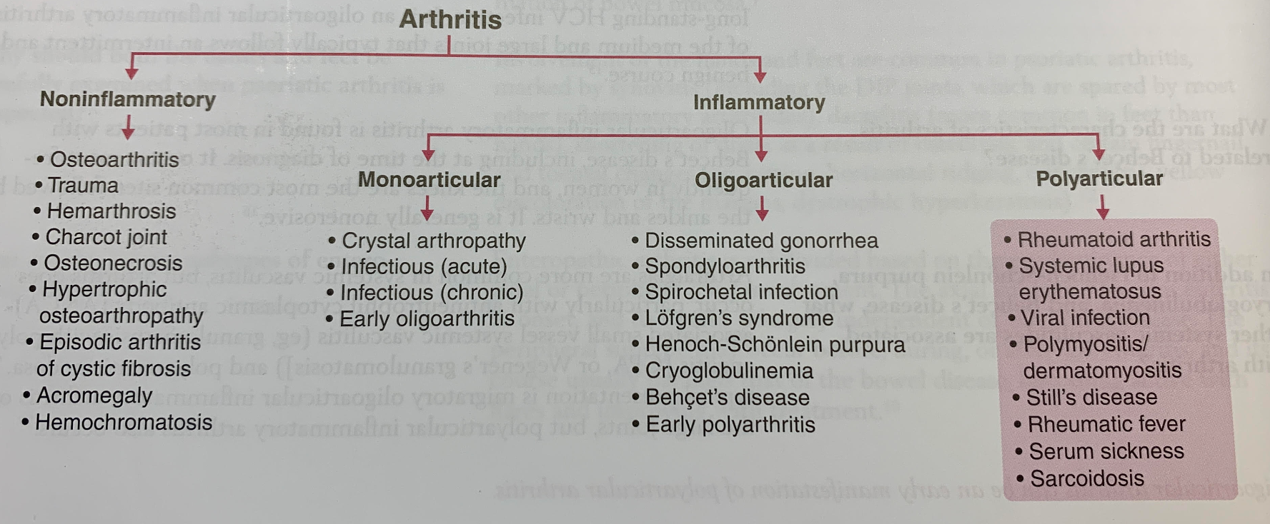
Cavitary Lung Lesion DDX

  • Mycobacteria
    • TB
    • M kansasii
  • Bacterial
    • Pseudomonas
    • Enterobacteriaceae
    • Klebsiella
    • Streptococci
    • Nocardia
    • Anaerobes
    • Staph aureus
  • Fungi
    • Crypto
    • Aspergillus
    • Histoplasma
  • Rare
    • Bacterial: Legionella, rhodococcus
    • Mycobacteria: MAC
    • Fungi: PCP/PJP, mucormycosis, blasto
    • Others: Vasculitis

PCP

Epidemiology

  • Immunocompromised patients
    • HIV with CD4 < 200
    • BMT, organ transplant patients
    • Leukemia/lymphomas
    • Rheumatology conditions
    • Primary immunodeficiencies
  • Rarely occurs in immunocompetent patients

Presentation

  • Typically subacute onset of pulmonary symptoms, non-productive cough, fever chills, malaise, SOB
  • Might have other clues of immunocompromised status

Diagnosis

  • CXR: Classic description is bilateral interstitial infiltrates
    • Rare instances: Lobar infiltration, nodules, cavitary lesions, pneumothorax
  • CT: Higher sensitivity but might not be necessary for the dx
  • LDH: Sensitive but not specific if elevated. An elevated LDH in an HIV patient without other medical co-morbidities that might inc LDH should raise suspicions for PCP
  • Beta D glucan: For HIV patients, good sensitivity, not that specific. If elevated, raises suspicions for PCP.
  • Gold standard: either visualize the organism via induced sputum (yield is variable, variable sensitivity but 100% specific), or via BAL.
  • Can also use 18S PCR

Management

  • First line: TMP-SMZ, 15-20 mg/kg. PO just as good as IV, duration 21 days
  • Sulfa allergy:
    • Mild to moderate: Desensitization
    • Severe: alternative agents
      • Clinda + primaquine
      • Trimethoprim + dapsone
      • Atorvaquone
      • Pentamidine
      • For severe disease, Clinda + primaquine or IV pentamidine is preferred.
    • Adjunctive steroids
      • ABG: If PaO2 < 70, or Aa gradient > 35, or hypoxemia on pulse ox, adjunctive steroid is indicated and has been shown to improve mortality
      • Pred 40mg BID x 5 days, then 40mg daily x 5 days, then 20mg daily x 11 days, total 21 days

Remember your Aa gradient equation!

Aa-gradient = PAO2 (calculated from the alveolar gas equation) – PaO2 (measured PaO2 on ABG)

PAO2 = FiO2 (baromeric pressure – water pressure) – PaCO2/(respiratory quotient)

Assuming sea level and a standard respiratory quotient of 0.8, this equation can be simplified to:

PAO2 = 150 – PaCO2/0.8

Culture Negative Endocarditis 5/7/2019

We recently had a case of a middle age man with SLE on chronic prednisone, ESRD on PD, presenting with acute on chronic shoulder pain x 10 days. Presentation was initially concerning for septic arthritis, and joint washout revealed gross purulence from the shoulder joint. Cultures were sent but no additional fluid studies were obtained.

A subsequent TTE, and later a TEE, confirmed a mitral valve vegetation concerning for concurrent infective endocarditis. However, multiple sets of blood cultures, fungal cultures, synovial fluid culture from the initial I&D/wash out, and even 16S PCR of the synovial fluid were all negative. This is a rare case of culture negative endocarditis which is later thought to be more likely Libman Sacs!


Septic Arthritis

  • Epidemiology
    • Risk factors: Advanced age, pre-existing joint dz, recent surgery or injection, SSTI, IVD, indwelling catheter, immunosuppression.
    • Most cases arise from hematogenous seeding, hence bacteremia is common.
    • Direct inoculation: usually due to trauma, surgery/injections, or wounds.
  • Microbiology
    • Usually mono-microbial, and Staph aureus is the most common cause of septic arthritis in adults.
    • GNR can be seen in older adults or in immunocompromised patients
  • Presentation
    • Monoarthritis is most common
      • Edematous, painful, warmth, limited ROM
      • Older patients may not be febrile
    • 20% of cases can present as oligoarticular or polyarticular infection. Polyarticular septic arthritis is more likely to occur in pts with RA
    • Most common affected joint is the knee
    • Could be a manifestation of infective endocarditis, esp amongst IVDU
  • Diagnosis
    • Synovial fluid analysis and culture, should be obtained prior to abx
    • Positive gram stain or culture is gold standard and diagnostic
      • PCR only required in rare cases since most non-gonococcal cultures obtained prior to antibiotics return positive. Negative cultures can result due to recent abx or atypical organism.
    • In pts with purulent synovial fluid (WBC 50k-150k) but culture negative, a presumptive dx can be made.
      • Likelihood of septic arthritis inc with inc leukocyte count
    • Blood cultures should be obtained
    • Should also evaluate for endocarditis given most common organism is staph aureus
    • Imaging:
      • Always get a radiograph to evaluate for concurrent bone/joint involvement
      • CT/MRI can be useful if looking for an effusion
  • DDx
    • Infectious
      • Gonococcal arthritis
      • Lyme disease
      • TB arthritis
      • Viral (usually polyarticular), i.e. Zika, Dengue, chikungunya, parvo, rubella, adenovirus
    • Non-infectious
      • Crystal dz
      • Reactive arthritis
  • Management
    • Joint drainage, severe infectious may require repeated aspiration or even wash out.
    • Abx
      • Most cases are staph, MRSA cases on the rise
      • Suspect pseudomonas if pt is immunocompromised or has h/o IVDU
      • Intra-articular abx: typically not used
      • Duration:
        • Staph aureus with bacteremia: At least 4 weeks
        • Staph aureus without bacteremia: at least 14 days IV, followed by 1-2 weeks PO
        • Bone involvement: 4-6 weeks
        • Any organisms, any bone involvement: 4-6 weeks
        • Other organisms: Typically at least 4 weeks

Culture Negative Endocarditis

  • Definition: Endocarditis without an identified organism in at least 3 independent blood cultures with negative growth after 5 days
  • Epidemiology
    • 2-7% of IE cases
    • 3 most common causes:
      • Previous abx
      • Inadequate samples
      • Atypical organisms (fastidious bacteria i.e. zoonotic microbes, fungal)
  • Microbiology
    • Farm animal exposure: Brucella, Coxiella (Q-fever)
    • Homeless: Bartonella Quintana
    • Cat: Bartonella hensale
    • Ingestion of unpasteurized milk: Brucella, Coxiella
    • Immunocompromised: Fungi, Coxiella
    • HACEK: Most common agents of culture negative endocarditis
      • Haemophilis aphrophulus
      • Actinobacillus
      • Cardiobacterum hominis
      • Eikenella corrdens
      • Kingella
  • Diagnosis
    • PCR, histology, special cultures are helpful.
    • PCR
      • 16S Ribosomal DNA: Bacteria
      • 18S Ribosomal DNA: Fungi
  • Non-infectious DDx
    • APLS, associated with Q fever
    • Acute rheumatic fever
    • Atrial myxoma
    • Libman Sachs endocarditis (non-bacterial thrombotic endocarditis or NBTE)
      • Seen in:
        • SLE
        • Advanced cancer
        • Hypercoagulable state
    • Vasculitis
    • Mural thrombus

NBTE (Non-bacterial thrombotic endocarditis)

  • Epidemiology
    • Rare affected all age group with no sex preference, most commonly 40s – 80s
    • Most commonly associated with pts with concurrent SLE or advanced malignancy (lung cancer, pancreatic cancer, gastric cancer)
    • Other associated conditions: APLS, rheumatic heart disease, RA.
  • Pathophysiology
    • A form of non-infectious endocarditis characterized by deposition of thrombi on halve valves, most commonly mitral or aortic
  • Presentation
    • Usually asx but high risk of thromboembolic events
    • May present with acute stroke or coronary ischemia
  • Diagnosis
    • Exclusion: Demonstration of vegetations on echo in absence of systemic infection in patients with risk factors.
  • Management
    • Systemic anticoagulation
      • Clinical experience and retrospective studies had shown this is beneficial due to high rate of emboli in pts with NBTE
    • Treat underlying condition
    • Surgery
      • Surgical excision for NBTE vegetation, can be considered in only selective cases and generally avoided.

Infective endocarditis

Thanks to Dr. Olivia Lee for letting us know of the case of this middle-aged woman with h/o endometrial cancer s/p TAH/BSO who was BIBA on a 5150 for GD after being found living in her yard.  Her medical clearance work up led to the diagnosis of endocarditis with a large abscess on the mitral valve leading to septic emboli to the brain, spleen, and kidneys as well as vitritis and endophthalmitis.  She was also noted to have an indwelling mediport with a vegetation at its tip, showering emboli into her lungs.  She successfully underwent urgent surgical replacement of her infected/destroyed valve.


Clinical Pearls

  • Use Duke’s criteria to help with your pre-test probability of endocarditis.  If patient meets criteria for definite endocarditis, consider going straight to a TEE.
  • TTE is not sensitive but highly specific for endocarditis.  However, in a patient with concerning clinical features (see next bullet point), a TEE is necessary to evaluate valve condition and plan for surgical intervention.  TTE is more useful if pre-test probability of endocarditis is low.
  • Indications for surgery
    • Valve dysfunction causing heart failure
    • Perivalvular extension with development of abscess, fistula, and/or heart block
    • Fungi or other highly resistant organisms that are difficult to treat with abx alone
    • Persistent bacteremia despite maximal treatment
    • Recurrent embolization with persistent vegetations
    • Large vegetations (>1 cm) with severe valvular regurg
    • S aureus prosthetic valve endocarditis
  • Indications for early surgery:
    • Heart failure
    • Uncontrolled infection
    • Prevention of embolic events
  • Most common cause of death in endocarditis is heart failure.

For a thorough review of endocarditis, please see our previous blog post here.

Bacteroides bacteremia

Some key learning points from our M&M case discussion today:

  • Ertapenem is a slow acting antibiotic and not an ideal empiric treatment in a patient who presents with sepsis or is acutely ill.  So if you want to use a carbapenem for empiric coverage, pick meropenem or imipenem instead.
    • Fun fact: ertapenem is actually more expensive than mero/imi.  The only use for ertapenem is in transitioning patients from hospitalization to home where its daily dosing is more favorable than the TID dosing of meropenem.
  • For diabetic foot ulcers, please refer to our SCVMC algorithm to help you figure out empiric antibiotics.  Simply open the HHSConnect browser and type in diabetic foot in the search bar to pull up the algorithm.
  • If blood cultures take a longer time to speciate (in our patient, over 5 days), expect anaerobes because anaerobes are difficult to culture and are sent out for speciation.  E coli is an organism that should speciate quickly and would grow in aerobic and anaerobic bottles.  If a species is only growing in anaerobic bottles, then it’s probably not E coli.
  • Levofloxacin is the only fluoroquinolone that has a role in outpatient treatment of GNR bacteremia, other fluoroquinolones (like cipro) are less effective.
  • Avoid using fluoroquinolones for empiric treatment of E coli bacteremia or pyelonephritis because our VMC antibiogram shows ~25% resistance with fluoroquinolones.