Tag Archives: Pulmonology

Pulmonary emergencies featuring Dr. Halley Tsai

  1. Attached a good and recent NEJM review on supplemental O2. There’s an video too that has everything in the text: https://www.nejm.org/doi/full/10.1056/NEJMvcm2035240
  1. ABG interpretation, comprehensive chapter on CHEST: https://www.thoracic.org/professionals/clinical-resources/critical-care/clinical-education/abgs.php
  1. Attached the CHEST cheat-sheet quick ABG I had in my talk. If people want very easy one-look cheat-sheets like that, the “All Resources” section has great ones to click through if people have time: https://www.chestnet.org/topic-collections/covid-19/clinical-resources

Pulmonary embolism

We discussed a case of a middle aged woman who presented with subacute to acute shortness of breath on exertion. We discussed her EKG with S1Q3T3 findings which is not specific for PE but indicative of cor-pulmonale. The differential for cor-pulmonale is COPD, ARDS, PNA, Pneumothorax and PE. More specifically, her CTA showed bilateral pulmonary embolus and RV enlargement and her labs indicated elevation in BNP and troponin. Due to her hemodynamic stability but clear evidence of RV strain, troponin and BNP elevation – she was classified as submassive PE (these days known as intermediate high risk classification). In addition to starting heparin infusion, with all submassive PE, it is important to consider systemic thrombolysis (low dose vs high dose) AND catheter directed thrombolysis / thrombectomy. This will be a nuanced discussion but admission to ICU and early consults to IR, Cardiology and Pulmonology are required.

Hemoptysis & Bronchiectasis

Today we learned about a patient who presented with hemoptysis in the setting of latent TB that was diagnosed as TB Bronchiectasis. We discussed the framework for hemoptysis:

Hemoptysis

We then reviewed the CT and determined it was bronchiectasis:

  • Bronchioectasis is defined as “Irreversible dilation and destruction of larger bronchi caused by chronic infection and inflammation”
  • Development of bronchiectasis always requires two factors
    • An infectious insult
    • Impaired drainage, airway obstruction or a defect in host defense
  • Etiologies
    • Any pulmonary Infections
      • Childhood infections (bacterial, viral or mycoplasma PNA)
      • Mycobacterial infections
    • Cystic Fibrosis
      • up to 7 percent of patients with cystic fibrosis (CF) are diagnosed at age 18 years or older
      • Sinusitis and bronchiectasis are the major respiratory manifestations of CF in adults
    • Airway obstruction
      • FBA or any other intraluminar obstructing lesion (such as a carcinoid tumor) or extraluminal compression
    • Tracheobronchomalacia/tracheobronchomegaly
    • Defective host defenses
      • Local: Ciliary dyskinesia
      • Systemic: hypogammaglobulinemia/prolonged immunosuppression
    • Young Syndrome
      • Bronchiectasis, sinusitis and obstructive azoospermia who have no evidence of cystic fibrosis
    • Rheumatic/Systemic Disease
      • RA/Sjogrens
    • Primary Ciliary Dysfunction
    • Allergic bronchopulmonary aspergillosis
      • should be suspected in patients with a long history of asthma that is resistant to bronchodilator therapy and associated with a cough often productive of sputum that is mucopurulent or contains mucous plugs.
    • Alpha-1 antitrypsin deficiencies
  • Signs and Symptoms
    • cough, mucopurulent sputum production, dyspnea, rhinosinusitis, hemoptysis (27%) and recurrent pleurisy
    • on exam crackles and wheezing are common
  • Radiographic Findings
    • The internal diameter of the bronchus is larger than that of its accompanying vessel
    • the bronchus fails to taper in the periphery of the chest
  • Treatment of Acute Exacerbation
    • Deciding when a patient has an acute exacerbation requires clinical judgement as there is no laboratory features specific for an exacerbation
    • Antibiotics decrease the existing bacterial burden and can decrease systemic inflammatory mediators
    • 10-14 day treatment course is appropriate (though the ERS 2017 guidelines suggest 14)

Malignant Pleural Effusion And White-Out!

Today we discussed a case of malignant pleural effusion causing complete opacification of a hemithorax. We learned the framework for subacute-chronic dyspnea, discussed the physical exam finding of clubbing, reviewed Light’s Criteria and discussed transudates and exudates. Here is a recap:

Subacute-Chronic Dyspnea DDx:

  • Pulmonary (malignancy falls into all of these categories)
    • Airway
      • bronchitis
      • COPD
      • Asthma
      • Bronchiectasis
      • Foreign body
    • Parenchyma
      • PNA
      • edema
      • atelectasis
      • ILD
    • Vasculature
      • PE
      • pHTN
      • AVMs (HPS can cause these)
    • Pleura
      • Pleural effusion
      • PTX
  • Cardiac
    • Arrhythmia
    • Ischemia
    • Valvular disease
    • Tamponade
    • Constrictive pericarditis
    • Myocarditis
  • Other
    • Anxiety
    • Anemia
    • Reduced PiO2
    • Hypoventilation
    • compensation for metabolic acidosis
    • pregnancy
    • Thyrotoxicosis

DDx for Clubbing:

  • 80% with underlying respiratory disorders
  • 10-15% with miscellaneous disorders
  • Congenital cyanotic heart disease, liver cirrhosis, chronic diarrhea, subacute endocarditis
  • 5-10% hereditary or idiopathic clubbing

Lights Criteria and Pleural Effusions (see this previous blog post for an excellent review)

https://scvmcmed.com/2017/04/17/am-report-32217-pleural-effusion/

DDx For Complete Opacification of a Hemithorax

  • Trachea pulled toward opacified side
    • Pneumonectomy
    • Total lung collapse
    • Pulmonary agenesis
    • Pulmonary hypoplasia
  • Trachea pushed away from the opacified side
    • Pleural effusion
    • Diaphragmatic hernia
    • Large pulmonary mass
  • Trachea remains central in position
  • Consolidation
  • ARDS/pulmonary edema
  • Pleural mass
  • Chest wall mass

Pneumocystis jiroveci pneumonia with a cavitary lesion 5/8/2019

Credit goes to Dr. Scott Burns from Roper Hospital in Charleston for his case on the Human Diagnosis Project.

Today we discussed a case of a young man with otherwise no medical history presenting with subacute dry cough, malaise, and weight loss. On exam he was septic on presentation with notable oral thrush. CXR revealed bilateral interstitial infiltrates and a RUL cavity with air fluid level which was confirmed on CT. LDH was elevated. He was confirmed HIV positive with a low CD4 count in the single digits, and BAL confirmed the diagnosis of PCP/PJP pneumonia!


Oral Thrush: NEVER assume normal, extremely rare in immunocompetent patients, so if you see this, consider whether the patient could be immunocompromised.


Cavitary Lung Lesion DDX

  • Mycobacteria
    • TB
    • M kansasii
  • Bacterial
    • Pseudomonas
    • Enterobacteriaceae
    • Klebsiella
    • Streptococci
    • Nocardia
    • Anaerobes
    • Staph aureus
  • Fungi
    • Crypto
    • Aspergillus
    • Histoplasma
  • Rare
    • Bacterial: Legionella, rhodococcus
    • Mycobacteria: MAC
    • Fungi: PCP/PJP, mucormycosis, blasto
    • Others: Vasculitis

PCP

Epidemiology

  • Immunocompromised patients
    • HIV with CD4 < 200
    • BMT, organ transplant patients
    • Leukemia/lymphomas
    • Rheumatology conditions
    • Primary immunodeficiencies
  • Rarely occurs in immunocompetent patients

Presentation

  • Typically subacute onset of pulmonary symptoms, non-productive cough, fever chills, malaise, SOB
  • Might have other clues of immunocompromised status

Diagnosis

  • CXR: Classic description is bilateral interstitial infiltrates
    • Rare instances: Lobar infiltration, nodules, cavitary lesions, pneumothorax
  • CT: Higher sensitivity but might not be necessary for the dx
  • LDH: Sensitive but not specific if elevated. An elevated LDH in an HIV patient without other medical co-morbidities that might inc LDH should raise suspicions for PCP
  • Beta D glucan: For HIV patients, good sensitivity, not that specific. If elevated, raises suspicions for PCP.
  • Gold standard: either visualize the organism via induced sputum (yield is variable, variable sensitivity but 100% specific), or via BAL.
  • Can also use 18S PCR

Management

  • First line: TMP-SMZ, 15-20 mg/kg. PO just as good as IV, duration 21 days
  • Sulfa allergy:
    • Mild to moderate: Desensitization
    • Severe: alternative agents
      • Clinda + primaquine
      • Trimethoprim + dapsone
      • Atorvaquone
      • Pentamidine
      • For severe disease, Clinda + primaquine or IV pentamidine is preferred.
    • Adjunctive steroids
      • ABG: If PaO2 < 70, or Aa gradient > 35, or hypoxemia on pulse ox, adjunctive steroid is indicated and has been shown to improve mortality
      • Pred 40mg BID x 5 days, then 40mg daily x 5 days, then 20mg daily x 11 days, total 21 days

Remember your Aa gradient equation!

Aa-gradient = PAO2 (calculated from the alveolar gas equation) – PaO2 (measured PaO2 on ABG)

PAO2 = FiO2 (baromeric pressure – water pressure) – PaCO2/(respiratory quotient)

Assuming sea level and a standard respiratory quotient of 0.8, this equation can be simplified to:

PAO2 = 150 – PaCO2/0.8

Hypersensitivity Pneumonitis Secondary to… Oyster Mushrooms 4/29/2019

Today we went over a case from the HumanDx Project (Credit goes to Dr. Maki Cronin, SSM Health St. Louis University Hospital). A young woman presents with 5 months of cough, dyspnea, and unintentional weight loss over the past 5 months in setting of working at an in-door oyster mushroom farm for the past 8 months. She was tachycardic and febrile on presentation, with crackles and clubbing on exam. CT revealed e/o fibrosis and GGO, and BAL revealed significant lymphocytosis. This presentation is consistent with a diagnosis of hypersensitivity pneumonitis, and more specifically, mushroom worker’s lung!


Hypersensitivity Pneumonitis (HP)

This condition has many faces/names:

  • Bird fancier’s lung (feathers, bird droppings)
  • Cheese-washer’s lung (Cheese Fancier per Sarasa)
  • Coffee worker’s lung
  • Compost lung (aspergillus)
  • Farmer’s lung (moldy hay)
  • Hot tub lung (hot tubs)
  • Mushroom worker’s lungs (mushroom)
  • Sauna worker’s lungs (contaminated sauna water)
  • Wine-grower’s lung (moldy grapes)

Epidemiology

  • 300 known antigens so far, most common (accounting for 75%) are:
    • Farming
    • Birds
    • Water contamination

Pathophysiology

  • Hypersensitivity to an environmental antigen leading to a type IV hypersensitivity reaction (they love asking these questions on tests for some reason) in genetically susceptible patients
    • Type 1: IgE mediated, immediate onset (min to hours)
      • Ex: Food allergies, PCN allergy, insect sting
    • Type 2: Cytotoxic hypersensitivity, Ab-mediated cell destruction
      • Drug induced cytopenias, Graves thyroiditis
    • Type 3: Immune complex formation
      • Ex: serum sickness, Arthus reactions, vasculitis, drug fever
    • Type 4: Cell-mediated delayed hypersensitivity
      • Activation of T-cells
      • Hours or days after antigen exposure
      • Ex: Tuberculin sensitivity, contact dermatitis, HP
  • Interstitial inflammation and infiltration with lymphocytes, later on granulomas and fibrosis develop over time

Presentation

  • Acute:
    • Occurs in sensitized pts with high level antigen exposure
    • Fever, chills, cough, chest tightness, dyspnea, nausea 4-8 hours after exposure
    • Exam: Tachypnea, inspiratory crackles, no wheezing
  • Chronic:
    • Occurs in pts with long-term low-level exposure
    • Months to years onset of exertional dyspnea, cough, fatigue, weight loss
    • Clubbing, fevers are uncommon but can happen
    • Over time: Pulmonary fibrosis, resp failure
  • Subacute:
    • Falls between acute and chronic forms

Diagnosis

  • A combination of clinical suspicions with exposure history, with assistance of imaging
  • CXR: Neither sensitive nor specific, may show reticular or nodular opacities
  • HRCT: typically shows profuse centrilobular nodules, predominantly GGO, more chronic exposure will lead to fibrosis, traction bronchiectasis
    • Mosaic pattern with areas of GGO is classic
    • More chronic picture leads to fibrosis, which can lead to traction bronchiectasis
    • Bronchiectasis is a chronic condition where the walls of the bronchi are thickened from inflammation and infection.
  • PFT: Can be obstructive, restrictive, or mixed. Obstruction more commonly seen in chronic.
  • BAL: Very sensitive but non-specific.
    • BAL lymphocytosis (often greater than 50%) is helpful but non-specific. Can also see lymphocytosis in COP and NIP but not this high.
  • Biopsy: Rarely done
  • Antigen-specific immunoassays: very high false positive rate, role unclear.

Management

  • Corticosteroids, usually pred 60 1-2 weeks, then tapered over 2-4 weeks for acute/subacute cases
  • Chronic: Longer course of prednisone
  • Remove from environmental exposure ASAP

Prognosis

  • Reversible if detected early and antigen exposure is eliminated
  • Chronic: leads to fibrosis, which is NOT reversible.

Sarcoidosis

Today, we talked about the fascinating case of a middle-aged man presenting with subacute cough, night sweats, and 15 pound weight loss, found to have bilateral hilar LAD on CXR and CT concerning for pulmonary sarcoidosis.  While awaiting LN biopsy, he developed L sided Bell’s palsy with MRI showing inflammation of CN5 and CN7 as well as nodular dural thickening of the trigeminal cave concerning for neurosarcoidosis.  LN biopsy showed non-caseating granulomas and he was subsequently started on high dose steroids for neurosarcoidosis.


Clinical Pearls

  • Sarcoidosis is a multisystem granulomatous disorder of unknown etiology.
  • The most common clinical presentation of sarcoidosis is pulmonary related.
  • In asymptomatic individuals with incidental hilar LAD, there is no indication for LN bx to diagnose sarcoidosis because 2/3 of cases resolve spontaneously without treatment.  Close follow up is needed to ensure that patients do not develop symptoms.
  • Similarly, Lofgren syndrome (arthritis, erythema nodosum, hilar LAD, and fevers) and Heerfordt’s syndrome (uveoparotid fever) are clinically consistent with sarcoidosis and there is no indication for LN biopsy.

DDx for unilateral facial droop

  • CNS lesion
    • Especially if forehead is spared.  However, keep in mind that a stroke involving the region of pons which houses the nucleus of CN7 would mimic Bell’s palsy!
  • PNS lesion
    • Infections
      • Zoster
      • HIV
      • Lyme
      • Syphilis
    • Malignancy
      • Parotid tumors
      • Acoustic neuroma/schwannoma
      • Lymphoma or other mass compressing CN7
    • Infiltrative/autoimmune
      • Sjogren’s syndrome
      • Sarcoidosis

Sarcoidosis

  • Multisystem granulomatous disorder of unknown etiology
  • 3-4 x more common in blacks
  • Overall prevalence is 10-20 per 100,000 people
  • Pathophys
    • Aberrant formation/accumulation of non-caseating granulomas
  • Clinical presentation
    • Age of onset is 20-60 years
    • Often discovered incidentally on a CXR
    • Most common organ involved is the lung (cough, SOB, CP) with ILD being the most common type of lung involvement. 30% of patients present with extrathoracic manifestations.
    • Fatigue, malaise, fever, and weight loss are common
    • Lofgren’s syndrome: arthritis, erythema nodosum, b/l hilar LAD
    • Heerfordt’s syndrome: parotid gland enlargement, facial palsy, fever, anterior uveitits
  • Diagnosis
    • No biopsy needed in the following
      • Asymptomatic with hilar LAD
      • Lofgren’s syndrome
      • Heerfordt’s syndrome
    • Otherwise, biopsy the easiest site to access.  Keep in mind that erythema nodosum does not have granulomas and would not help in diagnosing sarcoidosis.
    • Lab abnormalities that may be present:
      • Anemia
      • Leukopenia, eosinophilia, thrombocytopenia
      • ESR and CRP may be elevated
      • Hypercalciuria is more common than hypercalcemia
      • Moderate elevation in ALP suggests diffuse granulomatous hepatic involvement
      • Hypergammaglobulinemia and a positive RF (not usually part of routine work up)
      • ACE levels are elevated in 75% of untreated patients with sarcoid but has poor sensitivity and insufficient specificity (10% false positive rate with cocci, DM2, TB, hyperthyroidism, lung cancer, pneumoconiosis, etc.)
  • Organs that may be impacted in sarcoid
    • Pulmonary: occurs in over 90% of patients. Bilateral hilar LAD as well parenchymal disease. Can present with a restrictive spirometry pattern due to underlying fibrosis, pulmonary HTN
    • Cutaneous: can be disfiguring can be macules, papules, plaques and erythema nodosum
    • Liver/Spleen: a high alkaline phosphatase level suggests granulomatous liver disease
    • Neurologic: Noted in 5% of cases. MRI with contrast can help with diagnosis. Complications:
      • Cranial-nerve palsies (20-50%) → most common
      • Headache
      • Ataxia
      • Encephalopathy
      • Granulomatous meningitis
      • Weakness
      • Seizures
      • Neuroendocrine dysfunction
    • Optho: anterior uveitis most common manifestation
    • Cardiac: Typically cardiomyopathy, can also see arrhythmias (tachy or brady).
    • Renal: Can have hypercaliuria (more common than hypercalcemia) and renal calculi
    • Bone: chronic arthritis and cysts resembling rheumatoid, and diffuse granulomatous myositis.
  • Treatment/monitoring:
    • Asymptomatic? Follow up outpatient q3-4 months and annually thereafter to monitor for development of symptoms.
    • Symptomatic? Start steroids, re-evaluate q1-2 months
    • Refer to this NEJM article for organ specific treatments.
  • Chronic complications of pulmonary sarcoid
    • VTE is more common than the average patient population
    • Chronic pulmonary aspergillosis
    • Pulmonary HTN due to advanced fibrosis
  • Prognosis
    • Up to 80% of patients with hilar LAD spontaneously improve on their own!
    • With more symptomatic disease or more extrapulmonary manifestations, prognosis declines to less than 30% remission.
    • Overall mortality is <5%

Chronic Chest Pain and This X-ray… 2/11/2019

Gray Medicine had an interesting case of a 80yo F with history of treated TB (60 years ago), thoracic artery aneurysm s/p recent TEVAR, presenting with 3-4 months history of throbbing chest and back pain. She was admitted one month prior to the same complaint, CXR and CT Cx did not reveal significant pathology other than mild distal TEVAR graft dilatation. She presents 1 month later with worsening chronic chest pain, anorexia, weight loss.

This is the chest X-ray during this current hospitalization…

Miliary.png

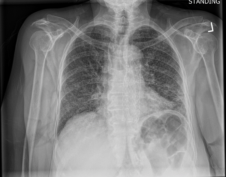
Burn this image into your head! This is a classic miliary pattern on a chest radiograph! The term miliary stems from millet seed, a term used to describe a group of small-seeded species of cereal crops or grains

Miliary2.jpg

Subsequent chest CT revealed innumerable bilateral pulmonary nodules, which were not present a month prior.

NewCTScan.gif

Let’s go through possible causes for a miliary pattern on a chest radiograph. In general it can be divided into three categories. The DDx can be quite wide!

Capture.JPG

In our case, given that our patient is an elderly woman with a remote history of treated TB, this presentation is highly concerning for miliary TB leading to reactivation. In general, miliary and disseminated TB are often used interchangeably. Disseminated TB refers to TB that affects at least two organ systems.

The most commonly affects organs are:

  • Lungs
  • Liver/GI
  • Spleen
  • Adrenals
  • CNS

Our patient was placed on airborne, and ultimately her sputum was MTB PCR positive! She has TB!

Presentation of Miliary TB

  • Very common: B-symptoms, FFT, typically subacute to chronic. 80-95% will have a fever.
  • Miliary, unlike typical TB, can present with acute sepsis or respiratory failure.
  • Pain/organ dysfunction based on location of the spread. Basically can affect anywhere. Hepatic TB, 79% of cases are due to miliary TB. Other commonly affected organs are spleen, adrenals, BM, lymphatics, and CNS.
  • Other manifestations: DIC, hyponatremia, pan-cytopenia, 50% cases will have normocytic anemia.

Risk Factors

  • Immunocompromised status
    • HIV
    • Extremes of age (infants, elderly)
    • Immunosuppressives
    • Post transplant
    • Other medical co-morbidities (CKD, cirrhosis, EtOH, etc)

Diagnosis

  • Chest radiograph: Classic faint reticulonodular infiltrate uniformly throughout lungs.
  • CT is more sensitive for miliary TB and usually is recommended. Typical finding might reveal numerous 2-3mm nodules but this is not specific.
  • Tissue, fluid, or lymph node biopsy
  • Gastric aspirate
  • Ultimately combination of clinical diagnosis with support labs/imaging.
  • Gold standard: AFB and culture + MTB PCR
  • All patient should have mycobacterial blood cultures
  • Urine mycobacterial cell wall glycolipid lipoarabinomannan (urine LAM) is a highly specific test with high sensitivity in HIV patients for disseminated TB.

Management

  • Intensive Phase: HREZ (aka RIPE) x 2 months
    • R: Rifampin
    • I: Isoniazid
    • P: Pyrazinamide
    • E: Ethambutol
  • Continuation Phase
    • After, 2 months of HREZ (RIPE), the continuation phase consists of 4 months of isoniazid and Rifampin.
    • Choice of medication and duration will change depending on resistance of the organism and location affected
  • Corticosteroids: Indicated if meninges or pericardium is involved.
  • Make sure to fill out a GOTCH form (not the GOAT form, as someone answered on Kahoot this morning) for Santa Clara County if you have a patient with active TB since a safe dispo will involve multiple disciplines and careful planning!


Cryptogenic Organizing Pneumonia 1/30/2019

Wendy presented a case of a middle age woman presenting with 4-6 weeks history of cough, shortness of breath, subjective fever and chills, non-improving after three courses of antibiotics. She was treated multiple times for presumed atypical CAP (bilateral infiltrates on CXR), and she presented again with worsening respiratory failure. Her infectious work up so far has been negative. CT Cx revealed bilateral infiltrates mainly in the peripheral lower lung zones.


Let’s go over non-resolving pneumonia and “typical pneumonia” for a little bit first.

“Typical” Pneumonia

  • Typically see sx improvement within 3-5 days of appropriate tx.
  • Vitals and O2 requirement expect to improve in 2 days
  • Fatigue and cough may take 2+ weeks to resolve.
  • Radiographic improvement usually takes weeks to months to clear up

If your patient is not improving within an expected time frame, then it’s time to broaden that differential! (The following are just some suggested ddx to consider)

Non-infectious causes (20% of the time)

  • Neoplasm:
    • Bronchogenic carcinoma, endobronchial obstruction secondary to mass effect, lymphoma
  • Inflammatory:
    • Vasculitis: GPA, pulmonary alveolar hemorrhage
    • Eosinophilic pneumonia
    • Acute interstitial pneumonia
    • Bronchiolitis obliterans organizing pneumonia (BOOP) or cryptogenic organizing pneumonia (COP)
      • Subacute, 75% of pts have sx < 2 months prior to diagnosis, flu like presentation initially mimicking an atypical pneumonia, patchy infiltrates also mimics pneumonia on chest radiograph.
    • Sarcoidosis
    • Connective tissue disease
    • Rare: Pulmonary alveolar proteinosis, plastic bronchitis
  • Drug-induced: Amiodarone, nitrofurantoin, chemo
  • PE
  • Pulmonary edema in abnormal lung architecture i.e. severe bullae seen in COPD patients.

Infectious causes

  • Streptococcus pneumoniae PNA: responsible for most cases of non-resolving infectious causes due to complications, i.e. multi-lobar involvement, drug resistance, co-morbidities.
  • Legionella
  • Mycoplasma pneumoniae
  • Chlamydia pneumoniae
    • Risk factors: SNF, military recruits
  • Haemophilus:
    • Risk factors: Elderly, immunocompromised
  • TB: Always on the DDx here.
  • Fungi: Always on the DDx here.
    • Aspergillus
    • Histo
    • Blasto
    • Cocci
    • Crypto
  • Nocardia
  • Actinomyces
  • PJP (HIV history)
  • Löffler’s syndrome
  • Complicated infection
    • Abscess (EtOH, poor dental hygiene at risk for anaerobes), might need prolonged course of abx.
    • Empyema: More likely in younger patients and those with illicit drug use

Diagnostic Approach in non-resolving cases of “pneumonia”

  • Assess for risk factors for delayed resolution, i.e. age, medical co-morbidities, pneumonia severity, and the pathogen involved.
  • If non-resolution, repeat history, assess for clues for atypical pathogen or non-infectious etiology. Ask if you’re treating the right bug if you’re sure that it’s an infectious cause (i.e. fungal?)
  • At this point, consider Chest CT and additional tests as needed. If CT is non-diagnostic, consider:
    • Bronchoscopy with BAL +/- transbronchial biopsy
    • CT-guided FNA if e/o LAD or lesion
    • Last resort: Consider surgical lung biopsy

Cryptogenic Organizing Pneumonia

Pathophysiology

  • Idiopathic diffuse interstitial process affecting distal bronchioles, alveolar ducts + walls leading to alveolar epithelial injury.

Epidemiology

  • Unknown! But pts are typically 40-60s, equally reported in M and F.

Risk Factors

  • Unclear, condition is not that well understood.

Presentation

  • Subacute to chronic cough, dyspnea, fever, malaise, may have an acute flu-like phase followed by a prolonged persistent of milder symptoms.
  • Typically diagnosed as CAP but fail to response to empiric abx.
  • Most common features:
    • Persistent non-productive cough (72%)
    • Dyspnea (66%)
    • Fever (51%)
    • Malaise (48%)
    • Weight loss (57%)
  • Lung exam: Ranging from normal to crackles

Diagnosis

  • Labs: Non specific but 50% of pts p/w leukocytosis, and elevated ESR (>100) and CRP are seen in 70-80%
  • CXR: Bilateral, patchy infiltrates
  • HRCT:
    • Usually reveals patchy air-space consolidations, GGO, small nodular opacities, and bronchial wall thickening. Patchy opacities occur more frequently in the peripheral and lower lung zones.
    • Mediastinal LAD might be present in rare cases
    • Closely resembles chronic eosinophilic pneumonia
  • PFT: Restrictive most commonly. DLCO is reduced in majority of cases, indicating gas exchange abnormalities.
  • Bronchoscopy + BAL:
    • Findings typically non-specific in COP but mainly done to rule out other etiology.
    • BAL: Might see increased lymphocytes, neutrophils, and eosinophils with lymphocytes predominance.
  • Trans-bronchial Lung biopsy: Usually done to ID other disease processes, non-specific findings in COP mimicking ILD.
  • Surgical Lung Biopsy: Will need a large sample

Management

  • No major RCTS so generally tx decisions are based on guidelines, experience, and case series.
  • Mild dz: Observe
  • Persistent symptomatic/worsening:
    • Oral glucocorticoids, usually up to 100mg/day but typically 60mg daily starting, x 4- 8 weeks, then taper over 3-6 months.
    • Serial radiographs
    • Failure to response to steroids:
      • Cyclophosphamide can be considered
      • Cyclosporine
      • Rituximab
    • Long term glucocorticoid dependence:
      • Can consider steroid sparing agents i.e. azathioprine (TPMT level!)
    • Severe, respiratory failure: High dose steroids initially then transition to orals.

Prognosis:

  • 2/3 of pts respond well to glucocorticoids with complete resolution of sx.
  • 1/3 have persistent symptoms and pulmonary abnormalities
  • Overall, better prognosis compared to ILD!

Take Home Points:

  • Typical illness script is a patient (men & women equally) in his/her 40-60s presenting with a chronic pneumonia like clinical picture not improving on antibiotics.
  • Chest radiograph with bilateral patchy infiltrates involving small airways/alveoli wall predominantly seen in the lower peripheral lung zones.
  • Responds well in most cases to corticosteroids, but most cases will need a prolonged course.
  • Check out this article from Chest for more learning!

Cavitary lung lesions and SJS/TEN

Today, we discussed the case of a Vietnamese man who presented with chronic cough, 40 pound weight loss, and joint pain, found to have cavitary lesions in his lungs with work up revealing pulmonary TB as well as tophaceous gout on urate-lowering therapy with allopurinol leading to SJS/TEN.


Clinical Pearls: 

  • Cavitary lung lesions have a broad differential (see below) aside from TB.
  • Risk factors for developing SJS/TEN include HIV (100x higher risk), genetics (especially Asians and South Asians), autoimmune diseases, malignancy, and high dose/rapid infusion of offending meds.  Consider genetic testing prior to starting meds associated with this allergy (allopurinol, sulfa drugs, PCNs, AEDs, etc.) in at risk populations.
  • Nikolsky sign can be positive in SJS/TEN, staph scalded skin syndrome, and pemphigus vulgaris
  • Time of onset is 1-3 after starting the offending drug
  • SCORTEN score is useful for determining prognosis
  • Early use of cyclosporine in patients with SJS/TEN has shown significant reduction in mortality.

DDx for cavitary lung lesions

  • Infection
    • Pyogenic (necrotizing pneumonia, septic emboli, lung abscess)
    • Atypical (MTB, fungi)
  • Autoimmune
    • GPA >> RA, sarcoid
  • Malignancy
    • Liquid (lymphoma, KS, lymphomatoid granulomatosis)
    • Solid (squamous, GU>GI)
  • Vascular
    • PE
  • Other
    • Foreign body granulomatosis

SJS/TEN (Steven Johnson vs toxic epidermal necrolysis)

  • < 10% = SJS, > 30% = TEN, in between = Overlap
  • Common causes
    • Sulfa drugs
    • Abx (PCN, quinolones)
    • AEDs
    • Allopurinol
    • Infx: Mycoplasma, graft-vs-host
    • Idiopathic
  • Risk factors
    • HIV (100x higher risk)
    • Genetics
      • There are a lot of them (check on uptodate for specific drugs) but a couple examples are:
        • HLA-B*58:01 (allopurinol)
          • Patients with this positive gene has higher risk for severe cutaneous hypersensitivity reaction to allopurinol including SJS and TEN. High risk Asian populations carrying this gene are Korean, Thai and Han Chinese.
        • HLA-B*15:02 is recommended before starting carbamazepine in Asians and South Asians
        • Cytochrome CYP2C19 polymorphism
    • Autoimmune disease
    • Malignancy
    • High doses and rapid infusion of medications
  • Clinical Presentation
    • 1-3 weeks after offending drug
    • Fever >39
    • Influenza-like symptoms (malaise, myalgias, arthralgias) x 1-3 days
    • Conjunctival itching or burning
    • Odynophagia
    • Cutaneous findings:
      • Acute onset macules over face, trunk, may form flaccid bullae
      • Nikolsky sign:
        • Positive when shear stress on the skin i.e. rubbing results in exfoliation. Indicates a pathology at the dermal/epidermal junction.
        • Positive in
          • SJS/TEN
          • Staphylococcal scalded skin syndrome
          • Pemphigus vulgaris
      • Asboe-Hansen sign (AKA bullae spread sign)
      • Mucous membrane involvement.
        • Eyes, mouth lesions
        • Respiratory sx
  • Prognosis:
    • SCORTEN score
    • Mortality with SJS is 10%, TEN 30%
  • Management
    • Supportive care for skin
    • Pain control
    • IV fluids
    • Prevention of vulvovaginal sequelae
    • Ocular management
      • Evaluate for loss of surface epithelium
      • Opthalmic therapy
        • Saline rinses to remove debris
        • Artificial tears
        • Topical steroids
        • If extensive sloughing, then amniotic membrane transplantation (prokera ring)
    • Adjunctive therapies
      • Steroids: may lead to higher rates of complications
      • IVIG: conflicting data
      • Cyclosporine: one large case series from Spain and two systematic reviews have shown that cyclosporine given at 3 to 5 mg/kg may slow the progression.  Inhibits T cell activation and thus prevents the production and release by cytotoxic T cell and natural killer cells of cytokines that could propagate SJS/TEN.
        • A study on 71 patients of whom 49 were treated with cyclosporine and 22 with other therapies found mortality rates were 10% and 32% respectively.  Expected mortality based on SCORTEN for the cyclosporine group was 24% and 29% in the other group.
        • A 2018 meta-analysis on 255 patients with TEN found that treatment with cyclosporine was associated with a 70% reduction in mortality risk
      • Plasmapharesis
      • Anti-TNF

Bonus info on gout:

  • Acute flare:
    • Steroids
    • NSAIDs
    • Colchicine (avoid in severe renal or hepatic impairment or with meds that inhibit CYP450 system)
  • Indications for urate-lowering therapy for chronic treatment
    • Frequent or disabling gout flares
    • Clinical or radiographic signs of joint damage
    • Tophaceous deposits in soft tissues or subchondral bone
    • Gout with renal insufficiency (CrCl<60)
    • Recurrent uric acid nephrolithiasis
    • Urinary acid excretion >1100 mg/day)
  • Goal uric acid is <6mg/dL
  • Agents for chronic management
    • Xanthine oxidase inhibitors
      • Allopurinol, lower dose for CKD3 or higher renal disease
      • Febuxostat, very expensive, cardiovascular and hepatic side effects
    • Uricosuric drugs: ineffective if CrCl<50. Can worsen kidney injury. Avoid use if GFR <30
      • Probenecid
      • Lesinurad
    • Uricase
      • Pegloticase, fast improvement of symptoms, contraindicated in G6PD deficiency