Tag Archives: Dermatology

Anti-HMGCR Immune-Mediated Inflammatory Myositis

Today we discussed a fascinating case of statin-related anti-HMGCR positive immune-mediated inflammatory myositis (also called necrotizing autoimmune myositis). The case highlighted the importance of a framework approach to diseases.

We first went over the framework for true muscle weakness, which can be anatomically divided as follows

Weakness

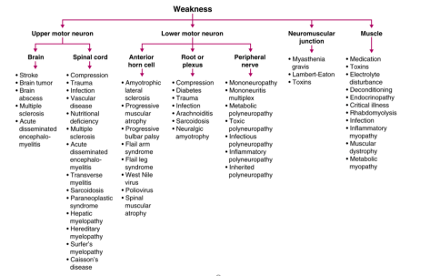
Source: Frameworks for Internal Medicine (Dr. Andre Mansoor from OHSU). Available on Amazon (highly recommended!)

To help us localize the lesion to a myopathy, we used the following framework to determine that it was likely a myopathy.

LocalizingTheLesion

The differential for myopathy is broad, and generally is the same for an elevated CK and non-traumatic, non-exertional rhabdomyolysis. The causes can be divided as follows. If you like mnemonics, think Drug-REGIIME for the various categories.

DrugRegiime

Once we narrowed the differential to an inflammatory myopathy, we utilized the following chart that guided us to the probable conclusion that it was an immune-mediated necrotizing myopathy (also known as necrotizing autoimmune myopathy). This was confirmed by a highly positive anti-HMGCR antibody

DifferentiatingInflammatoryMyopathy

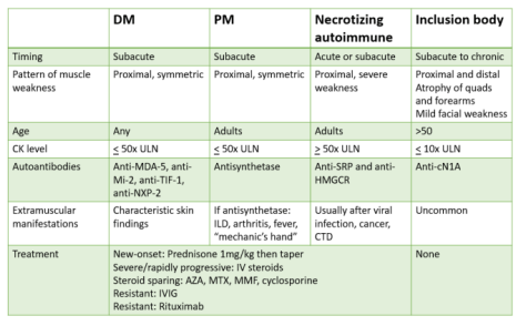
Adapted from a NEJM Article: https://www.nejm.org/doi/full/10.1056/NEJMra1402225

Clinical Pearls about INMN/NAM:

  • Can occur in association with viral infections, malignancy, in patients with CTD such as scleroderma or in patients taking statins
  • Can start acutely or sub-acutely with severe proximal muscle weakness and markedly elevated CK values of between 10-100k ULN (2K-20K)
  • A rare diagnosis, but some experts believe that polymyositis is overdiagnosed and that INMN may actually be more common!
  • Two antibodies are highly specific though not sensitive for the condition (anti-SRP and anti-HMGCR)
  • The majority of anti-HMGCR positive cases are related to known exposure to a prescribed statin (~70%)
  • Treatment is to discontinue the statin but most cases will require prompt immunosuppression
    • Statin-induced muscular events are on a spectrum, and include the following:
      • 1) Mildly elevated CK and myalgias
      • 2) Rhabdomyolysis
      • 3) Self-limited toxic myopathy
      • 4) IMNM
    • It is only IMNM that generally does not improve with merely cessation of the drug and it generally needs immunosuppression
  • Biopsy is required to make the diagnosis but the presence of the antibody will often result much more quickly and in our case, the patient started on immunosuppresion prior to the biopsy results because the clinical profile fit perfectly with this diagnosis

Shingles and Complications 3/11/2019

Thanks Elan for presenting a case of a 91 year old F presenting with a progressively painful and erythematous rash, 2 weeks after she was treated with presumed Shingles by her PCP. It turned out that she had superimposed cellulitis over her healing Shingles lesions and possibly elements of post-herpetic neuralgia, requiring a Dilaudid PCA for pain control.

Lame joke of the day: Shingles + Cellulitis = Shinglelitis, get it?


Shingles

Epidemiology

  • Risk inc with age, esp for pts > 50, but it can develop at any age
  • Fortunately, most people will only have one outbreak in their life time, < 4% recurrence

Pathophysiology

  • Reactivation of the varicella zoster virus in sensory ganglia after a long latency period following primary infection from varicella (chicken pox). When the virus activates, the virus travels down the nerve fibers to the skin, hence a dermatomal distribution.
  • Weakening of the immune system is associated with outbreaks, i.e. AIDS, lymphoma, immune-suppressives.

Presentation

  • 2-3 days prior to rash: pt might develop a tingling sensation, hypersensitivity, or itching over a particular dermatome. Later on vesicles on an erythematous base develop. Painful and very sensitive.
  • Blisters form over 3-5 days, then dry and crust over the next 5 days
  • Blisters are CONTAGIOUS until the vesicles scab over.
    • Keep affected area dry and clean!
  • Expanding rash or blisters that persist for > 2 weeks indicate immune-compromised status

Complications

  • Most common is post-herpetic neuralgia
    • 10% of patients, inc with age
    • Pain can be very debilitating, some patients need to be admitted for pain control.
  • Zoster ophthalmicus
    • Involves the eye, seen in 10-25% of cases when shingles hit V1
    • Antiviral should be administered ASAP, preferably within 72 hours of onset of sx.
    • Valacyclovir is recommended, 7-day course, 1000mg PO TID
    • Alternative: Acyclovir 800mg PO 5 times daily x 7-10 days, Famciclovir 500mg PO TID.
    • If e/o keratitis or uveitis, topical steroids can be used.
    • Can lead to vision loss, especially with corneal scarring. Some patients would require corneal transplant.
    • Post-herpetic neuralgia occurs in 36.6% of pts over 60, and 47.5% over age 70.
  • Disseminated zoster
    • If > 3 contiguous dermatomes or 2 separated dermatomes are affected.
  • Bacterial infection of the skin:
    • Risks inc with scratching
    • Inc risk of scarring
  • Ramsey Hunt Syndrome:
    • Reactivation of VZV at the geniculate ganglion.
    • Triad of Ipsilateral facial paralysis, ear pain, vesicles on face/ear or IN THE EAR. Can lead to deafness, tinnitis or vertigo due to vestibulocochlear nerve involvement.
    • Mgx: Anti-viral within 72 hours, steroids. Hearing loss is likely permanent so treat ASAP.

Diagnosis

  • Primarily clinical
  • Swabbing ulcer/vesicular fluid for HSV PCR has high sensitivity, quick turn around time.

Management

  • Acute management
    • Anti-viral: Valacyclovir, famiciclovir, acyclovir. Start ASAP and preferably even before blisters occur. Effectiveness is greatest if antiviral is started within 72 hours of onset of symptoms (even before vesicles appear if clinical suspicion is high enough!)
    • IV antiviral recommended for disseminated disease
    • Options: Acyclovir (5 times a day dosing), Valacyclovir (TID dosing), famiciclovir (TID as well).
    • Help shorten duration and complications
    • Pain control:
      • Lidocaine, capsaicin, gabapentin, Lyrica.
      • Use opioids if and only if necessary.
      • Antidepressants i.e. Cymbalta and Effexor have variable benefits for post-herpetic neuralgia.
    • Keep area dry and clean, DO NOT SCRATCH.
  • Infection Control:
    • Localized herpes zoster: Standard precautions, contact
    • Disseminated: airborne + contact
    • Immunocompromised patient: airborne + contact regardless
  • Post-exposure:
    • Previously received 2 doses of varicella vaccine: Monitor for 8-21 days for sx
    • Previously only received 1 dose of varicella vaccine: Should get the 2nd dose ASAP (minimum of 4 weeks apart from 1st dose). Monitor for sx.
    • No prior vaccination: Potentially contagious from days 8-21 post exposure, should be removed from patient care duties. Post-exposure vaccination should be provided ASAP. If varicella vaccination is contraindicated (i.e. pregnant), varicella-zoster immune globulin is recommended.
  • Vaccination/Prevention
    • Vaccinate children
    • Vaccinate adults > 50 regardless of whether they have had chicken pox or shingles and regardless of whether they had the older vaccine
      • Older: Weakened live virus, Zostavax
      • Newer: Recombinant Herpes Zoster vaccine, Shingrix, 2 doses IM, 2-6 months apart, at least 2 months after the older vaccine. Contains inactivated parts of the virus, not a live vaccine.
        • Effectiveness: 97% effective in preventing shingles for pts > 50, vs Zostavax which is 50-64% effective.
        • Reduces post-herpetic neuralgia if you get it shingles

Cavitary lung lesions and SJS/TEN

Today, we discussed the case of a Vietnamese man who presented with chronic cough, 40 pound weight loss, and joint pain, found to have cavitary lesions in his lungs with work up revealing pulmonary TB as well as tophaceous gout on urate-lowering therapy with allopurinol leading to SJS/TEN.


Clinical Pearls: 

  • Cavitary lung lesions have a broad differential (see below) aside from TB.
  • Risk factors for developing SJS/TEN include HIV (100x higher risk), genetics (especially Asians and South Asians), autoimmune diseases, malignancy, and high dose/rapid infusion of offending meds.  Consider genetic testing prior to starting meds associated with this allergy (allopurinol, sulfa drugs, PCNs, AEDs, etc.) in at risk populations.
  • Nikolsky sign can be positive in SJS/TEN, staph scalded skin syndrome, and pemphigus vulgaris
  • Time of onset is 1-3 after starting the offending drug
  • SCORTEN score is useful for determining prognosis
  • Early use of cyclosporine in patients with SJS/TEN has shown significant reduction in mortality.

DDx for cavitary lung lesions

  • Infection
    • Pyogenic (necrotizing pneumonia, septic emboli, lung abscess)
    • Atypical (MTB, fungi)
  • Autoimmune
    • GPA >> RA, sarcoid
  • Malignancy
    • Liquid (lymphoma, KS, lymphomatoid granulomatosis)
    • Solid (squamous, GU>GI)
  • Vascular
    • PE
  • Other
    • Foreign body granulomatosis

SJS/TEN (Steven Johnson vs toxic epidermal necrolysis)

  • < 10% = SJS, > 30% = TEN, in between = Overlap
  • Common causes
    • Sulfa drugs
    • Abx (PCN, quinolones)
    • AEDs
    • Allopurinol
    • Infx: Mycoplasma, graft-vs-host
    • Idiopathic
  • Risk factors
    • HIV (100x higher risk)
    • Genetics
      • There are a lot of them (check on uptodate for specific drugs) but a couple examples are:
        • HLA-B*58:01 (allopurinol)
          • Patients with this positive gene has higher risk for severe cutaneous hypersensitivity reaction to allopurinol including SJS and TEN. High risk Asian populations carrying this gene are Korean, Thai and Han Chinese.
        • HLA-B*15:02 is recommended before starting carbamazepine in Asians and South Asians
        • Cytochrome CYP2C19 polymorphism
    • Autoimmune disease
    • Malignancy
    • High doses and rapid infusion of medications
  • Clinical Presentation
    • 1-3 weeks after offending drug
    • Fever >39
    • Influenza-like symptoms (malaise, myalgias, arthralgias) x 1-3 days
    • Conjunctival itching or burning
    • Odynophagia
    • Cutaneous findings:
      • Acute onset macules over face, trunk, may form flaccid bullae
      • Nikolsky sign:
        • Positive when shear stress on the skin i.e. rubbing results in exfoliation. Indicates a pathology at the dermal/epidermal junction.
        • Positive in
          • SJS/TEN
          • Staphylococcal scalded skin syndrome
          • Pemphigus vulgaris
      • Asboe-Hansen sign (AKA bullae spread sign)
      • Mucous membrane involvement.
        • Eyes, mouth lesions
        • Respiratory sx
  • Prognosis:
    • SCORTEN score
    • Mortality with SJS is 10%, TEN 30%
  • Management
    • Supportive care for skin
    • Pain control
    • IV fluids
    • Prevention of vulvovaginal sequelae
    • Ocular management
      • Evaluate for loss of surface epithelium
      • Opthalmic therapy
        • Saline rinses to remove debris
        • Artificial tears
        • Topical steroids
        • If extensive sloughing, then amniotic membrane transplantation (prokera ring)
    • Adjunctive therapies
      • Steroids: may lead to higher rates of complications
      • IVIG: conflicting data
      • Cyclosporine: one large case series from Spain and two systematic reviews have shown that cyclosporine given at 3 to 5 mg/kg may slow the progression.  Inhibits T cell activation and thus prevents the production and release by cytotoxic T cell and natural killer cells of cytokines that could propagate SJS/TEN.
        • A study on 71 patients of whom 49 were treated with cyclosporine and 22 with other therapies found mortality rates were 10% and 32% respectively.  Expected mortality based on SCORTEN for the cyclosporine group was 24% and 29% in the other group.
        • A 2018 meta-analysis on 255 patients with TEN found that treatment with cyclosporine was associated with a 70% reduction in mortality risk
      • Plasmapharesis
      • Anti-TNF

Bonus info on gout:

  • Acute flare:
    • Steroids
    • NSAIDs
    • Colchicine (avoid in severe renal or hepatic impairment or with meds that inhibit CYP450 system)
  • Indications for urate-lowering therapy for chronic treatment
    • Frequent or disabling gout flares
    • Clinical or radiographic signs of joint damage
    • Tophaceous deposits in soft tissues or subchondral bone
    • Gout with renal insufficiency (CrCl<60)
    • Recurrent uric acid nephrolithiasis
    • Urinary acid excretion >1100 mg/day)
  • Goal uric acid is <6mg/dL
  • Agents for chronic management
    • Xanthine oxidase inhibitors
      • Allopurinol, lower dose for CKD3 or higher renal disease
      • Febuxostat, very expensive, cardiovascular and hepatic side effects
    • Uricosuric drugs: ineffective if CrCl<50. Can worsen kidney injury. Avoid use if GFR <30
      • Probenecid
      • Lesinurad
    • Uricase
      • Pegloticase, fast improvement of symptoms, contraindicated in G6PD deficiency

Varicella Pneumonia

Joe presented the case of a young man from Mexico with unknown immunization history who presented with acute onset of AMS, fevers, and a progressive vesicular rash, diagnosed with primary varicella infection (chickenpox!), now in the ICU with varicella pneumonia and likely varicella vasculitis induced stroke.


Clinical Pearls

  • Vaccinate your kids!
  • Two main VZV presentations are primary infection (chickenpox) and reactivation (shingles, disseminated zoster in immunocompromised individuals)
  • Varicella rash presents as vesicular lesions at varying stages.  Vesicular lesions at the same stage of development are concerning for smallpox.
  • The most common complication of primary VZV in adults is pneumonia.  Treatment is with IV acyclovir.
  • The most common neurologic complication of primary VZV is encephalitis.  No approved therapy exists.
  •  Isolation precautions for shingles is contact.  For disseminated zoster or chickenpox, make sure you patient is on contact and airborne precautions.

Differential for fever, rash, and pharyngitis:

  • Measles
  • Mono (due to EBV, CMV, toxo, HHV6)
  • Acute HIV
  • Parvovirus
  • Zoster
  • HSV
  • Mycoplasma

Fever and rash emergencies:

  • Meningococcemia
  • Subacute bacterial endocarditis
  • Rocky Mountain Spotted Fever
  • Necrotizing fasciitis
  • Toxic epidermal necrolysis
  • Toxic shock syndrome (staph aureus or GAS)

Varicella zoster (VZV)

  • Primary infection – chickenpox
    • Clinical manifestations:
      • Prodrome of fever, malaise, pharyngitis, loss of appetite
      • Rash is often pruritic and occurs in successive crops over days (new vesicle formation stops after 4 days). Vesicular lesions at varying stages on an erythematous base on the trunk, face, and extremities.
    • Diagnosis:
      • send swab (from ulcer base) for HSV PCR and DFA.  These have quick turn around time and high sensitivity.  Viral culture takes weeks and is less sensitive.
    • Most common complications
      • Children: skin infection
      • Adults:
        • Pneumonia (1/400 cases) with a mortality of 10-30%. In people requiring mechanical ventilation, mortality reaches 50%.
          • Risk factors for pneumonia development are cigarette smoking, pregnancy, immunosuppression, and male sex.
          • Develops 1-6 days after the appearance of rash
          • CXR usually with diffuse bilateral infiltrates with possible nodular component in early stages
          • Prompt administration of acyclovir has been associated with clinical improvement
        • Neurologic:
          • Encephalitis: acute cerebellar ataxia (more common in children), diffuse encephalitis (more common in adults)
            • No proven therapy once encephalitis occurs. Acyclovir has been used with anecdotal success
          • Transient focal deficits
          • Aseptic meningitis
          • Transverse myelitis
          • Vasculitis (medium to large vessel vasculopathy)
          • Hemiplegia
        • Hepatitis
          • More common in immunocompromised hosts and frequently fatal
        • Other
          • Diarrhea, pharyngitis, otitis media
    • Treatment
      • For healthy children <12 ⇒ nothing
      • For adults
        • if no complications, then oral valacyclovir (1g TID) or acyclovir (800 mg 5 times/day)
          • if immunocompromised ⇒ treat with IV acyclovir if active lesions present (10mg/kg q8h)
        • if complications
          • acyclovir IV 10mg/kg q8h for 7-10days
        • contact and airborne precautions!
  • Reactivation – shingles
    • Clinical manifestations –
      • Rash – most common location is thoracic and lumbar dermatomes
        • Localized, painful and restricted to a dermatome
        • Disseminated if > 3 contiguous dermatomes or 2 dermatomes on separate parts of the body, painful
      • Acute neuritis – 75% of patients have pain/burning/throbbing prior to onset of rash
    • Complications in immunocompetent hosts –
      • post-herpetic neuralgia (most common), superficial skin infections, ocular complications (acute retinal necrosis and zoster ophthalmicus), motor neuropathy, meningitis, Ramsay hunt syndrome (zoster oticus)
    • Treatment
      • For patient with localized disease presenting <72 hours after clinical symptom onset, treat with oral acyclovir, valacyclovir, or famciclovir
      • For patient with localized disease presenting >72 hours after disease onset, then monitor
      • Pregnant women, treat with acyclovir
      • Disseminated disease, treat with IV acyclovir


Leukocytoclastic vasculitis

Today, we talked about the case of a middle-aged man with history of diabetes, HTN, and A fib who presented with acute onset of progressive painful palpable purpura on his extremities, found to be cutaneous small vessel vasculitis on skin biopsy!


Clinical Pearls

  • Purpura implies problem at the level of vessel.  It can be divided into
    • Non-palpable purpura: petechiae (<3mm) or ecchymoses (>3 mm) and are usually associated with disorders of coagulation and platelets.
    • Palpable purpura: suggests inflammation and possible vasculitis.

Nomenclature:

  • Cutaneous small vessel vasculitis: disease limited to skin without any systemic vasculitis or glomerulonephritis
  • LCV: histopathologic term defining vasculitis of small vessels
  • Hypersensitivity vasculitis: small vessel necrotizing vasculitis
  • Immune complex small vessel vasculitis: associated with immune complex and/or complement deposition. If limited to skin, this is identical to cutaneous small vessel vasculitis. If not limited to skin, then other etiologies like cryo, SLE, Sjogren, RA, anti-GBM, IgA, etc.

Approach to purpura

Approach to purpura

  • Hypersensitivity (in the normal complement category of vasculitis) can result from medications/drugs as well as certain conditions such as HIV.
    • Numerous meds can cause LCV including some common ones such penicillins, cephalosporins, sulfonamides (including most loop and thiazide-type diuretics), phenytoin, and allopurinol have been most often implicated

Cutaneous small vessel vasculitis:

Clinical presentation:

  • Palpable purpura
  • + petechiae
  • Lesions can coalesce, ulcerate or be surrounded by hemorrhagic bullae
  • No visceral organ involvement in CSVV. However, it can occur later in the disease course.

Diagnosis:

  • Start with checking serum complement levels to guide your need for further laboratory work up!
  • Skin biopsy

Management and Prognosis:

  • Usually self limited and resolved within 2-4 weeks
  • If uncomplicated:
    • NSAIDs
    • Antihistamines
    • Rest, elevate, compression stockings
  • If complicated (presence of hemorrhagic blisters, cutaneous necrosis, or ulceration can lead to secondary infections, chronic wounds, and scarring)
    • Systemic glucocorticoids (oral steroids): pred 0.5 mg/kg of ideal body weight until new lesion formation ceases, then taper over 3-6 weeks
    • If relapse with prednisone: then colchicine or dapsone
    • If refractory: then azathioprine, methotrexate, and MMF

Example of palpable purpura with hemorrhagic blisters:

LCV skin example

Mycoplasma Induced Rash & Mucositis (MIRM!) 10/24/2018

Ernest presented a case of a young woman, with no medical history, presenting with acute onset severe mucositis (eyes, mouth, urogenital) after a few days of viral prodrome and one day after taking azithromycin prescribed by her PCP. Her skin findings were almost non-existent and the bulk of her symptoms were isolated to the mucosa. Her presentation is consistent with a diagnosis of MIRM!


MIRM (Mycoplasma Induced Rash and Mucositis)

Epidemiology

  • 25% of patients with mycoplasma pneumoniae experience extra-pulm manifestations
  • Coined different terms, incomplete SJS, Fuchs Syndrome, MIRM
  • Mean age: Young (median 11-12 yo), male predominance.

Presentation

  • Universally will have some sort of prodrome: cough, malaise, fever preceding eruption of lesions by ~ 1 week.
  • Manifestations: variable, mucositis alone, prominent mucositis with sparse skin involvement. Skin involvement tends to be very rare and on the milder side, presenting as vesiculobullous, targetoid, papules, macules. Rarely morbilliform.
  • Majority of cases are severe mucositis alone.
  • Involvement: Oral (100%), ocular (92%), urogenital (78%)

Diagnosis

  • Clinical Dx
  • Mycoplasma IgM/IgG helps but their sensitivity and specificity are highly variable.

Management:

Supportive care (especially pain control, hydration/nutrition, infection prevention) plus treat the underlying cause (mycoplasma)!

  • Systemic corticosteroids (mixed data so generally not recommended first line)
  • IVIG (has been used in very severe cases))

Prognosis

  • Better than SJS/TEN, 81% will make a full recovery.
  • Blindness/residual visual impairment is possible but less common vs SJS/TEN

Key distinguishing features:

MIRM: Young, slight male preference, usually 7 days after infection, predominantly mucosal involvement, very little cutaneous involvement, better prognosis vs SJS/TEN.

SJS/TEN: Any age, female preference, usually 1-3 weeks after drug exposure, diffused skin involvement (Nikolsky sign) + mucosal involvement, more severe ocular manifestation.

Please refer to this review article for more background on this condition.

92% blasts… Fever… AND a rash?! 9/19/2018

Narges presented a case of a middle age woman without any prior medical history, presenting with 1 week of bruising, epistaxis, and bleeding from her gums. Her initial lab work was notable for a WBC of 52.2 with 92% blasts, later confirmed to be AML. She developed a fever and a rash over the next few days… She had neutropenic fever, and around the same, time, developed AML-associated Sweet’s Syndrome!

AML: A quick overview

Accounts for 80% of acute leukemias in adults

Risk Factors

  • Benzene exposure
  • Radiation exposure, commonly 7-10 years after exposure
  • Prior tx with alkylating agents and topoisomerase II inhibitors like doxorubicin, etoposide.
  • Age: greatest risk factor, older = at high risk, median age 65
  • CML, MDS, and myeloproliferative syndromes have a chance to evolve into AML.

Initial presentation

  • Bruising, gum bleeding, epistaxis from thrombocytopenia
  • SOB, DOE, fatigue from anemia
  • Pyogenic infections of the skin
  • HSM found in 1/3 of pts
  • 50% might have gingival hyperplasia as first signs of the disease
  • Small subset might have concurrent HLH on presentation
  • If fever, almost always infection

Diagnosis

  • Buzz words: blasts on smears, Auer rods (peroxidase stain)
  • > 20% blasts cells
  • Flow cytometry
    • CD117, CD33 most common
    • CD19, if seen, suggests lymphoblastic origins
  • Subtypes
    • M3 (Acute promyelocytic leukemia), t(15; 17), prone to DIC, responsive to ATRA and potentially can be cured.
      • If pt receiving ATRA +/- Arsenic trioxide develop pulmonary sx think of an entity called Differentiation Syndrome, can be life threatening, stop treatment and give steroids.
    • Non APL: Everything else

Management:

  • Induction, consolidation (after complete remission, assess induction response via BM bx) via HiDAC high dose cytarabine, or autologous CT, or allogeneic HCT, maintenance (usually not needed but can be beneficial in some types of AML)
  • APL: ATRA +/- ATO
  • Non-APL: 7 day course of cytarabine and 3 day course of an anthracycline. For older pts with more comorbidities, can use a milder regimen with azacytidine or decitabine.


Neutropenic fever

Definition: T > 38.3 or > 38 sustained over 1 hour, with neutropenia (ANC < 500)

Determine high risk or low risk

  • Low Risk: Anticipated neutropenia < 7 days, clinically stable, NO medical comorbidities
    • IDSA: Can consider outpatient antibiotics, Cipro + Augmentin and able to tolerate PO, otherwise inpatient management
  • High Risk: Anticipated neutropenia > 7 days, clinically unstable, any medical comorbidities
    • Automatically inpatient management
    • Monotherapy with pseudomonal coverage initially is recommended by IDSA
      • Cefepime: 2g Q8H, higher dose than usual
      • Meropenem 1g Q8H
      • Imipenem
      • Zosyn 4.5g Q6-8
      • Ceftazidime increasingly avoided.
    • If history of MRSA, e/o catheter infection, skin infection, pneumonia, or unstable, add Vancomycin/MRSA coverage
    • PCN allergy: Can consider using Aztreonam, cipro
    • If recurrent of persistent fever after 4-7 days: Add an empiric antifungal, most of the time cover for candida since it’s the most common cause of invasive fungal infection.
      • Echinocandin is favored i.e. caspofungin, increasing azole resistance in candida.
      • Think aspergillus if e/o pulmonary nodules, ampho B and voriconazole then are preferred

Sweet’s Syndrome

Uncommon, inflammatory disorder, usually affects pts ages 30-60. Older = more likely malignancy associated

Presentation:

  • Abrupt, painful, edematous (juicy), erythematous papules/plaques/nodules + fever and leukocytosis.
  • Rare mucosal/oral involvement.
  • Can also rarely causes inflammation of a particular organ system, i.e eye, liver, heart, CNS, kidney, even bone.

Sweet.jpg
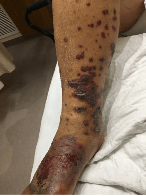
Image adapted from Derm 101

Types

  • Classic
    • Idiopathic, majority of cases
    • Associations: Infections (URI, GI) 1-3 weeks after infection
    • IBD
    • Pregnancy
    • HIV, TB, hepatitis, autoimmune conditions
    • Possible inc risk of malignancy
  • Malignancy associated
    • AML is the malignancy most associated with Sweet’s Syndrome.
    • Risk:
  • Drug-induced (long list but some of the potential ones we used more commonly are):
    • Bactrim, Macrobid, AED, hydralazine, clozapine, PTU, GCSF, Mirena, Lasix, Azathioprine, ATRA

Dx Criteria: both majors and 2 minors are required

  • Majors
    • Abrupt onset of painful erythematous plaques or nodules
    • Histopath evidence of dense neutrophilic infiltrate without evidence of leukocytoclastic vasculitis
  • Minor:
    • > 38C
    • Underlying malignancy, IBD, pregnancy, or recent upper resp, GI infection, or vaccination
    • Steroid responsive
    • Labs: ESR > 20, CRP elevated, leukocytosis > 8000 with > 70% neutrophils)

Biopsy: Dense, neutrophilic infiltrate in the dermis, w/o e/o vasculitis.

Catastrophic Antiphospholipid Syndrome, +/- Heparin induced thrombocytopenia & thrombosis, +/- SLE 8/1/2018

Wendy presented a fascinating (and confusing!) case of a patient with history of APS and DVT/PE on chronic warfarin presenting with painful, non-blanching, palpable purpuric rash on the left thigh and lower abdomen found to have thrombocytopenia,  and proteinuria. Skin biopsy revealed small vessel microthrombi. Work up positive SRA, positive ANA, positive DsDNA, low complements… Base on this presentation, the patient possibly has catastrophic antiphospholipid syndrome, with heparin induced thrombocytopenia, and newly diagnosed SLE!

CAPS1CAPS2CAPS3.JPG