Abdominal Pain Secondary to… Lots of Foreign Bodies 11/6/2018

Our doctor-in-training, Jacqueline, presented a case of a 46yo man with a complicated abdominal surgical history, as well as schizophrenia, who presents with acute onset vague abdominal pain. He could not provide any remarkable history (other than abd pain and losing a bag of coins), and his exam was otherwise benign except for mild diffused abdominal pain…

The mystery was resolved on a radiography.

Picture1


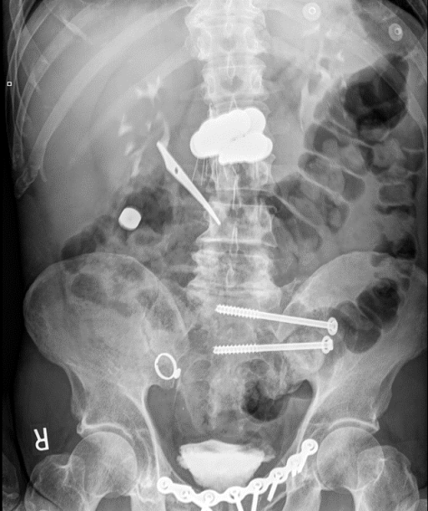
Foreign Body Ingestion

Epidemiology

  • Mostly in kids, peaks 1-2 years of age
  • Adults: Typically, accidental (95% of cases) usually related to fish, chicken bones, or toothpicks. More common in older adults, pts with mental illnesses, intoxicated, or inmates (drug trafficking, packers vs stuffers).
  • Most frequent cause of esophageal obstruction = food bolus on existing stricture

Presentation

  • Asymptomatic
  • Stridor/airway compromise/aspiration
  • Chest pain/abdominal pain
  • Fever, shock (perforation)
  • Hemoptysis, hematemesis
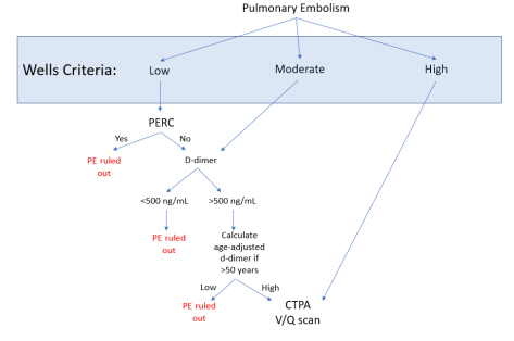
Diagnosis

  • Imaging, clinical history

Management:

  • Will depend on stability, the location, nature of the objects ingested, and progression.
  • Expectant management for most blunt objects, ~ 70-80% of objects will pass by day 4. Consider surgical/endoscopic intervention if failure to progress
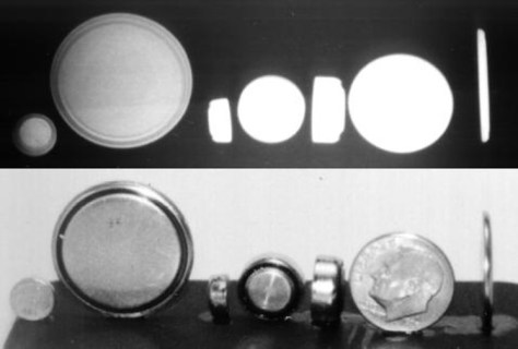
Battery

  • Presentation
    • Local necrosis secondary to pressure, electrical current, or caustic chemicals.
    • Ulceration can occur within 2-4 hours
    • Perforation can be seen as early as 4-8 hours
    • VERY IMPORTANT to distinguish between coin batteries (thicker, concentric circles) vs coins (thinner, confluent)!
    • Picture2.jpg
  • Complications
    • Vocal cord paralysis, esophageal perf, stricture, tracheal/esophageal fistula, aspiration pneumonia, mediastinitis, erosions into arteries, gastric hemorrhage, intestinal perf
  • Management
    • Esophagus: Emergent removal
    • Beyond esophagus: Depends, most (89%) will pass within 7 days
      • Surgical/Endoscopic option: consider if co-ingestion of magnets, or if remained in stomach for more than 48 hours.
      • GI symptoms
    • Cylindrical batteries: Relatively harmless and usually pass through GI tract without issues, but if stuck in stomach or esophagus, endoscopic removal is recommended

Magnets

  • Presentation
    • Fistula, perforation, volvulus, obstruction, localized necrosis (pressure)
    • Higher chance of complications if multiple magnets and/or metallic objects were ingested.
    • Can react with metal external of the body and cause injury
  • Complications
    • Localized bowel necrosis, obstruction
  • Management
    • Prompt removal endoscopically if in esophagus or stomach.
    • Beyond stomach: Surgery if symptomatic or failure to progress
    • Single magnet: Expectant management, serial XR, monitor progress, don’t be around anything ferromagnetic

Sharp

  • Presentation
    • High risk of perforation/injury if in esophagus, medical emergency
  • Complications
    • Esophageal perforation
    • Intestinal perforation
  • Management
    • Immediately endoscopic removal if in esophagus
    • Beyond:
      • Stomach/proximal duodenum: still consider urgent endoscopic removal, complication risk varies from as low as 10% to 40%
      • Beyond and failure to progress: Surgical intervention recommended.
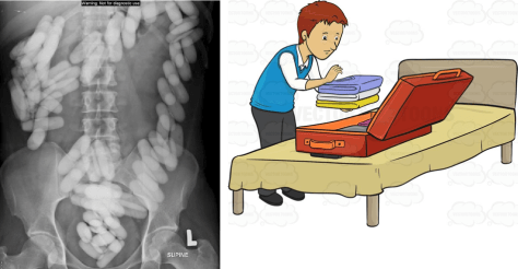
Packers vs Stuffers

  • Packers: Carefully PACKING illicit substances into packages, lower chance of leakage (image adapted from Vectortoons.com)
    • Picture3.png
  • Stuffers: Hastily STUFFING illicit substances to hide evidence from law enforcement (image adapted from Family Guy), higher chance of content leakage.
    • Picture4
  • Management:
    • Decontamination:
      • Packers: Whole-bowel irrigation safe and feasible
      • Stuffers: Controversial
    • Symptomatic:
      • Opioid (CNS depression, hypoventilation, pinpoint pupils): IV Naloxone 0.05 in nonapneic patients, 0.2 – 1mg in apneic patients. Larger doses may be required if pt ingested a large amount of heroin.
      • Sympathomimetic (agitation, hypertension, hyperthermia): Symptomatic management, airway monitoring, temperature control. AVOID pure beta blockers. Can consider GI decontamination but consult Poison Control.

If suspecting ingestion of potentially toxic substance, don’t hesitate to call Poison Control!

Picture5


Endobronchial Tuberculosis 11/5/2018

Sarasa presented a case of a young woman with recently diagnosed pulmonary TB on HREZ presenting with worsening dyspnea and voice changes. Her fiberoptic endoscopy of the upper airway was normal. She was found on CT to have tissue thickening and stranding in the mid/lower trachea as well as a small tracheal diverticula, very suspicious for endobronchial TB!


Endobronchial tuberculosis

Definition: TB that involves the tracheobronchial tree

Epidemiology

  • More common among patients with extensive pulmonary TB, especially with cavitary lesions, can occur in 10-40% of patients but less common now with anti-TB therapy.
  • For some reason more likely to occur in women in second to third decades of life.
  • Usually seen in main and upper bronchi, but in 5 % of cases: involves the lower trachea

Pathophysiology

  • Unclear but thought to be by either direct extension of pulmonary disease into the bronchi, spread via infected sputum, or hematogenous/lymphatic spread.

Presentation:

  • Cough, CP, hemoptysis, wheezing (low pitch), fatigue, fever, dyspnea. Can mimic foreign body aspiration, non-resolving pneumonia, or malignancy.
  • Can have significant sputum production, leading to bronchorrhea (> 500mL/day of sputum)
  • Complications: Atelectasis, obstruction, bronchiectasis, tracheal stenosis, fistula, hilar lymph node rupture

Diagnosis: CT and bronchoscopy are methods of choice for dx, with bronch being the most validated for confirming the diagnosis.

  • XR: Can be normal in 10-20% of cases
  • CT: Can demonstrate endobronchial lesions, stenosis (up to 2/3 of patients), or fistulas.
  • Bronchoscopy: able to visualize stenosis and biopsy. Can interview if severe symptomatic stenosis.

Management

  • Same as for other forms of TB but also prevent tracheobronchial stenosis
  • Medical Therapy
    • Intensive Phase: HREZ (aka RIPE) x 2 months
      • R: Rifampin
      • I: Isoniazid
      • P: Pyrazinamide
      • E: Ethambutol
    • Continuation Phase
    • Ex: 2HREZ/4HR = standard regimen, 2 months of HREZ (RIPE), followed by 4 months of isoniazid and Rifampin
  • Specific for endobronchial TB
    • Corticosteroids: Controversial.
      • Shown improvement in outcome (prevention of bronchial compression) in children.
      • Some data on shortening healing time and decrease severity of bronchial stenosis
    • Nebulized INH or streptomycin, mixed data
    • Surgery: Usually indicated for stenosis or stricture. Balloon dilatation, stenting, ablation, resection, cryosurgery.
    • Severe tracheobronchial stenosis sometimes requires pneumonectomy or lobectomy

Laryngeal TB:

Distinctive entity, also more prevalence in younger patients, most commonly presents with dysphonia (96%), odynophagia (25%), and stridor (9%). True vocal cords, epiglottis, and false vocal cords are most commonly involved.


Drug Resistance and TB

Definition:

  • Drug-resistance TB: resistant to one or more anti-TB drugs
  • Mono-resistant: single agent
  • Poly-resistant: resistant to multiple drugs, but susceptible to either INH or rifampin but not both
  • MDR: R to INH and rifampin + others.
  • XDR-TB: Extensively drug resistant TB: resistant to INH, rifampin, fluoroquinolones + either aminoglycosides or capreomycin or both.

1st Line Agents

  • Rifampin
  • Isoniazid
  • Pyrazinamide
  • Ethambutol
  • Streptomycin

2nd Line Agents

  • Fluoroquinolones (Levofloxacin, Moxifloxacin)
  • Injectable: Amikacin, Capromycin, Kanamycin
  • Other: Cycloserine, Linezolid, Ethionamide

Addon Agents/Tertiary

Bedaquiline, Para-aminosalicyclic acid, imipenem, meropenem + Augmentin, thioacetazone.

If suspecting resistant, strategy usually is to add at least 2 additional drugs. Adding single drug inc risk for resistance.

Management of drug-resistant TB:

Tx of MTB PCR will be based on susceptibility data

  • Conventional: 20-26 months treatment, with an intensive phase with at least 5 effective drugs for at least 6 months after negative sputum, followed by a continuation phase of at least 4 drugs for 18-24 months
  • Shorten version for 9-12 months if no extra-pulmonary manifestation and susceptible to quinolones.

Please refer to this informative article on a review of endobronchial TB.

All about PE – 11/1/18

Thanks to Barnie for presenting the case of a middle-aged woman who was admitted with acute onset of SOB, found to have submassive PE.


Clinical Pearls:

  • Risk stratification tools are helpful in estimating the pre-test probability of PE.  The best and most validated is Wells criteria.
    • YEARS items is a newer tool that was studied in an RCT in the Netherlands and found to lower the number of CTPA scans ordered by 14% without a significant impact on rates of missed PE diagnoses.
  • For patients at low risk of PE according to Wells, PERC is useful in ED or outpatient setting to rule out PE without ordering a d-dimer (see graphic below).
  • Age-adjusted d-dimer is age x 10 for patients older than 50 years.  This accounts for the increase in d-dimer baseline related to aging.  ADJUST-PE trial showed that age-adjusted d-dimer leads to higher specificity without subsequent VTE.
    • Studies have shown an 11.6% reduction in CTPA scans with the use of this correction factor without an appreciable increase in missed diagnoses of PE.
  • Think of PE in three broad categories:
    • Massive PE = hemodynamically unstable ⇒ anticoagulation + thrombolysis
    • Submassive PE = hemodynamically stable + RV strain ⇒ anticoagulation + thrombolysis
    • Low risk PE = hemodynamically stable, no RV strain ⇒ anticoagulation.  Use the PESI score to determine if your patient can be treated outpatient.
  • Remember that the most common EKG finding in PE is normal sinus rhythm!  The most common abnormal  EKG finding is sinus tachycardia.  S1Q3T3 pattern is only seen in 10% of patients with PE.

Diagnosis:

Suggested algorithm for diagnostic work up of suspected PE:

PE diagnostics

Remember that the scoring tools above are only there to add to your clinical judgment, not replace it!

Recent study in the Lancet looked at the utility of a different diagnostic algorithm, using the three most predictive items on Wells together with d-dimer.  Compared to Wells, this diagnostic tool led to a 14% reduction in unnecessary CTPA!

PE diagnostics 2

Treatment:

PE treatment.PNG

  • Remember that clot burden does not factor into the treatment categories of PE.  Low clot burden in a patient with baseline cardiopulmonary disease can still lead to hemodynamic compromise and would be considered massive PE.
  • Submassive PE treatment is an area of much debate.  A famous trial (PEITHO trial) in 2014 randomized 1006 patients to receive heparin + placebo vs heparin + tenecteplase (European version), and found a >50% reduction in combined death and cardiovascular collapse at 7 days but a > four-fold increase in risk of major bleed including intracranial hemorrhage.  Subsequent meta-analyses (and this one) found that the risk of major bleeding was highest in people >65 years of age.  So treatment decisions here are tricky and require consulting multiple services!

Signs of RV strain: 

  • EKG findings:
    • S1Q3T3: this is a sign of cor pulmonale and can be seen in a number of conditions in addition to PE
      • Bronchospasm (really bad asthma)
      • ARDS
      • Pneumothorax
  • Echo findings:
    • Elevated RVSP
    • Septal bowing
    • McConnell’s sign (regional wall motion abnormality sparing the RV apex)
      • Not sensitive but helpful in distinguishing RV strain due to chronic pulmonary HTN from RV strain due to acute PE
    • Increased RV size
    • Decreased RV function
    • Tricuspid regurgitation
  • Labs
    • Elevated troponin
    • Elevated BNP