Tag Archives: Toxicology

Caffeine Overdose! 5/1/2019

The world’s #1 addictive substance of choice… Caffeine! And yes you can OD on it!

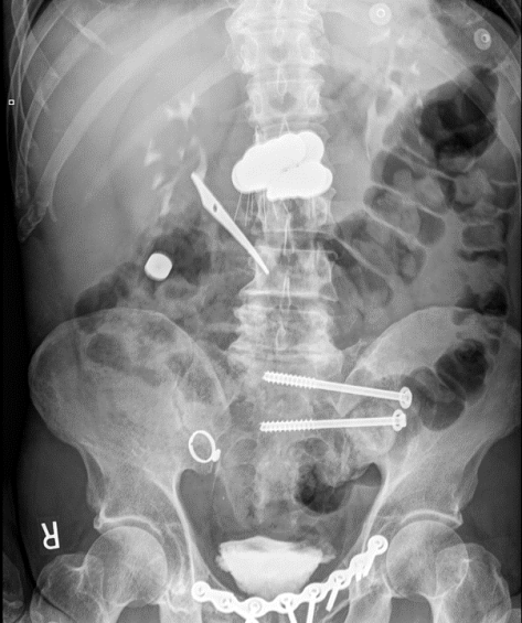
A 37yo F with history of anxiety presented with nausea and palpitations after ingesting 160 pills of Diurex in an attempt to fix her constipation for the past 2 weeks. Prior to arrival to the hospital, the paramedics administered activated charcoal. She was tachycardic, mildly hypotensive, hypokalemic, and acidotic (AG 20). Methamphetamine was found in her system as well. Fortunately she improved with fluids and supportive measures, but lethal cases of caffeine overdose, while rare, have been described in the medical literature!

Of note: An expresso doubt shot contains roughly 60-100mg of caffeine.

This patient took 16000mg (16g) of caffeine, equivalent to 160-260 shots of double expresso!


Caffeine Intoxication

  • Epidemiology
    • Caffeine = world’s #1 psychoactive compound consumed
    • Pure caffeine can be easily obtained, caffeine pills introduced in 2004.
    • Death from caffeine overdose is rare, only 92 cases have been described in literature.
    • Caffeine pills heavily advertised as weight loss supplements
  • Risk factors
    • Psychiatric conditions
    • Athletes (weight loss/work out supplements, these things are NOT regulated!)
    • Infants/young kids (accidental ingestion)
  • Pathophysiology
    • Dose-dependent MOA
    • CNS and cardiac stimulation, usually occurs at plasma concentration of 15mg/L or higher
    • Usually not food/beverages related. Most cases are related to caffeine-containing medications.
    • Lethal cases reported over 10g ingestion, highest reported ingestion is 100g (1000 double shots of Expresso)
    • Absorbed in the GI tract within 30 minutes
    • Half life 5-6 hours, metabolized in the liver
  • Presentation
    • Agitation, diaphoresis, anxiety, restlessness, insomnia, GI disturbances, tremors, psychomotor agitation
    • Complications
      • Serious cases: arrhythmias (SVT, VT), even V-fib (most common cause of death in caffeine intoxication)
      • Hypotension
      • Hypokalemia
      • Seizures
      • Lactic acidosis
      • Rhabdomyolysis
      • Renal failure
  • Diagnosis
    • Life threatening caffeine overdose more commonly associated with blood concentration > 80mg/L
    • Clinical history, serum measurement, ingestion history
  • Management
    • Activated charcoal for ingestions:
      • Effective only within a short time of ingestion, typically within 1-2 hour and patients have to be mentating
      • Interacts with caffeine and prevents it from being absorbed.
    • Hydration
    • Electrolyte repletion
    • Anti-arrhythmic agents (amiodarone, even lidocaine) in setting of arrhythmias, ACLS if unstable arrhythmias
    • Dialysis: Dialyzable

Source: GrepmedToxidrome Flow

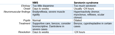
A summary of toxidromes:

Toxidrome.jpg

Severe Metabolic Acidosis Secondary to… Methanol Poisoning 4/3/2019

Credit goes to Eric for informing us about this case!

A 48yo M presents with acute onset encephalopathy. He has a distant history of alcohol abuse, and during the day of presentation he had complained about not being able to see. On presentation, his labs were notable for an anion gap of 35 with bicarb of 4, lactic acid of 11.9,  ABG of 6.56/52/336, and osm gap of 65. Volatile screen ultimately came back positive for methanol level of 145.56 mg/dL (yes this is very high). He was given bicarb pushes, fomepizole, and urgent dialyzed.

It turns out that he might have ingested Klean Strip denatured alcohol, which is 40-60% methanol!


Methanol Toxicity

Pathophysiology

  • Toxic metabolite of methanol poisoning is formic acid, which is formed from formic acid after methanol is metabolized in the liver by alcohol dehydrogenase and aldehyde dehydrogenase
  • Leads to retinal injury and eventual blindness (permanent)
  • Formic acid can also cause ischemic or hemorrhagic injury to the basal ganglia, hence in methanol poisoning you might see changes around the area (putamen is part of the basal ganglia).
  • Ingestion of 1g/kg is fatal, and toxicity has been reported in as little as one teaspoon
  • Ethylene glycol, on the other hand, mainly causes renal damage. (Flank pain, hematuria, oliguria). Buzz words = calcium oxalate crystals in urine.

Picture1.png

Presentation

  • Visual blurring, central scotomata (black spot in center of vision), and blindness are suggestive of methanol poisoning.
  • Eye exam might reveal: mydriasis, retinal edema, hyperemia of the optic disk
  • Co-ingestion of ethanol can delay presentation of toxicity.
  • Labs:
    • Profound AGMA, bicarb often < 8 mEqL
    • High serum lactate
    • Elevated plasma osmo gap
      • Calculated osmolality = 2 x [Na mmol/L] + [glucose mg/dL] / 18 + [BUN mg/dL] / 2.8 + [Ethanol/3.7]
      • OG = Measured osmo – calculated osmo
      • Nrl < 10
    • Typical agents that inc the osmolar gap:
      • Methanol
      • Ethylene Glycol
      • Diuretics (osmotic diuretics i.e. mannitol)
      • Isopropyl alcohol
      • Ethanol
    • Osmo gap takes Into account the quantity of uncharged molecules, hence it will only be elevated in presence of the parent alcohols.
    • In late presentation: most alcohols have been metabolized already into charged active metabolic, hence osmo gap is not very sensitive in late presentation.

Management

  • ABC comes first.
  • Sodium bicarb administration
    • Corrects systemic acidosis, which limits penetration of formic acid by converting it into negatively charged formate, which cannot cross cell membrane to cause damage.
      • Formic Acid <- -> H+  & Formate-, adding bicarb dec H+, hence driving the equilibrium equation to the right. See? College O-chem is still helpful.
    • HCO3 (mEq) required = 0.5 x weight (kg) x [24 – serum HCO3 (mEq/L)], and in general up front give 1-2 mEq/kg via IV bolus for any patients with pH < 7.3 followed by a sodium bicarb D5 gtt, at least 150-250cc/hr to correct acidosis
      • Goal of infusion is to keep pH above 7.35
    • Bicarb can be found in:
      • Most common: 8.4%, 1mEq/mL, 1Amp = 50mEq, you will see these in the crash cart, very helpful to familiarize yourself with setting the syringe up for injection in a code blue situation!
      • Gtt: 100-150 mEq/1000 mL in D5
      • Concentrated gtt: 1mEq/mL, might need pharmacy’s help in formulating this. You can only use this in the ICU!
      • PO tabs: Useful for CKD patients
  • Inhibition of alcohol dehydrogenase in the liver
    • Fomepizole
      • 1st line
      • Loading: 15mg/kg, followed by 10mg/kg Q12hr, continue until blood pH is normalized and serum methanol is less than 20mg/dL
    • Ethanol: ADH has better affinity for ethanol, leading to competitive inhibition.
      • Difficult to dose, sedating effect, cannot be used in cirrhosis patients, pregnant patients.
  • HD indicated if:
    • Severe acid-base derangements, or even high AGMA regardless of drug level
    • Severe levels of methanol
    • End organ damage
    • Interacts with fomepizole, hence if concurrent therapy, fomepizole should be dosed Q4H
  • Co-factor therapy
    • All methanol patients treated with ADH inhibition should also receive:
      • Leucovorin 50mg IV (folinic acid) or folic acid Q6H
      • Thiamine also commonly administered due to unclear nutritional status.
  • GI decontamination: Has no role whatsoever.

Aspirin toxicity

We discussed the case of a middle aged woman admitted with AMS, found to have AGMA and respiratory alkalosis with work up revealing ASA toxicity, managed with HD!


Clinical Pearls

  • In suspected ASA toxicity, check serum levels every 2 hours until two consecutive levels decrease from peak value
  • The goal in treatment of ASA overdose is to keep ASA in its charged and deprotonated state which has less end organ toxicity.
    • Give bicarb with the goal of maintaining urine pH of 7.5-8 and serum pH <7.60.
    • Treat hypokalemia aggressively (see below).
  • Patients with ASA overdose have a high minute ventilation so avoid intubation if possible to allow them to maintain their minute ventilation.
  • Call renal early for HD if indicated ⇒ AMS, cerebral/pulmonary edema, fluid overload, kidney injury, severe acidemia, ASA level >100 mg/dL, or clinical deterioration in spite of aggressive management

Management of ANY patient with suspected toxic ingestion:

  • ABCs (Airway, Breathing, Circulation)
  • Call Poison Control! (1800 222-1222)
  • Can patient get Activated Charcoal? (usually only within 1 hour of ingestion)
  • Check Utox, Salicylate screen, acetaminophen screen, +- alcohol and volatile screen if suspected. You don’t want to miss a potential co-ingestion! 

ASA overdose

  • Remember that ASA can be found in other compounds like topical salicyclic acid, herbal medications, bismuth subsalicyclate (part of Pepto-Bismol), and Oil of Wintergreen so don’t forget about those topical medications!
  • Most sensitive vital sign abnormality in early ASA overdose is tachypnea with hyperventilation. 
  • Classic acid/base abnormality is anion gap metabolic acidosis with respiratory alkalosis (see below)

How does ASA work?

  • Inhibition of cyclooxygenase results in decreased synthesis of prostaglandins, prostacyclin, and thromboxanes. This contributes to platelet dysfunction and gastric mucosal injury
  • Stimulates the chemoreceptor trigger zone to cause Nausea and Vomiting
  • Activates the respiratory center in the medulla leading to hyperventilation and respiratory alkalosis
  • Interferes with cell metabolism (Krebs cycle and decouples oxidative phosphorylation) leading to metabolic acidosis

Metabolism

  • Reaches peak concentration within 1 hour of ingestion. Takes longer with the enteric coated formulations
  • Detox occurs normally by the liver and then metabolites are excreted by the kidney. In OD, liver is overwhelmed so more of the drug becomes dependent on renal excretion (slow and can take up to 30 hours).

Clinical features

  • Tinnitus
  • Vertigo
  • Nausea and vomiting
  • Diarrhea
  • Hyperpnea (tachypnea and hyperventilation)
  • Hyperthermia (due to disturbances with oxidative phosphorylation)
  • Lethargy and confusion

Making the diagnosis

  • Check salicylate level and if elevated, check levels every two hours until two consecutive levels decrease from peak , value is less <40, and patient is asymptomatic.
    • <30 = therapeutic, >40 = toxic, >100 = absolute indication for HD regardless of symptoms
  • Check serum creatinine– ASA is renally excreted so significant renal failure will change management.
  • Check potassium level-need to treat hypokalemia aggressively (see below)

Other labs that can support diagnosis but not required

  • Coagulation studies (large overdose can cause hepatotoxicity and interfere with Vit K metabolism)
  • Lactate (can be elevated due to uncoupling of oxidative phosphorylation)
  • CXR if concern for pulmonary edema (potential complication of ASA overdose)

Treatment ASA overdose

  • Goal: keep salicylate (weak acid) in its charged and deprotonated form to prevent it from crossing into the blood brain barrier by maintaining alkalemia
  • ABCs
  • Fluids
  • Activated Charcoal if <1 hour from ingestion
  • AVOID intubation if possible (remember that these patients have high minute ventilation (RR x TV) due to ASA effect on the medulla and this can be hard to reproduce on the ventilator without causing significant auto-peep)
  • Volume resuscitation (be careful of pulmonary edema/cerebral edema)
  • Alkalinize urine with sodium bicarbonate
    • Sodium Bicarbonate 1-2 meQ/kg IV bolus followed by 100-150 meQ/D5W and titrated to maintain urine pH of 7.5 to 8.0 and continued until salicyclate level <30. It is OK to continue sodium bicarbonate even with alkalemia as long as pH<7.60Alkalinizing the urine keeps ASA in the non-acidic form (Sal-), thus avoiding a lot of the complications of ASA overdose.
  • Treat hypokalemia aggressively to maintain alkalinization. If hypokalemia is not corrected, the body will reabsorb potassium and acidify the urine, which is the opposite of what we want.
  • Consider giving glucose for neuro-glycopenic symptoms (controversial but patient can have neuro-glycopenic symptoms due to low CNS glucose even with a normal serum glucose)
  • Call renal early if patient may need hemodialysis 
    • Indications
      • AMS
      • Cerebral edema/pulmonary edema
      • Fluid overload
      • Acute or chronic kidney injury
      • Severe acidemia
      • ASA level >100 mg/dL
      • Clinical deterioration despite aggressive care

Status Epilepticus & Serotonin Syndrome

Today we talked about the case of a young woman who presented after suicide attempt by ingestion of multiple prescription medications, found to be obtunded, initially in status epilepticus and later with exam findings concerning for serotonin syndrome.


Clinical Pearls

  • Continuous EEG is indicated if a patient is not returning to baseline mentation 15 mins after a seizure to rule out non-convulsive status epilepticus.
  • Status epilepticus is defined as a seizure lasting > 5 mins or > 2 discrete seizures between which there is incomplete recovery of consciousness.
  • Treatment of status epilepticus involves acute management with IV/IM benzos, urgent long-term control with IV fosphenytoin (preferred), phenytoin, or valproic acid.  Remember that keppra is more useful in suppressing future seizures than treating an acute episode.
  • Serotonin syndrome is a clinical diagnosis and manifests with neuromuscular activation like tremors, hyperreflexia, and clonus (generally worse in lower extremities).  Use Hunter’s criteria to help with diagnosis.

Status Epilepticus

  • When is continuous EEG needed?
    • If patient is not returning toward baseline in 15 mins after a seizure, goal is to rule out nonconvulsive seizures
  • How to define status?
    • > 5 mins OR
    • > 2 discrete seizures between which there is incomplete recovery of consciousness
  • Non-convulsive status epilepticus
    • Suspect if LOC not improving by 10 mins after cessation of movement
    • Mental status remains abnormal for 30-60 mins after movement cessation
    • Dx requires a 24-hour EEG (we don’t have one ☹)
    • Treatment is the same as generalized status epilepticus, but prognosis is worse (mortality 65% vs 27%)
  • Treatment of status
    • Assessment and supportive treatment
    • Initial pharmacologic therapy
      • Ativan (2 mg IV q1-2 mins), total dose 0.1mg/kg
        • Watch out for respiratory depression and hypotension
        • Alternatives: versed 0.2mg/kg IM, valium 0.2mg/kg IV
    • Urgent long-term control
      • Fosphenytoin (preferred): 20 mg/kg at 150mg/min
        • Give extra 10 mg/kg if not responding
        • Dephosphorylates into phenytoin.  It’s more soluble in water and less likely to precipitate in the skin and vessels.
      • Phenytoin: 20mg/kg at 50mg/min (slower than fospheny)
        • If infused too fast, can irritate skin/vessels causing skin necrosis
      • Valproic acid: 20mg/kg at 5 mg/kg/min
        • Sometimes the preferred choice in patients with known generalized epilepsy b/c phenytoin can provoke absence seizures in that population.
      • What about Keppra?
        • Technically not FDA approved for status.  It has weak evidence to support its use.  More useful in suppressing subsequent seizures after status has been controlled.

Serotonin syndrome:

  • Clinical diagnosis. Serum serotonin concentrations do not correlate with clinical findings.
  • Severe disease can lead to DIC, rhabdo, renal failure, and ARDS.
  • Diagnostic criteria: use Hunter’s (84% sensitive, 97% specific)Hunter's decision rule
  • DDx
    • NMS
    • Anticholinergic toxicity
    • Malignant hyperthermia
    • Sympathomimetic intoxication
    • Sedative-hypnotic withdrawal
    • Meningitis
    • Encephalitis
  • Serotonin syndrome may be distinguished from other causes of agitated delirium on the basis of neuromuscular findings. Whereas patients with serotonin syndrome show signs of neuromuscular activation (eg, tremor, hyperreflexia and clonus that are greater in the lower extremities, ocular clonus, and increased muscle tone), patients with sympathomimetic toxicity or infections of the central nervous system lack these findings.
  • Treatment
    • Supportive care and sedation
      • Chemical sedation preferred over physical restraint
    • Autonomic instability
      • High BP: esmolol or nitroprusside (Short acting). Avoid longer acting agents
      • Low BP: neo or epi. Avoid idirect agents like dopamine because they are converted to epi and norepi.  When monoamine oxidase is inhibited, epi and norepi production at the cellular level is not controlled and could lead to an exaggerated HD response.
    • Hyperthermia
      • No benefit to Tylenol b/c increase in body temp is due to increased muscular activity rather than alteration in the hypothalamic temperature setpoint.
      • If temp >41.1, then sedate, paralyze, intubate
    • Antidote: cyproheptadine
      • Histamine receptor antagonist. Also has weak anticholinergic activity
      • 12 mg loading dose, then 2 mg q2h until clinical response is seen.
      • Is sedating (good) and can also cause transient hypotension.

Capture

A Beriberi Nice Case of… Thiamine Deficiency (1/9/2019)

Adam presented a case of a 32yo woman with an extensive alcohol history presenting with seizure in setting of recent cessation of alcohol. Pt has also been complaining of weakness in her legs to the point she could no longer walk, worsening vision, and urinary incontinence for the past few months. Per her family, she only ate one meal a day and she was quite picky in terms of her diet.

She was treated for alcohol withdrawal and delirium tremems. When she was stabilized, her neurological exam was concerning for significant weakness in proximal and distal upper and lower extremities, paresthesia, dysmetria, and hyporeflexia.  An EMG was done which revealed peripheral polyneuropathy. This constellation of symptoms (alcohol, poor nutrition, polyneuropathy) is consistent with… Beriberi!


Thiamine deficiency

Epidemiology

  • Developing countries
  • Alcoholics
  • Extreme poverty
  • Displaced populations, refugees

Common Risk factors

  • Poor nutrition
  • Alcohol
  • Weight loss surgery
  • Long term TPN

Presentation of Thiamine Deficiency

  • Wet beriberi
    • Heart failure due to thiamine deficiency (high out heart failure)
    • Vasodilation, tachycardia, widened pulse pressure, diaphoresis, lactic acidosis, peripheral edema
  • Dry beriberi
    • Peripheral polyneuropathy, affects predominantly lower extremities, both sensory and motor deficits, can lead to muscle wasting, loss of deep tendon reflexes, paralysis of the lower legs, mental confusion, speech difficulties, nystagmus
  • Wernicke Korsakoff
    • Wernicke Encephalopathy: triad of encephalopathy (disorientation, inattentiveness indifference), gait ataxia, and oculomotor symptoms (nystagmus, lateral rectus palsy, conjugate gaze palsies)
      • Triad only seen in 1/3 of patients, most only have around 2.
      • Diagnosis: Clinical but there is a proposed Caine Criteria
        • Dietary deficiency
        • Oculomotor abnrl
        • Cerebellar dysfunction
        • Encephalopathy or memory impairment.
        • 2/4
    • Korsakoff Syndrome: Memory loss, confabulation, +/-hallucinations

Pathophysiology

  • Chronic inadequate intake of thiamine (vitamin B1) leading to degeneration of the peripheral nerves, thalamus, mammillary bodies, and cerebellum.
  • Heart may become dilated, may lead to a high output heart failure
  • Vasodilation can occur causing edema

Diagnosis

  • Clinical history
  • Thiamine level
  • Clinical improvement with thiamine administration
  • CT: May see classic atrophy in the mammillary bodies in Wernicke Korsakoff, highly specific.

Management

  • DO NOT GIVE GLUCOSE 1st, thiamine must be repleted first or else glucose infusions may worsen symptoms. Alcoholics should receive IV thiamine, at least 100mg, before receiving any IV glucose solutions.
  • Nutritional support, thiamine replacement
  • Fix underlying cause (i.e. alcohol)
  • Thiamine initially is given in very high doses if treating, 500mg IV 3 times daily for 3 days, then 250mg daily for 3-5 days, then transition to 100mg PO daily.

Prognosis

  • Most will have a degree of neurological deficits despite treatment.

Abdominal Pain Secondary to… Lots of Foreign Bodies 11/6/2018

Our doctor-in-training, Jacqueline, presented a case of a 46yo man with a complicated abdominal surgical history, as well as schizophrenia, who presents with acute onset vague abdominal pain. He could not provide any remarkable history (other than abd pain and losing a bag of coins), and his exam was otherwise benign except for mild diffused abdominal pain…

The mystery was resolved on a radiography.

Picture1


Foreign Body Ingestion

Epidemiology

  • Mostly in kids, peaks 1-2 years of age
  • Adults: Typically, accidental (95% of cases) usually related to fish, chicken bones, or toothpicks. More common in older adults, pts with mental illnesses, intoxicated, or inmates (drug trafficking, packers vs stuffers).
  • Most frequent cause of esophageal obstruction = food bolus on existing stricture

Presentation

  • Asymptomatic
  • Stridor/airway compromise/aspiration
  • Chest pain/abdominal pain
  • Fever, shock (perforation)
  • Hemoptysis, hematemesis

Diagnosis

  • Imaging, clinical history

Management:

  • Will depend on stability, the location, nature of the objects ingested, and progression.
  • Expectant management for most blunt objects, ~ 70-80% of objects will pass by day 4. Consider surgical/endoscopic intervention if failure to progress

Battery

  • Presentation
    • Local necrosis secondary to pressure, electrical current, or caustic chemicals.
    • Ulceration can occur within 2-4 hours
    • Perforation can be seen as early as 4-8 hours
    • VERY IMPORTANT to distinguish between coin batteries (thicker, concentric circles) vs coins (thinner, confluent)!
    • Picture2.jpg
  • Complications
    • Vocal cord paralysis, esophageal perf, stricture, tracheal/esophageal fistula, aspiration pneumonia, mediastinitis, erosions into arteries, gastric hemorrhage, intestinal perf
  • Management
    • Esophagus: Emergent removal
    • Beyond esophagus: Depends, most (89%) will pass within 7 days
      • Surgical/Endoscopic option: consider if co-ingestion of magnets, or if remained in stomach for more than 48 hours.
      • GI symptoms
    • Cylindrical batteries: Relatively harmless and usually pass through GI tract without issues, but if stuck in stomach or esophagus, endoscopic removal is recommended

Magnets

  • Presentation
    • Fistula, perforation, volvulus, obstruction, localized necrosis (pressure)
    • Higher chance of complications if multiple magnets and/or metallic objects were ingested.
    • Can react with metal external of the body and cause injury
  • Complications
    • Localized bowel necrosis, obstruction
  • Management
    • Prompt removal endoscopically if in esophagus or stomach.
    • Beyond stomach: Surgery if symptomatic or failure to progress
    • Single magnet: Expectant management, serial XR, monitor progress, don’t be around anything ferromagnetic

Sharp

  • Presentation
    • High risk of perforation/injury if in esophagus, medical emergency
  • Complications
    • Esophageal perforation
    • Intestinal perforation
  • Management
    • Immediately endoscopic removal if in esophagus
    • Beyond:
      • Stomach/proximal duodenum: still consider urgent endoscopic removal, complication risk varies from as low as 10% to 40%
      • Beyond and failure to progress: Surgical intervention recommended.

Packers vs Stuffers

  • Packers: Carefully PACKING illicit substances into packages, lower chance of leakage (image adapted from Vectortoons.com)
    • Picture3.png
  • Stuffers: Hastily STUFFING illicit substances to hide evidence from law enforcement (image adapted from Family Guy), higher chance of content leakage.
    • Picture4
  • Management:
    • Decontamination:
      • Packers: Whole-bowel irrigation safe and feasible
      • Stuffers: Controversial
    • Symptomatic:
      • Opioid (CNS depression, hypoventilation, pinpoint pupils): IV Naloxone 0.05 in nonapneic patients, 0.2 – 1mg in apneic patients. Larger doses may be required if pt ingested a large amount of heroin.
      • Sympathomimetic (agitation, hypertension, hyperthermia): Symptomatic management, airway monitoring, temperature control. AVOID pure beta blockers. Can consider GI decontamination but consult Poison Control.

If suspecting ingestion of potentially toxic substance, don’t hesitate to call Poison Control!

Picture5