All posts by vmcimchiefs

SVC Syndrome with Complete Thrombotic Occlusion

This morning we presented a case of SVC syndrome with complete thrombotic occlusion.

SVC 101

  • What is it?
    • Obstruction of blood flow through the SVC
  • What are the three mechanisms by which this can happen?
    • Thrombosis
    • Invasion
    • Extrinsic Pressure
  • How does the body compensate?
    • Collateral veins develop to return blood to the heart

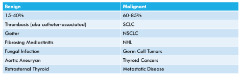
Causes of SVC Syndrome

CausesofSVCSyndrome.PNG

SVC Symptoms

  • Facial and neck swelling
  • Chest pain
  • Respiratory symptoms
  • Neurological manifestations such as head fullness, which may worsen by bending forward or lying own
  • Headaches, confusion, audiovisual disturbances
  • Cerebral edema can be fatal
  • Arm swelling
  • Onset of symptoms depends on whether collaterals had a chance to form

Physical Examination

  • May see distended chest wall veins
  • Pemberton’s sign may be positive
    • initially discovered in the context of a goiter, it can also be useful to identify other causes of SVC obstruction
    • have the patient raise their arms for two minutes and watch for increasing facial plethora (swelling and redness)

Treatment Options

  • Depends on urgency. If emergent, ABCs then straight to endovascular management with pharmacologic thrombolysis/balloon angioplasty/etc +/- stenting
  • If non-emergent, can obtain imaging and biopsy and plan treatment course with chemotherapy or radiation

Lastly, check out Radiopedia! It’s a great learning tool and really fun too.

https://radiopaedia.org/?lang=us

Hypercalcemia of Malignancy


Today we discussed an interesting case of acute encephalopathy due to hypercalcemia of malignancy due to bone metastases.

We first reviewed the importance of maintaining a broad differential for acute encephalopathy/delirium/acute confusional state/AMS.

We reviewed the following framework:

Neurologic / Toxic / Metabolic / Infectious / Other.

We then reviewed the differential for hypercalcemia. For those of you interested, please check out this awesome review video by Dr. Strong:

Our patient had hypercalcemia of malignancy and the following patients were made:

  • It is the most common cause of hypercalcemia in inpatients
  • The most commonly associated malignancies are breast, lung, multiple myeloma, and renal
  • There are three major mechanisms for hypercalcemia of malignancy
    • Secretion of PTHrP – 80% of malignancy related hyperCa
    • Osteolytic Metastases – ~20% of cases
    • Increased 1,25-OH-Vitamin D (calcitriol) 2/2 to increased 1-alpha-hydroxylase activity from activated macrophages in lymphomatous (and also granulomatous) tissue
  • We reviewed the PTH-based algorithm and determined that it was a PTH-independent process (PTH low or low-normal) and then used the PTHrP, calcidiol and calcitriol to guide to the correct diagnosis which was confirmed by MRI and bone scan

Finally, here is a way to distinguish between malignancy related hypercalcemia and primary hyperparathyroidism (most common cause in outpatients & higher co-occurrence in patients with malignancy)

primaryHyperPTH

For treatment options, please see the following blog post for more details!

Hypercalcemia of malignancy

Hyponatremia and B symptoms

Today’s case presented by Dr. Neharika Khurana was an elderly male with a history of tobacco use who presented with subacute/chronic cough, unintentional weight loss, night sweats, and hyponatremia.

Our general approach to hyponatremia:

Capture

*Diuretics create a high urine sodium regardless of volume status unless severe dehydration is present

*If renal disease or intracranial pathology (cerebral salt wasting syndrome) is present, urine sodium may be high when the volume status is actually low

Infection-Related GN

Today we discussed a case of a gentleman with a recent hospitalization for bacteremia who presented with hypertension, renal failure, nephrotic range proteinuria and dysmorphic RBCs consistent with a glomerulonephritis. We learned the following, pathological-based approach to classifying GNs.

GN

Courtesy: AccessMedicine

Infection-Related GN (IRGN)

  • Classically, IRGN begins 7-10 days after a RTI and 2-4 weeks after a skin infection but more commonly the GN coincides with the infection in adults
  • The presentation involves variable degrees of HTN, edema and AKI with a nephritic sediment (dysmorphic RBCs and RBC casts are highly specific though not sensitive for an acute GN)
  • variable degrees of proteinuria can occur
    • we reviewed that virtually any nephritic syndrome can present with nephrotic range-proteinuria (>3g/day)
  • treatment is supportive in most cases
  • Likelihood of full renal recovery is low, and and in some reports up to 50% of patients progress with ESRD
  • Bottom-line, infection-related kidney injury has a broad differential (sepsis-mediated, hypovolemia, medication-related ATN/AIN, ischemic ATN, AND infection-related GN). Even though IRGN is much, much less common than most of the other etiologies, keep it on the differential

Anti-HMGCR Immune-Mediated Inflammatory Myositis

Today we discussed a fascinating case of statin-related anti-HMGCR positive immune-mediated inflammatory myositis (also called necrotizing autoimmune myositis). The case highlighted the importance of a framework approach to diseases.

We first went over the framework for true muscle weakness, which can be anatomically divided as follows

Weakness

Source: Frameworks for Internal Medicine (Dr. Andre Mansoor from OHSU). Available on Amazon (highly recommended!)

To help us localize the lesion to a myopathy, we used the following framework to determine that it was likely a myopathy.

LocalizingTheLesion

The differential for myopathy is broad, and generally is the same for an elevated CK and non-traumatic, non-exertional rhabdomyolysis. The causes can be divided as follows. If you like mnemonics, think Drug-REGIIME for the various categories.

DrugRegiime

Once we narrowed the differential to an inflammatory myopathy, we utilized the following chart that guided us to the probable conclusion that it was an immune-mediated necrotizing myopathy (also known as necrotizing autoimmune myopathy). This was confirmed by a highly positive anti-HMGCR antibody

DifferentiatingInflammatoryMyopathy

Adapted from a NEJM Article: https://www.nejm.org/doi/full/10.1056/NEJMra1402225

Clinical Pearls about INMN/NAM:

  • Can occur in association with viral infections, malignancy, in patients with CTD such as scleroderma or in patients taking statins
  • Can start acutely or sub-acutely with severe proximal muscle weakness and markedly elevated CK values of between 10-100k ULN (2K-20K)
  • A rare diagnosis, but some experts believe that polymyositis is overdiagnosed and that INMN may actually be more common!
  • Two antibodies are highly specific though not sensitive for the condition (anti-SRP and anti-HMGCR)
  • The majority of anti-HMGCR positive cases are related to known exposure to a prescribed statin (~70%)
  • Treatment is to discontinue the statin but most cases will require prompt immunosuppression
    • Statin-induced muscular events are on a spectrum, and include the following:
      • 1) Mildly elevated CK and myalgias
      • 2) Rhabdomyolysis
      • 3) Self-limited toxic myopathy
      • 4) IMNM
    • It is only IMNM that generally does not improve with merely cessation of the drug and it generally needs immunosuppression
  • Biopsy is required to make the diagnosis but the presence of the antibody will often result much more quickly and in our case, the patient started on immunosuppresion prior to the biopsy results because the clinical profile fit perfectly with this diagnosis

Heat Stroke

We learned about a patient who presented with a heat stroke during our recent heat wave. Remember the importance difference between fever and hyperthermia

Fever: elevation of hypothalamic set-point leading to increased core temperature

Hyperthermia: elevation of core temperature independent of changes in the body’s thermoregulatory center.

In clinical practice it is often difficult to discern the two – and therefore for challenging cases, it may be useful to think about the differentials for both. Remember, everyone NEEDs MISO 🙂

NEED

MISO

Constrictive Pericarditis

Thanks to Phuong today for presenting the case of a young woman who presented with volume overload, found to have constrictive pericarditis!


Clinical Pearls

  • Constrictive pericarditis and restrictive cardiomyopathy can have similar clinical presentations.  Cardiac cath is generally needed to help distinguish between the two.
  • The most common cause of constrictive pericarditis is idiopathic!
  • Exam findings in constrictive pericarditis include volume overload, pulsus paradoxus, Kussmaul’s sign, pericardial knock, and occasionally (<20% of the time) pericardial friction rub.
  • ECG and CXR can be normal in constrictive pericarditis.
  • Treatment of early disease is supportive care.  Treatment of late stage constrictive pericarditis is pericardiectomy.

Etiologies of constrictive pericarditis:

  • Idiopathic (42-61%) ⇒ most common cause!
  • Post-cardiac surgery (11-37%)
  • Post-radiation therapy (2-31%) particularly after Hodgkin disease or breast cancer
  • Connective tissue disorder (3-7%)
  • Post-infectious – TB or purulent pericarditis (3-15%)
  • Miscellaneous causes (malignancy, trauma, drug-induced, asbestosis, sarcoidosis, uremic pericarditis) (1-10%)

Clinical Presentation

  • Symptoms related to fluid overload
  • Symptoms related to diminished cardiac output in response to exertion
  • Exam:
    • Elevated JVP
    • Pulsus paradoxus – drop in SBP >10 mmHg due to drop in stroke volume and cardiac output with inspiration (20%)
    • Kussmaul’s sign – lack of an inspiratory decline in JVP.  (Also present in people with severe tricuspid valve disease or R heart failure.
    • Pericardial knock – 47%
    • Pericardial friction rub – 16%
    • Stigmata of heart failure
  • ECG: can be normal
  • CXR: Majority of people do NOT have pericardial calcifications
    • Interestingly, calcifications are more common in people with idiopathic disease, a longer duration of symptoms, and those with TB!

Management of Constrictive Pericarditis

  • Early disease is usually managed with supportive care.  Diuretics can help mitigate symptoms of volume overload but must be used cautiously due to preload dependent physiology.
  • Late stage disease is treated with pericardiectomy.  Complication rates tend to be high and operative mortality can reach 12%!

Constrictive Pericarditis vs Restrictive Cardiomyopathy:

CP vs RCM

We also talked about a helpful way of breaking up new onset ascites to help generate a DDx:

Ascites fluid distribution

Hemolytic Uremic Syndrome

Today, we talked about the case of a middle aged woman with recent diagnosis of metastatic breast cancer on palliative Paclitaxel who was admitted with acute onset of bloody diarrhea found to have Shiga toxin and progression to HUS!  She developed neurologic manifestations for which she underwent PLEX and is now recovering in rehab.


Clinical Pearls

  • TTP and HUS present very similarly and are difficult to distinguish clinically.  HUS typically has worse renal failure than TTP and rarely has neurologic manifestations.
  • Because they are tough to tease apart, start PLEX early if TTP is on your differential for HUS because TTP has a high mortality rate.
  • Acute diarrhea requires work up in those with severe illness, inflammatory features, risk factors, persistent symptoms, or work in fields that are of public health related concern (food handlers, daycare workers, etc.)
  • The most common cause of acute bloody diarrhea worldwide is shigella.
  • Bloody diarrhea with a normal fecal leukocyte/lactoferrin count is highly suggestive of E. histolytica.
  • Majority of shiga toxin produced in adults is by E coli.
  • Avoid antibiotics if possible in a patient with bloody diarrhea due to shiga toxin as it can precipitate HUS.

Indications for work up of acute diarrhea:

  • Age >65
  • Immunocompromised
  • Significant volume depletion
  • Blood in stool
  • Fever
  • Severe abdominal pain
  • Recent antibiotics
  • Known or suspected IBD
  • Food handler, daycare worker, healthcare worker
  • Recent travel

DDx for acute bloody diarrhea:

  • IBD
  • Ischemic colitis
  • Invasive infections
    • Shigella (most common)
    • EHEC and EIEC (most commonly associated with shiga toxin)
    • Campylobacter
    • Nontyphoidal salmonella
    • Entamoeba histolytica
    • Schistosoma (more common in resource limited settings)

Work up for acute bloody diarrhea:

  • Enteric pathogen panel (NAAT):
    • Campylobacter, salmonella, shigella species, vibrio, yersinia, shiga toxins, norovirus, and rotavirus
  • Stool culture
    • Grows campylobacter, Shigella, Salmonella, and E coli strains. If suspecting other organism, must specifically request that culture from lab
  • Stool leukocytes or lactoferrin
    • More helpful if negative to rule in amebiasis.

Shiga toxin mediated hemolytic uremic syndrome

  • Characterized by the triad of MAHA, thrombocytopenia, and acute renal failure. Rare neurologic manifestations can occur as in our patient.
    • Other clinical symptoms ⇒ bloody stools, absence of fever, WBC>10k, and abdominal pain.
    • 23-47% require hospitalization
  • 6-9% of people infected with EHEC (O157:H7 and O104:H4) can go on to developing HUS and it is much more common in children.
  • Pathophys
    • Ingestion of undercooked beef and E coli
    • Shiga toxin produced by E coli binds to vascular endothelial cell surface, thereby inhibiting protein synthesis, generating lots of cytokines and chemokines, and causing end organ damage and thrombosis.
  • Clinical course
    • HUS develops 5-10 days after onset of diarrhea
    • Up to 50% of patients require dialysis and 39% have long term renal injury.
    • Mortality is 3-5%
  • Treatment:
    • Supportive care is the mainstay
    • Some data from an outbreak in Germany suggests there may be benefit to plasma exchange (PLEX) via removal of shiga toxin and prothrombotic factors from the body
    • Eculizumab ⇒ beneficial in patients with complement-mediated HUS (not shiga-toxin mediated)

TTP: 

  • Main distinguishing features from HUS
    • Renal failure can often be mild
    • Neurologic impairment is more common
    • Mortality rates are much higher
    • Confirmatory test is ADAMTS13
    • Mainstay of treatment is PLEX!

Moral of this story: start PLEX while you’re waiting to decide if it’s HUS or TTP!