Tag Archives: Pulmonology

Parapneumonic effusions

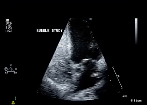
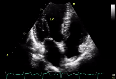
Thanks to Julie for presenting the case of a middle-aged man with recent CAP who presented with progressive SOB, pleuritic chest pain, weight loss, and anorexia, found to be septic with a large empyema, eventually requiring open decortication!


Clinical Pearls

  • Think of parapneumonic effusions in two broad categories: infected (complicated and empyema) and sterile (uncomplicated).
    • Infected (complicated and empyema) require chest tube placement and can be complicated by loculated effusions.
    • Uncomplicated resolve with the treatment of underlying pneumonia
  • Anaerobic organisms are a common cause of infected parapneumonic effusions.  Malodorous fluid at the time of thoracentesis is diagnostic!  But make sure to send anaerobic cultures to the lab to help with speciation.
  • pH of pleural fluid can be falsely elevated if not immediately stored on ice upon collection and processed in a blood gas analyzer.
  • Differential for pleural fluid that has low glucose/low pH is short: infection, TB, malignancy, rheumatoid pleurisy, and lupus pleuritis.
  • Remember that while ADA has high sensitivity (86%) and high specificity (87%) for TB, the study on which it is based was done in a high risk population so its utility in screening low risk patients is limited.

Parapneumonic effusions:

  • Form in 40% of bacterial pneumonia:
      • Uncomplicated: negative GS and Cx, pH>7.2, glucose >60, no loculations
      • Complicated: positive GS or Cx or pH <7.2, or glucose <60. LDH >1000 makes it more likely
      • Empyema: frank pus aspirated during thora, cell count with >50k WBCs

The latter two categories require chest tube placement to prevent formation of pleural “peels” that can lead to trapped lung and loss of lung function.
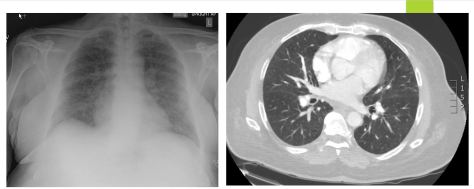
  • Imaging
    • Lateral decub or ultrasound, latter is more sensitive than CXR for diagnosing complicated parapneumonic effusions.
    • CT with contrast is the optimal imaging for empyema or loculated effusion
      • Look for the “split pleura sign”
  • Labs:
    • Serum procalcitonin >0.18 ng/mL is 83% sensitive and 81% specific for effusion having a bacterial infectious etiology
    • Bacteriology:
      • Anaerobic bugs are often the culprit!  So it is important to send pleural fluid for both aerobic and anaerobic cultures
      • Other bacteria: CAP organisms such as strep and staph as well as klebsiella in diabetic patients
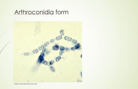
      • Fungi
      • TB
  • Treatment:
    • Tube thoracostomy (chest tube): first intervention
      • CT within 24 hours to ensure correct positioning and adequate drainage, left in place until drainage is <50 cc/day
    • Fibrinolytic agents
      • DNA is a main contributor to viscosity of empyema fluid.  However, based on this trial published in NEJM in 2011, tPA and DNAase combined is associated with significant radiographic improvement of empyema, reduction in hospital stay, and lower number of surgical referrals.
    • VATS
    • Decortication
      • To remove the thickened fibrin layer covering the pleura.
    • Open thoracostomy
      • Rib resection and opening the chest wall at the inferior border of empyema to allow for ongoing drainage.  High risk of infection and complications.

Hepatopulmonary Syndrome 10/10/2018

A 67 year old man with history of cirrhosis secondary to Hepatitis C and alcohol, hepatocellular carcinoma with recent TACE, presented with worsening dyspnea on exertion and positional shortness of breath. His breathing was worse when he sat upright, and better when he was supine. What’s going on?

Just to go over some terms:

  • Orthodoxia: Drop in PaO2 by 5mmHg or O2sat by 5% when moving from supine to upright.
  • Platypnea: Dyspnea that is induced by moving to an upright position, relieves when supine.

Hepatopulmonary syndrome

Triad

  • Chronic liver disease or portal hypertension
  • Intrapulmonary vascular dilations (IPVD)
  • Impaired oxygenation

Epidemiology:

Up to 25% of patients with chronic liver disease will have some degree of shunting, can occur at any stage (mild or severe)

Pathophysiology

  • Not well understood but the theory is due to increased nitric oxide production and reduced NO clearance, resulting in pulmonary vasodilation (IPVDs) mostly concentrated at the lung bases.
  • When upright, blood preferentially perfuse the lower lung  zones due to gravity.
  • Vasodilation leads to poor gas exchange.

Diagram

  • This leads to a VQ mismatch

Diagnosis:

  • CXR: Not helpful, might show e/o interstitial lung markings.
  • CT: Can reveal IPVDs
    • Dilated peripheral pulmonary vessels
    • Inc pulmonary artery to bronchus ratios
  • PFT: Not helpful
  • Transthoracic contrast echo (TTCE): Can be used to demonstrate presence of intrapulmonary shunts supportive of presences of IPVDs
    • Concept of bubble study: Shooting agitated saline (with bubbles into the vasculature
    • Bubbles visible in the R heart chambers, should not be visible in the left heart chambers.
    • If presence of bubbles in the left: This is indicative of a shunt:
      • Intracardiac shunt: bubbles seen within 1 beat
      • Intrapulmonary shunts: bubbles seen after 3-8 beats.


Normal Echo: Notice how the agitated saline bubbles remain on the right side of circulation and do not cross over. The bubbles were filtered out by the pulmonary vasculature.

Normal Echo


Echo demonstrating intrapulmonary shunting (see bubbles crossing over from the right to the left)

HPS


Management

  • Supplemental O2 indicated if O2 sats < 88%, PaO2 < 55mmHg
  • Mild to moderate: Monitor Q6-12 months
  • Severe to very severe: Referral for liver transplant
  • Insufficient data on other treatment options (garlic, pentoxifylline, NO synthase inhibitors, IPVD embolization, plasma exchange, oxtreotide).

HPS_diagnosis_algorithm

Image adapted from Uptodate

Check out this article if you’re interested in the data behind pentoxifylline!

Pleural effusions – 7/25/17

Symptoms: Dyspnea, cough, and pleuritic chest pain

Exam: Decreased breath sounds, dullness to percussion, decreased tactile fremitus

Indications for thoracentesis: Any new unexplained effusion

Light’s criteria:

  • Pleural protein/Serum protein > 0.5
  • Pleural LDH/Serum LDH > 0.6
  • Pleural LDH > 2/3 ULN

Examples of transudates:

  • Heart failure
  • Nephrotic syndrome
  • Hepatic hydrothorax
  • Low albumin

Examples of exudates:

  • Parapneumonic effusions
  • Malignancy
  • TB
  • PE
  • Autoimmune disease (RA, SLE)

Uncomplicated effusion – pH > 7.2, glucose > 60, free flowing, < 1/2 hemithorax – treat the underlying cause, no need for chest tube

Complicated effusion – pH < 7.2, glucose < 60, can be > 1/2 hemithorax or loculated – treat the underlying cause and would benefit from chest tube

Empyema – complicated effusion with positive gram stain and culture – place a chest tube

Malignant effusions – if re-accumulating rapidly, can place a long-term chest tube or do a pleurodesis with talc