Category Archives: Morning Report

Seizure Secondary to Neurocysticercosis 1/14/2019

Sorry for posting this late!

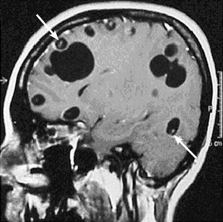
A 38yo M with history of asthma, but otherwise healthy, presents acute onset seizure as well as LLE, LUE weakness and paresthesia. He has never had seizures before prior to this. History revealed that patient was born in Honduras, and he grew up on a pig farm, and his brother actually had a history of Taeniasis. MRI revealed a cystic structure with a thin septae/soft tissue component within, consistent with neurocysticercosis!


Let’s talk about seizures for a little bit.

In developeED countries, the most common cause of  epileptic seizure is often idiopathic.

In developING countries, the most common cause of epileptic seizure in both kids and adults, is neurocysticercosis (NCC).


Neurocysticercosis: When larvae form of the Taenia solium (aka the tapeworm) move to non-GI tissues and into CNS.

Picture1.png

Presentation: Viable (asx, many years) -> degenerating (loses ability to evade immune system, localized inflammation) -> non-viable (calcified granulomas, can still cause szx)

  • Intra-parenchymal
    • Most common form of neurocysticercosis, > 60% of cases
    • 3-5 years after infection but can occur > 30 years after initial exposure.
    • Seizure = most common manifestation. Occur in setting of cysts degeneration or granulomas.
    • Endemic areas: NCC is the most common cause of adult onset seizure
    • Headaches, usually mild
    • Rare complications: vision, focal neuro, meningitis, encephalitis (higher parasitic load)
    • Most, however, are asx and diagnosed incidentally.
  • Extra-parenchymal
    • When NCC occurs in the spine, eyes, ventricles, subarachnoid space. More commonly in adults.
    • Can cause increased ICH, hydrocephalous
    • Sub-arachnoid form is most severe, esp in the basilar cisterns. 5% of cases.
    • Spinal: 1% of cases, can cause localized/dermatomal pain.
    • Ocular: 1-3% of cases, impaired vision, eye pain, diplopia, chlorioretinitis, retinal detachment.
  • Extra Neural
    • Most common: muscles, subcutaneous
    • Cardiac has been described…

Diagnosis

  • Stool O&P can help
  • Eosinophilia is uncommon
  • Imaging: Clinical history, endemic history, enhancing cystic lesion on MRI is very likely.
    • CT: Useful for IDing calcifications, parenchymal cysticerci, and eye involvement
    • MRI: Useful for small lesions, degenerative changes, edema, and visualizing scolices within calcified lesion
    • Scolex.JPG

(Mahale et al. Extraparenchymal (Racemose) Neurocysticercosis and Its Multitude Manifestations: A Comprehensive Review. J Clin Neurol. 2015 Jul;11(3):203-211)

  • Serology: Should be performed for confirmation Enzyme Linked Immunoelectrotransfer Blot Ab is highly sensitive and specific, but takes time and availability is limited.

Management

  • Latest IDSA Recommendation in 2018
    • Calcified lesions: symptomatic management only with anti-epileptics
    • Enhancing lesions
      • Patients who acquired NCC in a non-endemic area should have their household members screened.
      • Screen for latent TB, strongyloides infection given possibility of prolonged steroid use
      • Fundoscopic exam is recommended for all patients with NCC
      • Prior to anti-parasitic therapy, all patients should be treated with corticosteroids prior to initiation.
      • Anti-epileptics should be used in all patients with seizures.
      • Albendazole + praziquantel is better than monotherapy with albendazole if greater than 2 lesions. 1-2 lesions, monotherapy with albendazole should suffice.
    • Intraventricular neurocysticercosis
      • Minimally invasive surgical removal prior to antiparasitic therapy to minimize inflammatory response.
    • Subarachnoid neurocysticercosis in the basilar cisterns or Sylvian fissures
      • Prolong course of anti-parasitic until radiologic resolution, can last more than a year.
      • Corticosteroids recommended while on treatment but methotrexate can be considered a steroid-sparing agent in patients requiring prolonged anti-inflammatory.
      • Surgery case by case
    • Spinal neurocysticercosis
      • Combination surgery and anti-parasitics, start steroids first with evidence of spinal cord dysfunction and prior to antiparasitics
    • Ocular:
      • Surgery preferred over medical management.

A Beriberi Nice Case of… Thiamine Deficiency (1/9/2019)

Adam presented a case of a 32yo woman with an extensive alcohol history presenting with seizure in setting of recent cessation of alcohol. Pt has also been complaining of weakness in her legs to the point she could no longer walk, worsening vision, and urinary incontinence for the past few months. Per her family, she only ate one meal a day and she was quite picky in terms of her diet.

She was treated for alcohol withdrawal and delirium tremems. When she was stabilized, her neurological exam was concerning for significant weakness in proximal and distal upper and lower extremities, paresthesia, dysmetria, and hyporeflexia.  An EMG was done which revealed peripheral polyneuropathy. This constellation of symptoms (alcohol, poor nutrition, polyneuropathy) is consistent with… Beriberi!


Thiamine deficiency

Epidemiology

  • Developing countries
  • Alcoholics
  • Extreme poverty
  • Displaced populations, refugees

Common Risk factors

  • Poor nutrition
  • Alcohol
  • Weight loss surgery
  • Long term TPN

Presentation of Thiamine Deficiency

  • Wet beriberi
    • Heart failure due to thiamine deficiency (high out heart failure)
    • Vasodilation, tachycardia, widened pulse pressure, diaphoresis, lactic acidosis, peripheral edema
  • Dry beriberi
    • Peripheral polyneuropathy, affects predominantly lower extremities, both sensory and motor deficits, can lead to muscle wasting, loss of deep tendon reflexes, paralysis of the lower legs, mental confusion, speech difficulties, nystagmus
  • Wernicke Korsakoff
    • Wernicke Encephalopathy: triad of encephalopathy (disorientation, inattentiveness indifference), gait ataxia, and oculomotor symptoms (nystagmus, lateral rectus palsy, conjugate gaze palsies)
      • Triad only seen in 1/3 of patients, most only have around 2.
      • Diagnosis: Clinical but there is a proposed Caine Criteria
        • Dietary deficiency
        • Oculomotor abnrl
        • Cerebellar dysfunction
        • Encephalopathy or memory impairment.
        • 2/4
    • Korsakoff Syndrome: Memory loss, confabulation, +/-hallucinations

Pathophysiology

  • Chronic inadequate intake of thiamine (vitamin B1) leading to degeneration of the peripheral nerves, thalamus, mammillary bodies, and cerebellum.
  • Heart may become dilated, may lead to a high output heart failure
  • Vasodilation can occur causing edema

Diagnosis

  • Clinical history
  • Thiamine level
  • Clinical improvement with thiamine administration
  • CT: May see classic atrophy in the mammillary bodies in Wernicke Korsakoff, highly specific.

Management

  • DO NOT GIVE GLUCOSE 1st, thiamine must be repleted first or else glucose infusions may worsen symptoms. Alcoholics should receive IV thiamine, at least 100mg, before receiving any IV glucose solutions.
  • Nutritional support, thiamine replacement
  • Fix underlying cause (i.e. alcohol)
  • Thiamine initially is given in very high doses if treating, 500mg IV 3 times daily for 3 days, then 250mg daily for 3-5 days, then transition to 100mg PO daily.

Prognosis

  • Most will have a degree of neurological deficits despite treatment.

Anti-Synthetase Syndrome 1/8/2019

Jonathan presented a case of a 39yo M with no significant medical history, presenting with 1 month of non-improving dry cough, dyspnea on exertion, subjective fevers, and leg weakness. His CK was significantly elevated on admission to 27k, and CXR revealed peri-hilar lung base opacities which could represent pneumonia. His exam was significant for debilitating proximal muscle weakness (distal strength was intact!) with hyporreflexia. Ultimately Anti-Jo1 antibodies returned positive, and CT Cx revealed predominantly lower lobe GGO without evidence of honeycombing or traction atelectasis. This constellation of findings (myopathy, lung pathology, anti-synthetase antibody positivity) is consistent with Anti-synthetase syndrome!


Anti-synthetase syndrome

Epidemiology

  • Up to 30% of patients with DM or PM will have this constellation of clinical findings, terms Anti-Synthetase syndrome.
  • More acute onset of the following:

Presentation: Often acute

  • Constitutional symptoms i.e. fever
  • Myositis
  • Raynaud’s phenomenon
  • Mechanic hands
  • Non-erosive arthritis
  • ILD: Often severe and rapidly progressive, frequently predominates other symptoms
  • Cardiac arrhythmias or ventricular dysfunction

Diagnosis

  • Poorly defined as a condition, but in general, diagnostic criteria based on expert consensus is positive antisynthetase antibodies plus at least 1 feature
  • Antibodies:
    • Antibodies to aminoacyl-rRNA synthetases (antisynthetase antibodies), i.e. Anti-Jo1 (most common)
    • Anti-PM-Scl
    • Anti-U1 RNP
    • Anti PL-7 & PL12 (seen in pts with predominantly ILD sx, often very severe)
    • If antiRo or ANA present, suspect more of an myositis associated ILD
  • CT:
    • Most common findings are traction bronchiectasis, GGO
  • Diagnosis usually is made by combination of CT findings, serology, PFT, and clinical findings.

Antibodies.png

Management

  • Often requires multiple immunosuppressives for symptomatic control.
  • First line: Corticosteroids, monotherapy associated with more frequent lung disease recurrence
  • Other agents often added i.e. azathioprine, mycophenolate, tacrolimus, rituximab, cyclophosphamide.

meds

Monitoring

  • Chronic steroids: osteoporosis, PJP prophylaxis if > 20mg > 1 month
  • Hep B reactivation
  • Azathioprine: Check TPMT levels!


Prognosis

  • Not associated with inc risk of malignancy
  • Anti PL7 and PL12 are associated with more aggressive ILD, and worse prognosis.
  • Presence of Anti-Jo1 and arthritis/myositis are actually good prognostic indicators.
    • Single center study: 10 year survival for Anti-Jo1 was 70%, vs 49% for non-Jo1

Community Acquired Bacterial Meningitis Secondary to Streptoccocus pneumoniae 1/7/2019

Jihong presented a case of a 57yo woman with no medical history, who was in her usual state of health until 2 weeks ago when she started complaining of a sore throat. The day prior to presentation, the patient’s roommate saw her “sleep walking” and not acting like herself. When her cousin saw her, she immediately brought her to the hospital because the patient was becoming increasingly more confused and agitated. She was ultimately diagnosed with bacterial meningitis secondary to strep pneumo.


Acute Bacterial Meningitis

Epidemiology

  • Developed Countries
    • Community acquired
      • Streptococcus pneumoniae (most common)
      • Neissseria meningitidis
      • Listeria monocytogenes (age > 50, immunodeficiency)
      • H. influenzae Type B: less likely given HiB immunization but rates of vaccination are declining…
    • Healthcare acquired
      • Associated with neurosurgery (i.e. drains), skull trauma.
      • Usually staphylococci and GNR
    • Risk factors
      • HIV
      • Immunosuppression
      • Diabetes
      • EtOH
      • Asplenia (encapsulated organisms, H.influ, strep)

Presentation

  • Usually quite acute and patients are usually ill appearing
  • Headache (usually generalized, severe, seen in most patients) + Classic Triad:
    • Fever (95%, often > 38, others might be hypothermic)
    • Nuchal rigidity (Also present in most patients, 88%)
    • Encephalopathy (78%)
  • Only 44% of patients will have all 3 triad
  • More variable and subtle presentation the older you get
  • Other findings
    • Seizures (inc risk in Listeria)
    • CN palsies (inc risk in Listeria)
    • Cerebral infarction
    • Papilledema
    • Petechial rash, arthritis (Neisseria meningitidis)

Diagnosis

  • Physical Exam: Brudzinski and Kernig: Not sensitive at all but quite specific. Jolt accentuation is more sensitive but less specific (horizontal rotation of the head 2-3 times per second causing a headache) but overall still quite poor in terms of both.
  • Clinical history
  • LP
    • Low glucose
    • High WBC, usually 1k – 5k, typically > 80% neutrophils
    • Protein > 200mg/dL
    • Glucose < 40mg/dL, or CSF:serum glucose ratio of < 0.4, the lower the worse)
    • 99% PPV if any one of these are present: CSF glucose < 34, protein above 220, and WBC > 2000
    • Some data suggesting using CSF lactate
      • WBC can be falsely elevated in traumatic tap or underlying head trauma (i.e. prior SDH)
      • Correction for traumatic tap: subtract 1 WBC for every 500-1500 RBC in the CSF, just use 1000 to remember easier.
    • Blood + CSF cultures
  • When to CT first
    • Do CT first if the patient has the following:
      • Focal neuro deficits
      • Papilledema
      • Immunocompromised (HIV, transplant, etc)
      • H/o CNS mass, stroke, focal infection
    • Otherwise, ok to go straight for the LP, but LP is contraindicated (but not absolute) under these circumstances:
      • PLt < 50
      • INR > 1.4
      • Lovenox, please hold at least 12 hours in advance
      • Paraspinal abscess

Management

  • Empiric antibiotics, should not wait after LP to start!
    • Start ASAP, don’t wait for the LP
    • Most community acquired meningitis: Vancomycin and Ceftriaxone (2g Q12H) should suffice
      • Age > 50 adults or immunocompromised: Add ampicillin 2g Q4H for Listeria coverage
      • Community and immunocompromised: substitute CTX with cefepime
      • Healthcare associated: Vancomycin, cefepime or carbapenem class with pseudomonal coverage
      • PCN allergy: Can use Vanc, moxifloxacin, and Bactrim (Listeria coverage)
  • Dexamethasone
    • Studies have shown improved neurological outcome with dexamethasone prior to abx, give dex 10mg IV q6H for 4 days.
    • Start before Abx
    • Also potentially reduces risk of post-infectious neurological complications.
    • Most of the data is from management of pneumococcal meningitis, some have suggested that dex can be DC if another cause is diagnosed.

Prognosis

  • < 60 yo: ~ 17% mortality
  • 37% mortality in > 60
  • Staph aureus: 43% mortality
  • Seizures, focal neuro deficits, coexisting medical conditions, high CSF pressure, older age, coma, low CSF:serum glucose ratio tend to be associated with poor prognosis
  • Post-infectious neurological complications
    • As high as 15-22% in kids
    • Up to 1/3 in adults

Hot Topics of 2018!

Today, we reviewed some of the hottest and potentially practice changing articles of 2018.  This is by no means an exhaustive list and meant to encourage debate and tickle your fancy for more!


1. Aspirin for primary prevention of cardiovascular disease?

Bottom line: no net benefit in primary prevention of cardiovascular disease.

  • ARRIVE: 12000 middle aged (mean age 64), non-diabetic participants with moderate ASCVD risk (>20%) randomized to receive aspirin 100 mg or placebo for primary prevention and followed for 5 years. Found that ASA showed no reduction in major adverse cardiovascular events or mortality, but a 2-fold higher risk of bleeding.
  • ASCEND (aspirin): 15000 middle aged (mean age 63) diabetic participants randomized to aspirin 100 mg vs placebo for primary prevention of CVD and followed for 7 years. Authors found a 12% reduction in major adverse cardiovascular events with ASA but a 29% higher risk of bleeding.
  • ASPREE: 19000 older patients (median age, 74) regardless of other risk factors randomized to ASA 100 mg or placebo and followed for 5 years.  Study found that patients who received ASA had a 14% higher all cause mortality, no decrease in the rate of adverse CVD, and no change in disability-free survival.

2. Omega-3 for primary prevention of cardiovascular disease?

Bottom line: potentially beneficial at really high doses in patients with CV risk factors

  • VITAL: 26000 middle aged (mean age, 67) people without CV disease were randomized to receive fish oil (1g) or placebo and followed for ~5 years. Study found similar rates of primary endpoint (nonfatal MI, stroke, or CV-related death) and all cause mortality in the two groups and a small but significant decrease in the incidence of MI in the fish oil group (1.1% vs 1.5%).
  • ASCEND (fish oil): same study population as ASCEND for aspirin, also looked at using 1g fish oil or placebo and followed patients for ~7 years and found no difference in the risk of major adverse cardiac events.
  • REDUCE-IT: 8000 participants with controlled LDL but elevated triglycerides, randomized to receive 2g of a different fish oil (icosapent ethyl as opposed to the eicosapentaenoic acid plus docosahexaenoic acid used in the previous two trials) vs placebo and followed for 5 years.  Study found a 25% reduction in risk of major cardiovascular events. Caveat is high dose used at this formulation is very expensive and the study was funded by Amarin Pharma.

3. VTE prophylaxis in hospitalized patients

Bottom line: High rates of inappropriate use of pharmacologic VTE prophylaxis.  Use padua score before prescribing VTE prophylaxis.

  • Grant et al. JAMA Intern Med 2018: retrospective study of 45000 non-ICU patients hospitalized for > 2 days found that prophylaxis (pharmacologic or mechanical) was prescribed for 78% of low-risk patients. 27% of high risk patients with contraindications to pharmacologic prophylaxis still received it, and 22% of high risk patients did not receive prophylaxis.

4. Is there such a thing as too much oxygen?

Bottom line: higher rates of mortality associated with liberal use of oxygen in hospitalized patients.

  • Chu et al. Lancet 2018: Meta-analysis of 25 randomized trials on 16000 hospitalized patients treated with liberal (median FiO2 0.52) vs conservative (median FiO2 0.21) supplemental oxygenation found that at 30 days, the relative risk of death was significantly higher in the liberal oxygenation group.

5. Plavix + ASA for TIA or minor stroke?

Bottom line: Starting DAPT within 12 hours of symptom onset (likely for 30 days) in patients with high risk TIA or minor ischemic stroke reduces 90 day stroke incidence but increases bleeding rates.

  • POINT:  Followed the earlier CHANCE trial in a Chinese population that showed DAPT for 21 days after TIA or minor stroke reduced stroke recurrence at 90 days without a difference in bleeding rates.  POINT randomized ~5000 patients to DAPT for 90 days vs ASA alone in a primarily white patient population and found lower rates of recurrent stroke but higher rates of bleeding. Majority of stroke reduction occurred during the first 7 days after stroke and extended for 30 days whereas the bleeding rates were stable throughout the 90 day follow up period.

6. Steroids in septic shock?

Bottom line: Steroids might be beneficial in high risk patients with refractory septic shock.

  • Rochwerg B et al. Crit Care Med 2018: Meta-analysis of 42 randomized trials with >10000 patients receiving steroids vs none in septic shock found a 2% relative reduction in 30-day mortality with steroids which was not statistically significant, and a similar reduction in mortality at 60 days to 1 year which reached significance (NNT 50). Reversal of shock at 7 days occurred more frequently in the steroid group (NNT 10) but mild-to-moderate adverse events also occurred more frequently in this group (hyperglycemia, hypernatremia, and neuromuscular disease).

7. Is it safe to discharge to home from the ICU?

Bottom line: patients admitted to the ICU for substance-related disorders, seizures, or metabolic derangements may be ok to go home from the ICU.

  • Stelfox et al. JAMA 2018: retrospective cohort study of 6700 adult patients admitted to ICUs in Canada, 14% of whom were discharged to home, found that 30 day hospital readmissions and ED visits and 1 year mortality rates were similar in those discharged from the ICU vs wards.  Those discharged home were typically younger and more likely to have been admitted due to overdose, seizure, substance withdrawal, or metabolic derangements.

8. NS vs LR?

Bottom line: balanced crystalloids (like LR) are associated with fewer adverse events than normal saline in hospitalized patients.

  • SMART: 16000 patients admitted to the ICU were randomized to NS or a balanced crystalloid (majority received LR). Study found that more patients in the NS group reached the composite outcome of major adverse kidney events (death, renal replacement therapy, or doubling of creatinine at discharge) vs those who received balanced crystalloids.
  • SALT-ED: 13000 patients admitted from the ED to non-ICU beds were randomized to NS vs a balanced crystalloid (majority received LR).  Study found similar rates of primary outcome of hospital-free days but a higher rate of adverse kidney events within 30 days than the NS group.

Necrotizing Fasciitis of the Lip Secondary to… Hypermucoid variant Klebsiella pneumoniae! 1/2/2019

Richard presented a patient from China with a history of diabetes not on medications who presents to the hospital with 4 days of lip swelling. He had a pimple just inferior to his nostrils, which he popped the day prior to onset of symptoms. He was feeling fine and he initially thought it was an allergic reaction after his lips became swollen and pruritic. His vitals were normal, but notable upper lip swelling and surrounding erythema were noted. Ultimately he underwent I&D after CT revealed gas within the soft tissues consistent with a necrotizing soft tissue infection. Cultures turned out to be hypermucoid klebsiella!


Risk Factors for necrotizing soft tissue infection

  • Diabetes
  • Chronic disease
  • Immunosuppressive drugs (eg, prednisolone)
  • Malnutrition
  • Age > 60 years
  • IVDU
  • Peripheral vascular disease
  • Renal failure
  • Underlying malignancy
  • Obesity
  • Precipitating events (both traumatic and non-traumatic)
    • Traumatic: Surgery, procedures, acupuncture, IVDU, penetrating injuries
    • Non-traumatic: Soft tissue infection, childbirth

Presentation

  • Pt in general are acutely ill.
  • Most cases involve the limbs, perineum, or trunk. Head & neck involvement are only seen in 5% of cases

Organisms

  • Type A Nec Fas: Most common, polymicrobial
  • Type B Nec Fas: Less common, monomicrobial
  • In general, the organisms involved in nec fas are:
    • Strep (group A) & staph (most common
    • Anaerobes
    • Gram negatives i.e. pseudmonas
    • Enterococcus
    • Vibrio vulnificus (especially in patients with cirrhotic with sea water exposure!!!)
    • Clostridium perfringens
    • Candida spp

How about hypermucoid klebsiella? A single center study in Taiwan in 2012 found that klebsiella accounted for 17% of monomicrobial nec fas, with 2/3 of them being the hypermucoid variant. In Asian countries, HvKP necrotizing infection is as common as those caused by staph and strep.

In North America though? The first case report of community acquired HvKP associated  was reported in 2015. See this article for details.


Hypermucoid variant Klebsiella (HvKP)

Epidemiology

  • Usually community acquired, highly virulence strain of KP that is typically pan-sensitive.
  • Primarily in SE and E Asian countries but seeing this strain increasingly in the US.
  • From a necrotizing soft tissue infection stand point, it has been reported in SE Asia beginning in 1996. Most info published in the literature are case reports,
  • KP itself, in a single center study done in Taiwan 2012 found that KP in general accounted for 17% of monomicrobial nec fas, and 2/3 of these cases are HvKP. vs 22% staph vs 18% strep.
    • Associated with higher mortality
    • Main risk factors are baseline immunocompromised status, and 80% of pts with HvKP had DM

Risk factor

  • Primary risk factor seems to be just diabetes! But this can affect completely healthy patients in the community.

Presentation

  • Pyogenic liver abscess
  • Metastatic infection, likes to travel and stick to places.
  • Associated infections
    • Primary liver abscess, usually in the right lobe for some reason
      • HvKP is the culprit organism in 9-12% in pts with primary liver abscesses
    • Splenic abscess, SBP, PNA, soft tissue infection, osteo, UTI
    • Associated endophthalmitis especially if bacteremic or presence of abscess in up to 50% of pts
      • Not so much if pulmonary or urine

Diagnosis

  • String Test! > 5mm = diagnostic.
  • Fig-1-String-test-result-for-Klebsiella-pneumoniae-Stretching-of-K-pneumoniae.png
  • Capsular subtypes: some are more virulent, K1, K2, rmpAvirulence-associated gene

Management

  • Fortunately, most are pan-sensitive
  • Penicillin or cephalosporins = main stay, but if treating for a liver abscess, should cover for anaerobes as well.
  • Surgical drainage if e/o abscess
  • If concerned for metastatic infection, combination of surgical and medical therapy depending on location of the infection

Dieulafoy lesion causing obscure overt GI bleed!

Today we talked about an elderly man with recent ACS on DAPT, HFrEF 25%, and h/o colonic angiodysplasia induced LGIB who presented with acute onset of obscure overt upper GI bleed, found to have a dieulafoy lesion on repeat EGD!


Clinical Pearls

  • Overt GI bleed refers to bleeding that is clinically evident (i.e. hematemesis, hematochezia, melena, etc.)
  • Occult GI bleed refers to slow bleed that primarily manifests as iron deficiency anemia and/or positive guaiac stool.
  • Obscure GI bleed refers to evident GI bleed without a clear source on EGD/colo.
    • Most common cause is angiodysplasia
    • First step in the work up of obscure GI bleed is to repeat EGD/colo.  Up to 50% of cases are successfully diagnosed this way.
  • Tachycardia is the first sign of blood loss and suggests up to 30% total blood volume loss.  Hypotension develops once blood loss >30%.
  • For people at high risk of thromboembolic events requiring anticoagulation, restart anticoagulation/antiplatelet therapy as soon as possible after acute bleeding is resolved (prior to discharge!). Be sure to discuss risk of rebleeding with endoscopist prior to restarting these agents.

Obscure GI bleed

Etiology:

The following is a simplified breakdown of diagnoses to consider in obscure GI bleed.  Keep in mind that many diagnoses can present as overt or occult GI bleed.  We have listed them here under the more common way in which they present:

Obscure GI bleed breakdown

  • Remember that NSAID induced ulcers can occur anywhere in the GI tract as far distally as the splenic flexure!
  • Angiodyplasia is the most common cause of obscure GI bleed.

Work up:

  • Repeat EGD/colo is the first step.  Up to 50% of the cases are diagnosed in this way.
  • If EGD/colo inconclusive, then
    • Obscure active bleed
      • Tagged RBC scan: technetium 99m-labeled RBC or sulfur colloid nuclear scans. Can detect slow bleeds with accuracy varying from 24-91%.  They can only identify a general area where bleeding is occurring (not accurate) and a follow up separate intervention is indicated if a source is identified.
      • Angiography: Can identify faster bleeds, more effective at localizing bleed, but less sensitive than tagged RBC (27-77%). Allows intervention at the same time.
      • Enteroscopy: push, single/double balloon enteroscopy are sensitive (up to 80%) and allow for intervention but are operator dependent and may not always be available
      • Intraoperative endoscopy: laparotomy or laparoscopy (sensitivity 58-80%). Last resort.
    • Obscure occult bleed
      • Capsule endoscopy: allows for imaging of the small bowel and can has a high sensitivity (83%). May be difficult to localize lesion based on imaged. Not a good tool in active bleed.
      • Enteroscopy as noted above

Management:

  • Treat the underlying etiology!
  • In the case of dieulafoy lesions, treatment with electrocautery, hemoclips, argon plasma coagulation, injection therapy, or a combination is effective.
  • For people with DAPT or on anticoagulation with warfarin, the current recommendation is to start these agents as soon as hemostasis is achieved.  The decision on when to start depends on the type of lesion, risk of bleed (based on edoscopist’s opinion), and risk of thromboembolic event (based on this cohort study and this meta-analysis).

Hemodynamics in GI bleed:

  • Tachycardia is the first vital sign abnormality in GI bleed and is noted with 15-30% blood volume loss.  With lower amounts of blood loss, tachycardia may be present upon standing.  If a patient is tachycardic while laying supine, blood loss is closer to 30%.
  • Blood loss >30% total body volume results in hypotension.  This typically begins as a widening of pulse pressure (drop in diastolic BP) followed by a drop in systolic BP.


Myasthenia Crisis Secondary to… a Thymoma! 12/26/2018

Our case today is a 49 year old woman with no medical history, presenting with 1 month of difficulty swallowing, voice changes, and more recently dysphagia with liquids and solids, and shortness of breath. Her symptoms are worse during the night time to the point that she couldn’t swallow her own spit/secretions. She presented with respiratory failure requiring intubation, and on CXR/CT she was found to have an anterior mediastinal mass concerning for a… thymoma!


Let’s first briefly review Myasthenia Gravis before moving onto Myasthenia Crisis, and lastly, Thymomas.

Myasthenia Gravis

Epidemiology

  • Bimodal: Early peak in 2-3rd decades (female predominance) and late peak 60-80s (male predominance).
  • F in post-partum period have inc risk.
  • Possible association with: neuromyelitis optical, autoimmune thyroid disorders, SLE, RA.

Diagnosis

  • Bedside
    • Ice-pack test: Improvement of ptosis after application of an ice pack = positive. Sensitivity around 80ish %, limited to patients with ptosis and not helpful for those with extraocular muscle weakness.
      • myastheniagravis-myasthenia-diagnosis-clinical-icepack-original.jpeg Source: NEJM, Grepmed
    • Edrophonium test is no longer used very often, in a nut shell, it is a Acetylcholinesterase inhibitor with rapid onset (within 30-45 seconds), produces improvement of affect muscles after injection.
      • 80-90% sensitivity but high rates of false positive. Not very specific.
  • Serology (seropositive in 90% of MG patients).
    • AChR Ab
      • Titers do not correlate with disease activity
      • 85% positive in generalized MG
      • Highly specific, extremely low false positives (LE, certain motor neuro dz, polymyositis)
    • MuSK-Ab
      • Seen in 38-50% with generalized MG who are AChR Ab negative.
    • Thymoma patients with MG: 98-100% will have positive AChR-Ab.
      • NPPV for thymoma in the absence of AChR-Ab is 99.7%
    • Seronegative: 6-12%, more likely to have purely ocular myasthenia.
  • EMG: Can help confirm diagnosis
    • Single fiber EMG
      • Abnormal in > 90% of those with generalized MG, less so in ocular MG
      • Most sensitive diagnostic test for MG, 90-95% sensitivity (les for ocular MG), 91% specific.
    • Repetitive nerve stimulation
      • Readily available but less sensitive vs SFEMG
      • Nerve is stimulated multiple times, and the compound muscle action potential is recorded, test is considered positive if progressive decline in CMAP readings with the first 4-5 stimuli.
      • Sensitivity 75-80%

Management

  • Symptomatic: Pyridostigmine, max daily dose 7mg/kg
    • Too much pyridostigmine can cause cholinergic crisis, leading to—weakness. Chances of this dec by limit daily dose of pyridostigmine to less than 960mg daily
  • Chronic immunotherapy: Required for those with sx on pyridostigmine or recurrence of sx on pyridostigmine after initial improvement.
    • Steroids or immunosuppressives i.e. azathioprine, mycophenolate, cyclosporine.
  • Thymectomy: Recommended for age < 60, has been considered beneficial even without presence of a thymoma.

Myasthenic Crisis

Definition: Weakness severe enough to impair muscles of respiratory requiring mechanical ventilation.

  • 10-20% of pts with MG will experience at least one crisis, annual risk 2-3%
  • For 13-20% of pts with MC, the crisis is their first clinical manifestation of MG and initial diagnosis.
  • Most occur in the first few years after diagnosis of MG.

Presentation

  • Progressive generalized or bulbar weakness leading to respiratory failure.
  • Variable presentation in terms of degree of weakness (general vs respiratory)
  • May be precipitated by: infection, surgery, pregnancy, childbirth, medication tapering, certain drugs (beta blockers, antibiotics), magnesium

Evaluation

  • Airway/Breathing: Monitor respiratory muscle strength frequently, should be admitted to MICU
  • Indications for intubation:
    • FVC < 15 – 20 mL/kg
    • NIF < -25 to -30 cmH2O (i.e. 0 to -24)
    • Respiratory fatigue
    • PCO2 > 50
    • Difficulty with secretions

Management

  • Intubation if signs of impending respiratory failure.
    • Elective intubation, rather than emergent, is preferred.
  • Rapid IVIG or plasma exchange, FAST
    • Plasmapheresis directly removes acetylcholine receptor ab in the circulation
  • High dose glucocorticoids, azathioprine, cyclosporine, or mycophenolate
  • Wean as respiratory muscle strength improves after completing or IVIG or plasma exchange.
  • Aggressive pulmonary toilet.
  • Pyridostigmine generally avoided after intubation temporarily since it might increase secretions, leading to more complex pulmonary care.
    • Can be resumed after extubation.

Thymoma

Epidemiology

  • Median age 40-60
  • Men ~ Women
  • No known risk factors but strong association with myasthenia gravis

Presentation

  • Local thoracic symptoms
  • Asx
  • Paraneoplastic symptoms
  • Up to ½ of pts with thymoma will have MG like sx.
  • MG is common with thymomas but rare in thymic carcinoma

Paraneoplastic presentation

  • Neuro: MG, polymyositis, Lambert Eaton, Isaac’s syndrome, stiff person syndrome
  • Heme: Pure red cell aplasia, hemolytic anemia, pernicious anemia, agranulocytosis
  • Derm: Alopecia areata, pemphigus, scleroderma, vitiligo, oral lichen planus
  • Endo: Addison’s disease, Cushing syndrome, panhypopit, thyroiditis
  • Other: Nephrotic syndrome, RA, sarcoid, hepatitis, hypogammaglobulinemia, myocarditis

Diagnosis

  • CT and/or MRI
  • Carcinoma findings: Necrotic, cystic, or calcified, irregular contour
  • Definitive dx requires tissue biopsy

Staging

  • Masaoka staging system vs American Joint Committee on Cancer (AJCC), with the former being more commonly used.

Capture.JPG

Management

  • Surgical resection: as much as possible, including complete resection of the thymus. Potentially curative.
  • If extensive disease, can consider chemo followed by radical resection +/- RT for potentially resectable cases.
  • If complete resection cannot be done, maximal debulking followed by post-op RT.
  • Potential phrenic nerve damage due to tumor expansion or surgery. Can sacrifice one for surgical resection but if both are involved, then it’s a more complicated discussion.

Prognosis

  • Main determinant = staging and complete resectability of the tumor
  • Most commonly used staging system = Masaoka Staging System
  • Masaoka stage I and II: Favorable
  • Masaoka stage III: 27% recurrence after complete resection, 62% with incomplete resection. 10yr survival 83%
  • Masaoka stage IV: 10-yr survival is 47%

Cardiac arrest & anoxic brain injury

Today we discussed the case of a young man with Duchenne’s Muscular Dystrophy complicated by chronic respiratory failure and dilate cardiomyopathy who was found down at home in asystolic arrest.  Though ROSC was achieved en route to the hospital, patient suffered significant sequelae of anoxic brain injury.


Clinical Pearls

  • Most common causes of out-of-hospital cardiac arrests are
    • Acute MI
    • Cardiomyopathy
    • Primary arrhythmia
  • Most immediate threat to survival post cardiac arrest is cardiovascular collapse.
  • Most common cause of death in out-of-hospital cardiac arrests is neurologic injury.
  • Post cardiac arrest hemodynamic targets
    • SpO2 > 40%
    • PaCO2 > 40
    • MAP >65 (preferably 80-100 mmHg)
  • Read below for more info on the ongoing battle between therapeutic hypothermia (TH) and targeted temperature management (TTM) to reduce brain injury.

Post cardiac arrest management

Goals

  • Determining and treating cause of arrest
  • Minimizing brain injury
  • Managing cardiovascular dysfunction
  • Managing problems that arise from global ischemia and reperfusion injury

Most immediate threat to survival post ROSC is cardiovascular collapse.

  • Correct hypotension to maintain end-organ perfusion
  • Optimize oxygenation and ventilation
  • Correct electrolyte abnormalities

Determining cause and extent of injury:

  • Focused history
  • Exam:
    • Remember ABCs
    • Baseline neurologic exam
      • Make sure patient is off sedation or neuromuscular blocking agents
      • Brainstem reflexes:
        • Pupillary
        • Corneal
        • Oculocephalic
        • Gag
        • Cough
      • GCS ⇒ with special attention to motor score as it correlates with neurologic recovery
    • Work up
      • Causes
        • Most common are acute MI, cardiomyopathy, and primary arrhythmia ⇒ check ECG!
        • Labs
          • ABG
          • Basic electrolytes and liver function studies
          • CBC
          • Troponin q8-12 hours for 24 hours
          • Trend lactate
          • Toxicology studies
        • Management
          • Ventilator:
            • Target SpO2 >94% and PaCO2 > 40
              • Avoid hyperventilation because it leads to cerebral vasoconstriction and worsening cerebral perfusion
              • Avoid hyperoxia ⇒ a systematic review of 14 observational studies found that those with PaO2>300 mmHg had a higher in-patient mortality following cardiac arrest
          • Hemodynamics
            • Keep MAP >65 mmHg and preferably 80-100 mmHg to optimize cerebral perfusion
            • Prevent arrhythmia with meds only if patient has recurrent or ongoing unstable arrhythmia. No data on routine prophylactic use of these agents in other patients.
            • Coronary revascularization if indicated
          • Decrease brain injury
            • Targeted temperature management (TTM) and therapeutic hypothermia (TH)
              • Rationale
                • Neurologic injury is the most common cause of death in patients with out-of-hospital cardiac arrest
              • Indications
                • Anyone not following commands or showing purposeful movements following resuscitation from cardiac arrest
              • Contraindication
                • Active non-compressible bleeding
                • TH is associated with higher risk of bleeding in patients undergoing coronary cath or those who received thrombolytics
              • Timing
                • To be achieved as soon as possible and maintained for at least 48 hours
                  • Rate of good functional outcome is higher with 48 hours rather than 24
                • Avoid fever at all costs in the first 48 hours
              • Goal temperature
                • This is an area of much debate amongst neurologists and intensivists.  There are two main goal temperatures:
                  • 33ºC (TH)
                    • The studies in support of cooling to a temperature of 33 come from two landmark NEJM papers published back in 2002 (HACA and OHCA) which found that mild to moderate hypothermia improved neurologic outcomes post cardiac arrest.  The caveats were that all these patients had VF/VT arrest (not PEA/asystole), no baseline brainstem function was reported before randomization, and the sample sizes were small. Based on these findings, TH is recommended for anyone with the following:
                      • Deep coma (loss of motor response or brainstem reflexes)
                      • Malignant EEG patterns
                      • Early CT changes suggesting development of cerebral edema
                    • Adverse effects:
                      • Increased rates of infection
                      • Coagulopathy and bleeding
                      • Cold diuresis
                      • Bradycardia and QT prolongation induced cardiac arrhythmias
                  • 36ºC (TTM)
                    • The TTM trial published in 2012 is the largest study on the topic that randomized 939 patients with out-of-hospital cardiac arrests to 33 or 36 degrees temp regulation and found no difference in all cause mortality or neurologic recovery between the two groups.  They included all patients regardless of type of arrest (VF/VT/asystole/PEA).  In subgroup analyses, they found no difference in outcomes based on type of initial rhythm, shock on presentation, age, gender, or time from cardiac arrest to ROSC.
                • So what to do?
                  • General consensus seems to be that avoiding fever at all cost in the first 48 hours is the most important intervention.  Beyond that, keeping patients at 36 might be better as further cooling is associated with risks and no clear indication of benefit based on our best available evidence.
            • General critical care
              • Elevate HOB to 30 degrees
              • Stress ulcer ppx
              • DVT ppx
              • Early PT/OT
              • Seizures and myoclonic jerks
                • Common and a marker of more severe brain injury
                • Continuous EEG recommended if available
                • No benefit in prophylactic treatment

Anoxic brain injury

Nomenclature

  • Brain death: irreversible cessation of cerebral and brainstem function
  • Persistent vegetative state: subgroup that suffers from severe anoxic brain injury and progresses to a state of wakefulness without awareness
  • Minimally conscious state: can have some purposeful movements or interactions with the environment.

Clinical parameters associated with an unfavorable prognosis

Clinical parameters Unfavorable prognosis
Duration of anoxia >8-10 minutes
Duration of CPR >30 minutes
Pupillary light reaction Absent on day 3
Motor response to pain Absent on day 3
Brainstem reflexes Absent
Blood glucose on admission >300 mg/dL
Glascow coma score on day 3 <5
Table above adapted from UpToDate

Work up:

  • Somatosensory evoked potentials (SSEPs): absent response at 24-72 hours appears to be most useful in identifying those with poor prognosis
  • EEG: can be affected by sedative drugs, metabolic derangements, and sepsis so careful with interpreting its findings!
  • Lab test for neuron-specific enolase (NSE)
    • Markedly elevated levels are associated with poor outcomes but no clear cut-off established
  • Imaging:
    • CT/MRI: look for edema and inversion of gray-white densities associated with poor outcome.

Emphysematous Cystitis Secondary to Proteus mirabilis 12/18/2018

Becky presented a case of a middle age man with NIDDM2, HTN, and history of phimosis s/p slit procedure 4 years prior, who presents with 3 months of dysuria, hematuria, urgency, frequency, and suprapubic pain. He was seen in the ED 2 months prior and his symptoms initially improved, but they gradually recurred until the pain was unbearable. Pt also started noticing bubbles in his urine, suspicious for pneumaturia. Given the amount of pain he was in, a CT AP was performed, which revealed a diagnosis of emphysematous cystitis!


Emphysematous UTI

Epidemiology

  • Rare, a few hundred case reports, one of the largest publication on current experience with this only has a sample size of 48.
  • Prior to 2006, 135 cases reported in the English literature

Risk Factors

  • Diabetes (main risk factor, median A1c > 9.9)
  • Elderly (Age > 60-70), women (2-6:1)
  • Immunocompromised
  • Neurogenic bladder
  • Obstructive uropathy (2nd most common risk factor)
  • Recurrent UTI


Presentation of Emphysematous Cystitis

  • Highly non-specific, presents similar to uncomplicated cystitis (dysuria, hematuria, abdominal pain, urgency/frequency. Pneumaturia, however, is unique to emphysematous cystitis.
  • Can progress rapidly, fatal if not recognized early on

Diagnosis

  • Abd/Pelvic imaging showing presence of gas in the bladder wall and/or lumen. CT has higher sensitivity.

Etiology

  • Infection
    • 2/3 cases = E.coli
    • ¼ Klebsiella
    • Rare = others, i.e. candida, clostridium, enterococci, staphylococcus, proteus
  • Vaginal fistula
  • Colovesical fistula (fecaluria might be seen)
  • Crohn’s disease
  • Malignancy of the colon or cancer
  • Instrumentation, obstruction, or trauma

Management

  • Early recognition and initiation of IV antibiotics, at least 10-14 days
  • Catheter drainage, bladder rest
  • Surgical debridement or cystectomy may be required for patients with poor response
  • 10% of patients required combined medical and surgical therapy.

Prognosis

  • Mortality up to 7-10% especially if not recognized early.
  • Early medical therapy decreases need for surgical intervention.


Presentation of Emphysematous Pyelonephritis

    • Critically ill, similar to complicated and severe pyelonephritis.
    • May be abrupt or develop over 2-3 weeks
    • 54% have concurrent bacteremia
  • Diagnosis/Prognosis: Based on CT scan findings
    • Class 1: Gas in the collecting system only
    • Class 2: Gas in the renal parenchyma without extension to the extrarenal space
    • Class 3A: Extension of gas or abscess to the perinephric space (between renal capsule and renal fascia)
    • Class 3B: Extension of gas or abscess to the pararenal space (between renal fascia and adjacent tissues)
    • Class 4: Bilateral involvement or one functional kidney with emphysematous pyelo
  • Management
    • IV antibiotics
    • Percutaneous Catheter drainage, bladder rest
    • Surgical debridement, nephrectomy
  • Prognosis
    • Mortality up to 25%, mainly in class 3 & 4 where incidence of thrombocytopenia, acute renal failure, encephalopathy, and septic shock.

This rare condition has been featured on NEJM Images in Clinical Medicine

Check out this article for more information on this condition.