Tag Archives: Infectious Disease

Neurosyphilis? Wait… reactive arthritis!

Thanks to Tim for presenting the interesting case of a middle-aged man with h/o inadequately treated syphilis who presented with neck stiffness worse in the mornings, back pain, and blurry vision, admitted for presumed neurosyphilis.  Exam revealed inflammation of T2/T3 joints, L SI joint tenderness, and an inflamed R foot with dactylitis of the 3rd and 4th digits.  Further history revealed a recent gonorrhea/chlamydia for which he was treated and HLA B27 positivity consistent with reactive arthritis!  He was started on NSAIDs with significant improvement of symptoms.


Clinical Pearls:

  • Neurosyphilis is most commonly seen in HIV positive patients and can present at any time after infection.
  • Early neurosyphilis occurs within the first year after infection and involves the CNS, meninges, and vasculature
    • Neurosyphilis presents with posterior uveitis or pan-uveitis whereas reactive arthritis presents with anterior uveitis
  • Late neurosyphilis occurs >10 years after infection and involves the brain and spinal cord parenchyma
  • The four main spondyloarthropathies are ankylosing spondylitis, psoriatic arthritis, reactive arthritis, and IBD-related arthritis.
  • The genital pathogen most commonly associated with reactive arthritis is chlamydia trachomatis.
    • HLA B27 is positive in 30-50% of patients
    • Mainstay of treatment is NSAIDs
    • Disease typically lasts 3-5 months.


Syphilis

Clinical manifestations and treatment of different stages of syphilis

Neurosyphilis manifestations

  • Refer to this prior post
  • Early (w/n first year of infection)
    • CSF, meninges, vasculature
    • Symptomatic meningitis
    • Ocular syphilis (posterior uveitis, panuveitis)
    • Meningovascular syphilis
      • Arteritis of any sized vessel which can lead tostroke or spinal cord infarction
  • Late
    • Brain and spinal cord parenchyma
      • General paresis (10-25 years after initialinfection)
        • Progressive dementia
        • Psychiatric symptoms
      • Tabes dorsalis (>20 years after initialinfection)
        • CSF may be completely normal
        • Affects dorsal columns
        • Symptoms
          • Sensory ataxia
          • Argyll-Robertson pupil
          • Lancinating pains
  • Diagnosis
    • Non-treponemal tests (poor sensitivity but highspecificity)
      • VDRL
      • RPR
    • Treponemal tests
      • FTA-ABS
      • Syphilis EIA
    • In an HIV negative patient with suspectedneurosyphilis and a non-reactive CSF-VDRL, one can establish the diagnosis with
      • CSF lymphocytes >5 cells/microL
      • CSF protein concentration >45

Reactive Arthritis

  • Epimiology
    • Young adults, M:F equal
  • Typically follows GI or urogenital infections (several days to weeks after infection)
    • Chlamydia trachomatis (most common genital infection associated)
    • Yersinia
    • Salmonella
    • Shigella
    • Campylobacter
    • E coli
    • C diff
    • Chlamydia pneumoniae
  • Manifestations
    • Mono- or oligoarticular pattern of arthritis,often involving the lower extremities, sometimes associated with dactylitis and enthesitis
    • The triad of arthritis, urethritis, andconjunctivitis is only present in a subset of patients (formerly called Reiter’s syndrome)
    • Ocular manifestions: conjunctivitis, less frequently anterior uveitis, episcleritis, and keratitis.
    • Other: 
      • Skin: keratoderma blennorhagica, erythema nodosum
      • Circinate balanitis 
      • Nail changes resembling psoriatic arthritis
  • Lab
    • E/o of antecedent or concomitant infection
    • Elevated acute phase reactants
    • Positive HLA-B27 (present in 30-50% of patients)
    • Inflammatory synovitis
    • Imaging consistent with enthesitis or arthritis
  • Treatment
    1. Treat any ongoing concurrent infection
    2. NSAIDs (first line)
    3. Steroids (if refractory to NSAIDs)
    4. DMARDS (for chronic reactive arthritis)
    5. Anti-TNF (last resort)
  • Prognosis
    • Duration is typically 3-5 months
    • >6 months duration is considered chronic reactive arthritis
    • Most remit completely or have little active disease w/n 6-12 months after presentation
    • 15-20% may experience more chronic persistent arthritis

Spontaneous Bacterial Peritonitis secondary to… Acinetobacter? 11/14/2018

Our doctor-in-training Jacqueline presented a middle man with infrequent medical care, with a history of heavy alcohol use, who presents with generalized swelling and anorexia. He was septic on presentation with a distended abdomen. Ascitic fluid anlysis was concerning for bacterial peritonitis, and blood cultures (4/4 bottles) were positive for acinetobacter with OXA resistance marker!


Spontaneous bacterial peritonitis

Important to distinguish between Spontaneous bacterial peritonitis (SBP) vs Secondary bacterial peritonitis (also SBP but for the sake of clarity, SBP from this point on will refer to spontaneous bacterial peritonitis)

  • Secondary: Bacterial peritonitis secondary to something else besides spontaneous, i.e. bowel perforation, surgery.
    • 100% mortality without surgical intervention. Surgical risk is high but patient mortality is almost guaranteed without surgery!
    • If Spontaneous BP, mortality can approach 80% with abdominal surgery.
  • Diagnosis: history, fluid analysis
  • Cultures from peritoneal fluid usually polymicrobial (gut flora)
  • Tertiary bacterial peritonitis: Persistence of peritonitis or abscess following adequate treatment of primary or secondary peritonitis

Epidemiology

  • Pts with cirrhotic ascites, suspect SBP in all these patients, and also pts with ascites suffering from a GIB.
  • Organisms: E.coli, Klebsiella, strep pneumo are most common, usually single organism
  • Less common: Acinetobacter, pseudomonas, proteus
  • If polymicrobial of anaerobes, suspect secondary bacterial peritonitis
  • Rarely fungal but they have been described, poor prognosis.

Presentation

  • S/S of ascites
  • May have fever, malaise, encephalopathy, decompensated liver cirrhosis, peritoneal signs sometimes.
  • Frequently an instigator for hepatorenal syndrome in cirrhotic patients.

Diagnosis:

  • PMN > 250 cells/mL
  • Positive cultures/Gram-stain
  • Absence of secondary causes

Management:

  • Antibiotics:
    • Cefotaxime 2g Q8H
    • Ceftriaxone once daily is an alterative with some evidence trending toward improved survival and less ICU stay with 2g daily dosing vs 1g.
    • Cefepime 1-2g Q8H is an alternative as well esp for resistant pathogens.
    • Fluoroquinolones: Consider alternative if pt already on a quinolone for prophylaxis prior to developing SBP. Can use Cipro, Levo, or Moxi.
    • Carbapenems
    • Beta lactam/Beta lactamase inhibitors i.e. Zosyn
    • Duration: At least 5 days
  • Albumin: Recommended, RCT published in NEJM in 1999 established the administration of albumin decreases the incidence of renal failure with albumin + antibiotics as well as decrease in mortality.
    • Patients in the study who were most likely to benefit from albumin had:
      • Bili > 4
      • Cr > 1
      • BUN > 30
    • 1.5g albumin /kg on day 1, the 1.0g/kg on day 3. Dose limited to max of 100g
  • HRS (Hepatorenal syndrome): 1.0g/kg albumin days 1 & 2 and see if renal function improves (albumin challenge)

Prognosis

  • Renal failure can be seen in 30-40% of patients with SBP
  • Prognosis tends to be poor once HRS sets in
  • HRS
    • Type 1: Rapid progressive decline, 50% 1 month mortality
    • Type 2: More subacute/chronic, not associated with an inciting event, median survival 6 months

 Prophylaxis

  • Primary
    • Cirrhotics presenting with GIB should get primary prophylaxis, total duration of therapy x 7 days
    • Ascitic protein < 1.0 g/dL can also be considered (RCT published in Journal of Hepatology in 2008)
    • Ascitic protein <1 and Childs Pugh > 9 or T.bili > 3 or renal dysfunction: can also consider long-term primary prophylaxis, based on an RCT from Gastroenterology in 2007, drug of study was norfloxacin.
  • Secondary
    • Indicated after first episode of SBP, one year recurrence rate of 40-70%, mortality rate of 50-70%
    • Meds: Norfloxacin or cipro daily, Bactrim also an equivocal alternative
    • Life-long or until transplant

Please refer to this article for an overview of SBP.


Acinetobacter

Epidemiology:

  • Nosocomial, ICU
  • Tropical/humid environments
  • Water and soil
  • Certain strains can survive in a desiccated environment for weeks.

Presentation

  • Most commonly in ventilator associated pneumonia and blood stream infection (1.5% – 2.4%)
  • Others: Central line, catheters, surgical site infection
  • Can be contamination, pts and health care workers can be colonized
  • Can also present as endocarditis or meningitis or ocular infection (contact lens)
  • Peritonitis secondary to acinetobacter usually more common in peritoneal dialysis patients.

Risk Factors

  • Prior antibiotics, especially beta lactams, carbapenems, fluoroquinolones
  • Presence of catheters, ICU
  • PD (especially in setting of peritonitis secondary to actinobacteria)
  • Trauma, burn, immunosuppression

Resistance

  • Increasingly resistant, both acquired and inherent
  • ESBL phenotype also emerging

Management

  • 1st line: cephalosporin (ceftaz, cefepime), beta-lactam/beta lactamase inhibitor, carbapenems are highly effective, ampicillin0sulbactam is also very effective.
  • Sometimes combination therapy is used i.e. with a fluoroquinolone or aminoglycoside due to concerns of emerging and acquired resistance but limited data on combo therapy vs emergences of resistance or whether clinical outcome is improved.
  • Other possible options: minocycline, tigecycline, polymyxins

Prognosis

  • 2x more likely to die from a carbapenem resistant strain

Endobronchial Tuberculosis 11/5/2018

Sarasa presented a case of a young woman with recently diagnosed pulmonary TB on HREZ presenting with worsening dyspnea and voice changes. Her fiberoptic endoscopy of the upper airway was normal. She was found on CT to have tissue thickening and stranding in the mid/lower trachea as well as a small tracheal diverticula, very suspicious for endobronchial TB!


Endobronchial tuberculosis

Definition: TB that involves the tracheobronchial tree

Epidemiology

  • More common among patients with extensive pulmonary TB, especially with cavitary lesions, can occur in 10-40% of patients but less common now with anti-TB therapy.
  • For some reason more likely to occur in women in second to third decades of life.
  • Usually seen in main and upper bronchi, but in 5 % of cases: involves the lower trachea

Pathophysiology

  • Unclear but thought to be by either direct extension of pulmonary disease into the bronchi, spread via infected sputum, or hematogenous/lymphatic spread.

Presentation:

  • Cough, CP, hemoptysis, wheezing (low pitch), fatigue, fever, dyspnea. Can mimic foreign body aspiration, non-resolving pneumonia, or malignancy.
  • Can have significant sputum production, leading to bronchorrhea (> 500mL/day of sputum)
  • Complications: Atelectasis, obstruction, bronchiectasis, tracheal stenosis, fistula, hilar lymph node rupture

Diagnosis: CT and bronchoscopy are methods of choice for dx, with bronch being the most validated for confirming the diagnosis.

  • XR: Can be normal in 10-20% of cases
  • CT: Can demonstrate endobronchial lesions, stenosis (up to 2/3 of patients), or fistulas.
  • Bronchoscopy: able to visualize stenosis and biopsy. Can interview if severe symptomatic stenosis.

Management

  • Same as for other forms of TB but also prevent tracheobronchial stenosis
  • Medical Therapy
    • Intensive Phase: HREZ (aka RIPE) x 2 months
      • R: Rifampin
      • I: Isoniazid
      • P: Pyrazinamide
      • E: Ethambutol
    • Continuation Phase
    • Ex: 2HREZ/4HR = standard regimen, 2 months of HREZ (RIPE), followed by 4 months of isoniazid and Rifampin
  • Specific for endobronchial TB
    • Corticosteroids: Controversial.
      • Shown improvement in outcome (prevention of bronchial compression) in children.
      • Some data on shortening healing time and decrease severity of bronchial stenosis
    • Nebulized INH or streptomycin, mixed data
    • Surgery: Usually indicated for stenosis or stricture. Balloon dilatation, stenting, ablation, resection, cryosurgery.
    • Severe tracheobronchial stenosis sometimes requires pneumonectomy or lobectomy

Laryngeal TB:

Distinctive entity, also more prevalence in younger patients, most commonly presents with dysphonia (96%), odynophagia (25%), and stridor (9%). True vocal cords, epiglottis, and false vocal cords are most commonly involved.


Drug Resistance and TB

Definition:

  • Drug-resistance TB: resistant to one or more anti-TB drugs
  • Mono-resistant: single agent
  • Poly-resistant: resistant to multiple drugs, but susceptible to either INH or rifampin but not both
  • MDR: R to INH and rifampin + others.
  • XDR-TB: Extensively drug resistant TB: resistant to INH, rifampin, fluoroquinolones + either aminoglycosides or capreomycin or both.

1st Line Agents

  • Rifampin
  • Isoniazid
  • Pyrazinamide
  • Ethambutol
  • Streptomycin

2nd Line Agents

  • Fluoroquinolones (Levofloxacin, Moxifloxacin)
  • Injectable: Amikacin, Capromycin, Kanamycin
  • Other: Cycloserine, Linezolid, Ethionamide

Addon Agents/Tertiary

Bedaquiline, Para-aminosalicyclic acid, imipenem, meropenem + Augmentin, thioacetazone.

If suspecting resistant, strategy usually is to add at least 2 additional drugs. Adding single drug inc risk for resistance.

Management of drug-resistant TB:

Tx of MTB PCR will be based on susceptibility data

  • Conventional: 20-26 months treatment, with an intensive phase with at least 5 effective drugs for at least 6 months after negative sputum, followed by a continuation phase of at least 4 drugs for 18-24 months
  • Shorten version for 9-12 months if no extra-pulmonary manifestation and susceptible to quinolones.

Please refer to this informative article on a review of endobronchial TB.

Septic dural sinus thrombosis – 10/29/18

Thanks to Arathi for presenting the case of a middle-aged man with poorly controlled diabetes who presented with blurry vision and ear pain, found to have multiple cranial nerve palsies, diagnosed with skull base osteo, septic dural sinus thrombosis, and orbital cellulitis!


Clinical Pearls

  • Septic thrombophlebitis is venous thrombosis with inflammation in the setting of bacteremia and can impact any vein.  Most common cause of this condition in the hospitalized patient is indwelling lines and catheters.
    • Septic thrombophlebitis of the jugular vein is called Lemierre’s syndrome and is frequently preceded by pharyngitis.
  • Septic dural sinus thrombosis is extremely rare (only several hundred cases diagnosed in recent history).  The most common presenting symptom is headache.  There are three types:
    1. Cavernous sinus thrombosis
    2. Lateral (transverse) sinus thrombosis (rare)
    3. Superior sagittal sinus thrombosis (very rare)
  • Cavernous sinus thrombosis can present with CN III, IV, V1, V2, VI palsies.  Of these, CN VI is the first one to get affected.  So for patients presenting with lateral gaze palsy and headache, think cavernous sinus thrombosis!
  • The mainstay of treatment for septic dural sinus thrombosis is antibiotics.  Most common organism involved is staph aureus.
    • The role of anticoagulation is controversial.  The few retrospective studies done have shown a potential reduction in mortality/morbidity without a significant increase in risk of ICH.  Common practice currently is to start anticoagulation with heparin early on especially in patients with unilateral symptoms.

Septic dural sinus thrombosis

  • Uncommon disease with only several hundred cases reported in the antibiotic era. So you might only see one in your whole career!
  • Difficult to diagnose and often diagnosis and treatment are delayed.
  • Encompasses three basic syndromes: manifestations of each are unique
    1. Cavernous sinus thrombosis
    2. Lateral sinus thrombosis
    3. Superior sagittal sinus thrombosis
  • All three manifest as severe headaches which are often the presenting symptom.

Septic cavernous sinus thrombosis

  • Most common. Lots of trabeculae to trap bacteria.
  • Clinical manifestations
    • Headache and cranial nerve palsies should raise your suspicion!
    • Fever, periorbital edema. Pain is usually unilateral, retroorbital and frontal in nature with radiation to the occiput.
    • Diplopia
    • Altered mental status especially in older people
    • Less common: photophobia, tearing, and ptosis
  • Exam:
    • Fever
    • B/l ptosis, proptosis, chemosis, and ocular muscle paralysis but can be subtle
    • Fundoscopic exam with papilledema
    • Ophthalmoplegia
      • Lateral gaze palsy (isolated CN VI) is the first manifestation because of the location of the nerve in the cavernous sinus.
    • Loss of visual acuity from papilledema
    • Compression of optic nerve by mycotic aneurysm of the intercavernous segment of the internal carotid/ophthalmic artery can lead to blindness.
  • Labs:
    • CSF can show inflammatory cells in 75% of cases.
    • Micro
      • Staph aureus is the most common (70%) followed by strep and anaerobes.
    • Imaging:
      • CT venogram or MR venogram
    • Treatment
      • Antibiotics: IV and prolonged for at least 3 weeks b/c thrombus may prevent abx penetration
      • Anticoagulation: No prospective data. One retrospective study showed a significant reduction in mortality in patients with unilateral involvement who presented early and received heparin. A second showed no change in mortality but decreased morbidity.  No increased risk of ICH.  Based on these small studies, experts suggest heparin early on in patients with unilateral CST.  Duration of anticoagulation is at the discretion of the clinician (no data).
      • Surgery: Drainage of sinus infection if present, otherwise no benefit.
      • No role for steroids
  • Outcomes
    • Mortality is 30%
    • Infection can spread to meninges and the pituitary and morbidity can reach 50%.
    • 30% suffer serious sequelae:
      • Persistent oculomotor weakness
      • Blindness
      • Hemiparesis
      • Pituitary insufficiency

Septic lateral sinus thrombosis

  • Rare due to early treatment of otitis media. Generally results from untreated OM à mastoiditis à lateral sinus thrombosis.
  • Earache is generally the first symptom for several weeks

Septic superior sagittal sinus thrombosis

  • Extremely rare
  • Usually due to bacterial meningitis
  • Complete thrombosis is universally fatal.

Differential for cavernous sinus obstruction:

  • Infectious
    • fungal
    • TB
    • septic thrombosis
    • intra-orbital abscess
  • autoimmune/rheum
    • Tolosa-Hunt syndrome (granulomatous inflammation of the superior orbital vein and cavernous sinus)
    • Polyarteritis nodosa (Cogan syndrome)
    • sarcoid
    • IgG4 dz
    • GPA
  • malignant
    • Lymphoma
    • Nasopharyngeal tumor
  • Vascular
    • Thrombus

Skull base osteomyelitis:

  • Frequently seen in elderly patients with poorly controlled diabetes or immunocompromise
  • Most commonly a complication of malignant otitis externa.
  • Results in multiple cranial nerve palsies VII through XII due to involvement of the stylomastoid, jugular, and hypoglossal foramens.
  • Treatment requires antibiotics for at least 4-6 weeks.

Rat Bite Fever – 10/23/18

Thanks to Joe for presenting the fascinating case of a middle-aged man who presented with AMS and SIRS, found to have a LP findings concerning for aseptic/viral meningitis with a negative work up and persistent fevers, eventually found to have a swollen joint which was tapped and synovial fluid sample sent for 16s rDNA showing streptobacillus moniliformis, also known as Rat Bite Fever!


Clinical Pearls

  • The predominant cell type found in CSF of patients with viral or aseptic meningitis is monocytes and PMNs
  • Traumatic LPs result in blood seeping into the CSF fluid sample.  To correct for a traumatic LP, subtract 1 WBC for every 1000 RBCs in the tube.
  • Most common causes of aseptic meningitis are enteroviruses (Coxsackievirus and echovirus).
  • Most common cause of proven viral encephalitis in the US is West Nile Virus (remember that it presents with flaccid paralysis first and is often confused for GBS).
  • The most commonly used OTC medications associated with aseptic meningitis are NSAIDs.
  • While >90% of rodents are estimated to carry streptobacillus moniliformis (bacteria causing Rat Bite Fever), rate of transmission to humans after a bite is ~10%.
  • Streptobacillus moniliformis does not grow on routine cultures.  16s rDNA gene sequencing (AKA universal PCR) can be sent on synovial fluid or tissue to establish the diagnosis.

CSF fluid findings in various diseases

Capture

Figure above adapted from the UpToDate and the UCSF Hospitalist Handbook.

Fever of unknown origin (FUO): Our patient did not meet this definition, but just for review

  • Defined as T >38.3 on several occasions over 3 weeks and failure to diagnose after 3 days of hospitalization or three outpatient visits
  • Etiologies
    • Infectious (~1/3): intra-abdominal, abscesses (dental, pelvic), culture-negative endocarditis, TB, lyme, C diff, prostatitis, EBV, CMV, HIV
    • Autoimmune (~1/3): Still’s, seronegative spondy, lupus, cryo, PAN, GPA
    • Malignancy (~20%): lymphoma, leukemia, sarcoma, mets, MDS, pancreatic, colon, RCC
    • Other: drug reaction, alcoholic hepatitis, ETOH w/d, toxic ingestion, central or factitious fever
    • Undiagnosed (~20%)
    • Nosocomial: PE, line infection, transfusion related, C diff, drug fever

Rat Bite Fever (RBF)

  • Epidemiology
    • Only 17 cases between 2000 and 2012 identified in California
    • Many cases go undiagnosed since it’s not a nationally notifiable disease and b/c the bacteria responds to empiric antibiotic therapy.
    • Risk of getting the infection after a rate bite is 10% (20,000 rat bites occur in the US/year)
    • At risk populations are pet store workers, lab techs, and people living in poverty
    • Fun fact: in Asia, RBF is known as sodoku.
  • Micro
    • Streptobacillus moniliformis is the most common cause in the US
    • Pleomorphic fastidious branching gram negative bacillus
      • Stains irregularly so can be mistaken for gram positive
    • Bacteria require specific media for isolation and incubation in a high CO2 environment
    • Grows slowly so if suspicious, hold onto cultures for at least 7 days
  • Transmission:
    • Found in the nasal and oropharyngeal flora of rats and other rodents that are asymptomatic.  High rate of carriage.
    • Can result from bite or scratch or from ingestion of food or water contaminated with infected rat feces (latter is called Haverhill Fever, named after town in Massachusetts where the illness developed after consumption of contaminated unpasteurized milk)
  • Clinical manifestations:
    • Ranges from mild flu-like illness (more common) to fulminant sepsis in children and adults
    • Mortality is 13% in untreated patients
    • Incubation period is <7 days
    • Abrupt onset of fever, myalgias, migratory arthralgias (typically of the knees but can go to elbows, wrists, shoulders, and hips as well), vomiting, pharyngitis, and headache
    • Bite wound has usually resolved by the time of presentation
    • No regional adenopathy
    • Can be accompanied by a maculopapular rash on extensor surfaces and palms/soles
    • Vomiting and pharyngitis are more common with ingestion
    • Arthritis may relapse or persist for years and sometimes pathogenic bacteria can persist in joints for months despite clearance of the organism from blood and other sites.
  • Complications
    • Bacteremia
    • Meningitis (as with our patient)
    • Endocarditis
    • Myocarditis
    • Pneumonia
    • Focal abscesses
    • Septic arthritis (as with our patient)
    • Multiorgan failure
  • Diagnosis
    • No serologic test is available
    • Analysis of 16s rDNA gene sequence has been used for diagnosis on appropriate specimens
      • Not available for blood but can be done on synovial fluid or tissue.
    • Dx is empirically made in patients with unexplained febrile illness or sepsis and a history of rat exposure, especially if relapsing or intermittent fever pattern and joint involvement.
  • Treatment
    • PCN
    • No good studies on the disease to help us determine duration because of paucity of cases
    • For severe cases, treatment mimics endocarditis
  • Prevention:
    • Avoid rats
    • If rat bite, then give a three-day course of oral PCN V 500 mg QID.  Efficacy of this approach however is unknown

Parapneumonic effusions

Thanks to Julie for presenting the case of a middle-aged man with recent CAP who presented with progressive SOB, pleuritic chest pain, weight loss, and anorexia, found to be septic with a large empyema, eventually requiring open decortication!


Clinical Pearls

  • Think of parapneumonic effusions in two broad categories: infected (complicated and empyema) and sterile (uncomplicated).
    • Infected (complicated and empyema) require chest tube placement and can be complicated by loculated effusions.
    • Uncomplicated resolve with the treatment of underlying pneumonia
  • Anaerobic organisms are a common cause of infected parapneumonic effusions.  Malodorous fluid at the time of thoracentesis is diagnostic!  But make sure to send anaerobic cultures to the lab to help with speciation.
  • pH of pleural fluid can be falsely elevated if not immediately stored on ice upon collection and processed in a blood gas analyzer.
  • Differential for pleural fluid that has low glucose/low pH is short: infection, TB, malignancy, rheumatoid pleurisy, and lupus pleuritis.
  • Remember that while ADA has high sensitivity (86%) and high specificity (87%) for TB, the study on which it is based was done in a high risk population so its utility in screening low risk patients is limited.

Parapneumonic effusions:

  • Form in 40% of bacterial pneumonia:
      • Uncomplicated: negative GS and Cx, pH>7.2, glucose >60, no loculations
      • Complicated: positive GS or Cx or pH <7.2, or glucose <60. LDH >1000 makes it more likely
      • Empyema: frank pus aspirated during thora, cell count with >50k WBCs

The latter two categories require chest tube placement to prevent formation of pleural “peels” that can lead to trapped lung and loss of lung function.

  • Imaging
    • Lateral decub or ultrasound, latter is more sensitive than CXR for diagnosing complicated parapneumonic effusions.
    • CT with contrast is the optimal imaging for empyema or loculated effusion
      • Look for the “split pleura sign”
  • Labs:
    • Serum procalcitonin >0.18 ng/mL is 83% sensitive and 81% specific for effusion having a bacterial infectious etiology
    • Bacteriology:
      • Anaerobic bugs are often the culprit!  So it is important to send pleural fluid for both aerobic and anaerobic cultures
      • Other bacteria: CAP organisms such as strep and staph as well as klebsiella in diabetic patients
      • Fungi
      • TB
  • Treatment:
    • Tube thoracostomy (chest tube): first intervention
      • CT within 24 hours to ensure correct positioning and adequate drainage, left in place until drainage is <50 cc/day
    • Fibrinolytic agents
      • DNA is a main contributor to viscosity of empyema fluid.  However, based on this trial published in NEJM in 2011, tPA and DNAase combined is associated with significant radiographic improvement of empyema, reduction in hospital stay, and lower number of surgical referrals.
    • VATS
    • Decortication
      • To remove the thickened fibrin layer covering the pleura.
    • Open thoracostomy
      • Rib resection and opening the chest wall at the inferior border of empyema to allow for ongoing drainage.  High risk of infection and complications.

Wound botulism – 9/26/18

Thanks to Alison for presenting the case of a middle aged man who presented with acute onset of ptosis, dysphagia, and dysarthria, with an evolving exam found to have botulism secondary to IV injection of black tar heroin.


Clinical Pearls

  • Botulism is extremely rare! Only 110 cases were reported last year in the US with 70-75% of them being related to infant botulism, 20-25% foodborne, and 5-10% wound botulism.  The latter category has been on the rise especially in California.
  • Most common form of botulism is infant botulism
  • Botulism presents with bulbar symptoms; progressive, descending paralysis; absent reflexes; and autonomic dysfunction
  • Management involves the following
    • Notify the Department of Public Health ASAP if botulism is on your ddx
    • Obtain wound/blood cultures (special tube to be sent to DPH)
    • Administer antitoxin EARLY (do NOT wait for culture confirmation)

Capture

For more information, check this prior post on our blog.

Acute Epiglottitis 9/24/2018

Yves-Paul presented a case of a middle age man with poorly controlled diabetes presenting with acute onset sore throat, which quickly progressed to dyspnea and dysphagia. He was noted to have stridor on evaluation and he was urgently intubated for airway protection. Subsequent endoscopic exam revealed a grossly purulent and inflamed epiglottis consistent with an abscess.

Stridor vs Wheezing

  • Stridor: Upper airway, inspiration, single pitch (remember the sound quiz we did?)
  • Wheezing: Relatively lower airway, expiratory musical sounds, but in bad cases can see wheezing in both

Picture1

Epiglottitis

Epidemiology

  • Kids more common (H. influenzae type B) but less common now due to vaccination.

Risk Factors

  • Kids: Immune deficiency, incomplete immunization
  • Adults: Immune deficiency, diabetes

Presentation: Drooling, dysphagia + odynophagia, dyspnea/distress

  • Acute in adults over 24-48 hours Kids can be hyperacute (< 12 hours) leading to airway compromise.
  • Obstruction less acute in adults due to larger AW diameter
  • Sore throat and odynophagia in most cases
  • Fewer have airway compromise; signs to look for are:
    • Drooling
    • Muffled voice
    • Respiratory distress
    • Stridor (impending obstruction)
    • Tripoding, extended neck (maximizes airway diameter)

Diagnosis:

  • Lateral X-ray with thumb sign demonstrates an enlarged epiglottis.

Picture2

  • Direct visualization (fiber optic), beefy red, stiff, edematous epiglottis is diagnostic.

Picture3

As you can see, the surrounding structures were grossly edematous and inflamed to the point we cannot identify the vocal cords at all.

Pathophysiology

  • Organisms: Strep, staph, non-type H.influenzae, beta hemolytic strep, Klebsiella
  • Viral: HSV, EBV, Para/influenza, VZV
  • Non-infectious: Foreign body

Management:

  • Secure airway FIRST. No not manipulate or remotely touch. Can be nasotracheal or orotracheal. If unable to intubate, tracheostomy can be done.
  • Abx: Beta lactamase resistant class (usually 3rd gen cephalosporin) in general unless also suspecting staph/strep, then add vancomycin as well
  • I&D if abscess

Picture4


92% blasts… Fever… AND a rash?! 9/19/2018

Narges presented a case of a middle age woman without any prior medical history, presenting with 1 week of bruising, epistaxis, and bleeding from her gums. Her initial lab work was notable for a WBC of 52.2 with 92% blasts, later confirmed to be AML. She developed a fever and a rash over the next few days… She had neutropenic fever, and around the same, time, developed AML-associated Sweet’s Syndrome!

AML: A quick overview

Accounts for 80% of acute leukemias in adults

Risk Factors

  • Benzene exposure
  • Radiation exposure, commonly 7-10 years after exposure
  • Prior tx with alkylating agents and topoisomerase II inhibitors like doxorubicin, etoposide.
  • Age: greatest risk factor, older = at high risk, median age 65
  • CML, MDS, and myeloproliferative syndromes have a chance to evolve into AML.

Initial presentation

  • Bruising, gum bleeding, epistaxis from thrombocytopenia
  • SOB, DOE, fatigue from anemia
  • Pyogenic infections of the skin
  • HSM found in 1/3 of pts
  • 50% might have gingival hyperplasia as first signs of the disease
  • Small subset might have concurrent HLH on presentation
  • If fever, almost always infection

Diagnosis

  • Buzz words: blasts on smears, Auer rods (peroxidase stain)
  • > 20% blasts cells
  • Flow cytometry
    • CD117, CD33 most common
    • CD19, if seen, suggests lymphoblastic origins
  • Subtypes
    • M3 (Acute promyelocytic leukemia), t(15; 17), prone to DIC, responsive to ATRA and potentially can be cured.
      • If pt receiving ATRA +/- Arsenic trioxide develop pulmonary sx think of an entity called Differentiation Syndrome, can be life threatening, stop treatment and give steroids.
    • Non APL: Everything else

Management:

  • Induction, consolidation (after complete remission, assess induction response via BM bx) via HiDAC high dose cytarabine, or autologous CT, or allogeneic HCT, maintenance (usually not needed but can be beneficial in some types of AML)
  • APL: ATRA +/- ATO
  • Non-APL: 7 day course of cytarabine and 3 day course of an anthracycline. For older pts with more comorbidities, can use a milder regimen with azacytidine or decitabine.


Neutropenic fever

Definition: T > 38.3 or > 38 sustained over 1 hour, with neutropenia (ANC < 500)

Determine high risk or low risk

  • Low Risk: Anticipated neutropenia < 7 days, clinically stable, NO medical comorbidities
    • IDSA: Can consider outpatient antibiotics, Cipro + Augmentin and able to tolerate PO, otherwise inpatient management
  • High Risk: Anticipated neutropenia > 7 days, clinically unstable, any medical comorbidities
    • Automatically inpatient management
    • Monotherapy with pseudomonal coverage initially is recommended by IDSA
      • Cefepime: 2g Q8H, higher dose than usual
      • Meropenem 1g Q8H
      • Imipenem
      • Zosyn 4.5g Q6-8
      • Ceftazidime increasingly avoided.
    • If history of MRSA, e/o catheter infection, skin infection, pneumonia, or unstable, add Vancomycin/MRSA coverage
    • PCN allergy: Can consider using Aztreonam, cipro
    • If recurrent of persistent fever after 4-7 days: Add an empiric antifungal, most of the time cover for candida since it’s the most common cause of invasive fungal infection.
      • Echinocandin is favored i.e. caspofungin, increasing azole resistance in candida.
      • Think aspergillus if e/o pulmonary nodules, ampho B and voriconazole then are preferred

Sweet’s Syndrome

Uncommon, inflammatory disorder, usually affects pts ages 30-60. Older = more likely malignancy associated

Presentation:

  • Abrupt, painful, edematous (juicy), erythematous papules/plaques/nodules + fever and leukocytosis.
  • Rare mucosal/oral involvement.
  • Can also rarely causes inflammation of a particular organ system, i.e eye, liver, heart, CNS, kidney, even bone.

Sweet.jpg
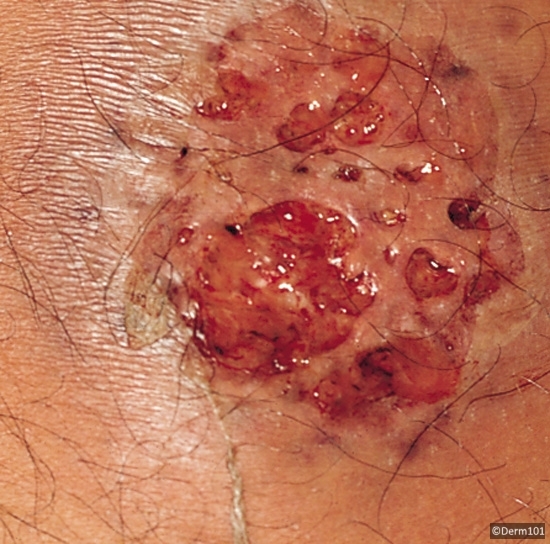
Image adapted from Derm 101

Types

  • Classic
    • Idiopathic, majority of cases
    • Associations: Infections (URI, GI) 1-3 weeks after infection
    • IBD
    • Pregnancy
    • HIV, TB, hepatitis, autoimmune conditions
    • Possible inc risk of malignancy
  • Malignancy associated
    • AML is the malignancy most associated with Sweet’s Syndrome.
    • Risk:
  • Drug-induced (long list but some of the potential ones we used more commonly are):
    • Bactrim, Macrobid, AED, hydralazine, clozapine, PTU, GCSF, Mirena, Lasix, Azathioprine, ATRA

Dx Criteria: both majors and 2 minors are required

  • Majors
    • Abrupt onset of painful erythematous plaques or nodules
    • Histopath evidence of dense neutrophilic infiltrate without evidence of leukocytoclastic vasculitis
  • Minor:
    • > 38C
    • Underlying malignancy, IBD, pregnancy, or recent upper resp, GI infection, or vaccination
    • Steroid responsive
    • Labs: ESR > 20, CRP elevated, leukocytosis > 8000 with > 70% neutrophils)

Biopsy: Dense, neutrophilic infiltrate in the dermis, w/o e/o vasculitis.

Encephalitis and CJD! – 8/22/18

Thanks to Joe for presenting the case of an elderly man presenting with subacute onset of AMS, vision changes, and ataxia, found to have creutzfeldt jakob disease (CJD).


Clinical Pearls

  • Rapidly progressive encephalitis should trigger prion disease, paraneoplastic encephalitis, or Whipple’s!
  • Most common malignancies associated with paraneoplastic encephalitis are SCLC, testicular tumors, thymomas, breast cancer, and hodgkin lymphoma
  • >90% of cases of CJD are sporadic
  • Definitive diagnosis of CJD is made by brain biopsy.  CSF testing of 14-3-3 protein marker and the RT-QuIC protein assay combined have sensitivity and specificity >90%.
  • If prion diseases are on your differential, be sure to let infection control know before doing an LP because strict precautions are required to prevent spread of infection!

Encephalitis:

Defined as AMS > 24 hours plus 2 of the following:

  1. Fever
  2. Focal neurologic deficit
  3. Seizure
  4. CSF pleocytosis
  5. Abnormal findings on EEG or neuroimaging

Differential

Capture

Prion diseases:

  • AKA transmissible spongiform encephalopathies
  • Rare, closely related, fatal, neurodegenerative conditions
  • Occur in humans and mammals
  • Result of accumulation of aggregated forms of the prion protein in the CNS
  • >90% are sporadic, the rest are infectious (kuru, variant CJD, and iatrogenic CJD)
    • Iatrogenic mostly resulting from receipt of growth hormone prepared from cadaveric pituitaries and contaminated cadaveric dura mater allografts
    • Sporadic is not transmissible by blood
  • Kuru was the first one recognized to be transmissible and linked to cannibalism among tribes in New Guinea

CJD: 

  • Most prominent clinical feature is disordered cognition
  • Typically, patients also have motor signs, such as ataxia or spasticity, vague sensory problems, or changes in visual perception
  • Myoclonus is common
  • Progressive neurologic decline resulting in death within 6-12 months
  • One in a million
  • Mean age of onset 57 – 62
  • More common in white people (may be ascertainment bias)

Diagnosis:

  • Elevated CSF levels of 14-3-3 are not very sensitive or specific.  Adding RT-QuIC protein assay to the test increases both sensitivity and specificity to >90%.
  • CDC requires the following criteria for diagnosis:
    • Progressive dementia AND
    • 2 of the following: myoclonus, visual or cerebellar disturbance, pyramidal/extrapyramidal dysfunction, akinetic mutism AND
    • Atypical EEG and/or positive 14-3-3 CSF assay  with clinical duration to death <2 years and or typical MRI abnormalities (see nice example here)

Prognosis:

  • Poor, majority die within 1 year
  • No treatment available