Wound botulism – 9/26/18

Thanks to Alison for presenting the case of a middle aged man who presented with acute onset of ptosis, dysphagia, and dysarthria, with an evolving exam found to have botulism secondary to IV injection of black tar heroin.


Clinical Pearls

  • Botulism is extremely rare! Only 110 cases were reported last year in the US with 70-75% of them being related to infant botulism, 20-25% foodborne, and 5-10% wound botulism.  The latter category has been on the rise especially in California.
  • Most common form of botulism is infant botulism
  • Botulism presents with bulbar symptoms; progressive, descending paralysis; absent reflexes; and autonomic dysfunction
  • Management involves the following
    • Notify the Department of Public Health ASAP if botulism is on your ddx
    • Obtain wound/blood cultures (special tube to be sent to DPH)
    • Administer antitoxin EARLY (do NOT wait for culture confirmation)

Capture

For more information, check this prior post on our blog.

Ophthalmoplegic migraine – 9/25/18

Thanks to Joe fore presenting the case of a middle aged woman with a history of migraines who presented with acute onset of painless binocular diplopia, found to have an isolated CN3 palsy concerning for ophthalmoplegic migraine after an extensive work up.


Clinical Pearls:

  • Types of diplopia:
    • Binocular diplopia refers to diplopia that is only present when both eyes are open (goes away when one eye is closed) and results from ocular misalignment.
    • Monocular diplopia refers to diplopia that is present even when one eye is closed and is more consistent with a local eye disease (globe related processes involving the cornea or the lens).
  • Approach to diplopia is similar to any neurologic deficit in which you would localize the lesion!
    • Upper motor neuron (brain)
    • Peripheral nerve (cranial nerves)
    • Neuromuscular junction
    • Muscle (extra-ocular muscles)
    • Globe (local eye disease or refractive error)
  • CN3 palsy usually presents with the eye in the “down and out” position and can also impact the levator palpebrae muscle resulting in ptosis.  These result from ischemia at the center of the nerve (secondary to diabetes/HTN).
    • In a patient with CN3 and a dilated pupil, you must rule out a PCA aneurysm!  Impingement of the CN3 by an enlarging aneurysm cuts off the parasympathetic fibers running on the outside of the nerve, resulting in a dilated pupil.  This is the only aneurysm that gives a warning sign before rupture!

Commonly tested gaze palsies:

Capture

Ophthalmoplegic migraine:

  • Rare condition, often manifests in children and young adults
  • Diagnosis of exclusion
  • Most commonly affects CN3 (but can go to CN4 and CN6 as well)
  • Can sometimes precede the headache
  • Permanent nerve damage has been reported and some believe that it is a demyelinating neuropathy (for more info, refer to this review article)


Acute Epiglottitis 9/24/2018

Yves-Paul presented a case of a middle age man with poorly controlled diabetes presenting with acute onset sore throat, which quickly progressed to dyspnea and dysphagia. He was noted to have stridor on evaluation and he was urgently intubated for airway protection. Subsequent endoscopic exam revealed a grossly purulent and inflamed epiglottis consistent with an abscess.

Stridor vs Wheezing

  • Stridor: Upper airway, inspiration, single pitch (remember the sound quiz we did?)
  • Wheezing: Relatively lower airway, expiratory musical sounds, but in bad cases can see wheezing in both

Picture1

Epiglottitis

Epidemiology

  • Kids more common (H. influenzae type B) but less common now due to vaccination.

Risk Factors

  • Kids: Immune deficiency, incomplete immunization
  • Adults: Immune deficiency, diabetes

Presentation: Drooling, dysphagia + odynophagia, dyspnea/distress

  • Acute in adults over 24-48 hours Kids can be hyperacute (< 12 hours) leading to airway compromise.
  • Obstruction less acute in adults due to larger AW diameter
  • Sore throat and odynophagia in most cases
  • Fewer have airway compromise; signs to look for are:
    • Drooling
    • Muffled voice
    • Respiratory distress
    • Stridor (impending obstruction)
    • Tripoding, extended neck (maximizes airway diameter)

Diagnosis:

  • Lateral X-ray with thumb sign demonstrates an enlarged epiglottis.

Picture2

  • Direct visualization (fiber optic), beefy red, stiff, edematous epiglottis is diagnostic.

Picture3

As you can see, the surrounding structures were grossly edematous and inflamed to the point we cannot identify the vocal cords at all.

Pathophysiology

  • Organisms: Strep, staph, non-type H.influenzae, beta hemolytic strep, Klebsiella
  • Viral: HSV, EBV, Para/influenza, VZV
  • Non-infectious: Foreign body

Management:

  • Secure airway FIRST. No not manipulate or remotely touch. Can be nasotracheal or orotracheal. If unable to intubate, tracheostomy can be done.
  • Abx: Beta lactamase resistant class (usually 3rd gen cephalosporin) in general unless also suspecting staph/strep, then add vancomycin as well
  • I&D if abscess

Picture4


Elevated Alk Phos… Only Clue to Breast Cancer 9/20/2018

Sarah presented a middle age woman with a history of schizophrenia, HFpEF, possibly COPD, who presented from her Board and Care facility due to inability to ambulate. She was able to provide much of a history but her exam was normal. Her labs were notable for alk phos in the 700-1000 range, and mildly elevated AST/ALT in the 60-70s. An abdominal US revealed hepatic steatosis but really nothing else… Her medical history was also obscure since she receives her care from multiple institutions.

She was incidentally found to have a breast mass on exam, and subsequent work up for her elevated alk phos unfortunately revealed metastatic breast cancer.

Elevated Alk Phos

Background: Alk Phos is derived from mainly bone and liver, higher in men, varies with age (higher in kids, thought to be due to physiologic osteoblastic activity)

GGT (gamma glutamyl transpeptidase): liver specific, can be used to verify if alk phos elevation is due to biliary disease if GGT is also elevated.

Alk Phos


Breast Cancer

Most common tumor in women

Risk

  • > 50
  • Personal Hx
  • Strong family Hx of pre-menopausal breast cancer
  • Genetic BRCA 1 & 21
  • Personal hx or ovarian or endometrial cancer
  • Dense breasts
  • OCP use for > 15 years
  • Late menopause

Screening:

  • Screen F > 50 or < 5-10 yrs prior to age in 1st degree relative with breast cancer, and then Q2yr
  • More frequent screening recommended for specific mutations, i.e. BRCA, TP53, then MRI Q year
  • How about F with breast implants? MRI, CT, or mammogram? The recommendation is still mammogram but with multiple views


Common Scenarios:

Breast

Local disease in situ No malignancy beyond basement membrane Lumpectomy + RT, or mastectomy. If ER +, use tamoxifen/aromatase inhibitors
Lobular carcinoma in situ (LCIS) Isolated to lobule, within basement membrane, not exactly cancer yet but high risk ER+ use tamoxifen/AI to dec risk of development into invasive breast cancer.

NSABP-P1 trial: Pt with LCIS tx with tamoxifen dec risk of invasive breast cancer, but inc risk of endometrial cancer esp in > 50yo

Infiltrating ductal carcinoma, LN negative Spread beyond BM, need to sample sentinel LN, if negative, no further need for dissection. Wide excision of mass with free margins + RT, adjuvant chemo for size > 1cm. Tamoxifen/AI if ER +
Infiltrating ductal carcinoma with LN positive Same as above but LN +, warrants further LN dissection, automatic adjuvant chemo Wide excision (modified radical mastectomy), RT + adjuvant chemo + Tamoxifen/AI if ER+.
Local invasive dz involving skin or chest wall   Chemo followed by mastectomy, + tamox/AI if ER+

Lymph node positivity is the most important prognostic factor, followed by tumor size, then receptors, and then grade.

For diagnosis, always get excision biopsy for dx, FNA has low sensitivity, hence even if negative, always follow by excisional bx.

Receptors and Management

  ER+ ER- HERR2+
Pre-menopausal Chemo + tamoxifen Chemo + trastuzumab
Post-menopausal Aromatase inhibitors + chemo Chemo + trastuzumab

Tamoxifen: Use for 10 years if ER/PR+. Dec risk of new breast cancer and effective for metastatic dz of receptor positive.

Down side: inc risk of endometrial cancer 3x, inc thromboembolic risk.

Aromatase inhibitors: Watch out for osteoporosis

92% blasts… Fever… AND a rash?! 9/19/2018

Narges presented a case of a middle age woman without any prior medical history, presenting with 1 week of bruising, epistaxis, and bleeding from her gums. Her initial lab work was notable for a WBC of 52.2 with 92% blasts, later confirmed to be AML. She developed a fever and a rash over the next few days… She had neutropenic fever, and around the same, time, developed AML-associated Sweet’s Syndrome!

AML: A quick overview

Accounts for 80% of acute leukemias in adults

Risk Factors

  • Benzene exposure
  • Radiation exposure, commonly 7-10 years after exposure
  • Prior tx with alkylating agents and topoisomerase II inhibitors like doxorubicin, etoposide.
  • Age: greatest risk factor, older = at high risk, median age 65
  • CML, MDS, and myeloproliferative syndromes have a chance to evolve into AML.

Initial presentation

  • Bruising, gum bleeding, epistaxis from thrombocytopenia
  • SOB, DOE, fatigue from anemia
  • Pyogenic infections of the skin
  • HSM found in 1/3 of pts
  • 50% might have gingival hyperplasia as first signs of the disease
  • Small subset might have concurrent HLH on presentation
  • If fever, almost always infection

Diagnosis

  • Buzz words: blasts on smears, Auer rods (peroxidase stain)
  • > 20% blasts cells
  • Flow cytometry
    • CD117, CD33 most common
    • CD19, if seen, suggests lymphoblastic origins
  • Subtypes
    • M3 (Acute promyelocytic leukemia), t(15; 17), prone to DIC, responsive to ATRA and potentially can be cured.
      • If pt receiving ATRA +/- Arsenic trioxide develop pulmonary sx think of an entity called Differentiation Syndrome, can be life threatening, stop treatment and give steroids.
    • Non APL: Everything else

Management:

  • Induction, consolidation (after complete remission, assess induction response via BM bx) via HiDAC high dose cytarabine, or autologous CT, or allogeneic HCT, maintenance (usually not needed but can be beneficial in some types of AML)
  • APL: ATRA +/- ATO
  • Non-APL: 7 day course of cytarabine and 3 day course of an anthracycline. For older pts with more comorbidities, can use a milder regimen with azacytidine or decitabine.


Neutropenic fever

Definition: T > 38.3 or > 38 sustained over 1 hour, with neutropenia (ANC < 500)

Determine high risk or low risk

  • Low Risk: Anticipated neutropenia < 7 days, clinically stable, NO medical comorbidities
    • IDSA: Can consider outpatient antibiotics, Cipro + Augmentin and able to tolerate PO, otherwise inpatient management
  • High Risk: Anticipated neutropenia > 7 days, clinically unstable, any medical comorbidities
    • Automatically inpatient management
    • Monotherapy with pseudomonal coverage initially is recommended by IDSA
      • Cefepime: 2g Q8H, higher dose than usual
      • Meropenem 1g Q8H
      • Imipenem
      • Zosyn 4.5g Q6-8
      • Ceftazidime increasingly avoided.
    • If history of MRSA, e/o catheter infection, skin infection, pneumonia, or unstable, add Vancomycin/MRSA coverage
    • PCN allergy: Can consider using Aztreonam, cipro
    • If recurrent of persistent fever after 4-7 days: Add an empiric antifungal, most of the time cover for candida since it’s the most common cause of invasive fungal infection.
      • Echinocandin is favored i.e. caspofungin, increasing azole resistance in candida.
      • Think aspergillus if e/o pulmonary nodules, ampho B and voriconazole then are preferred

Sweet’s Syndrome

Uncommon, inflammatory disorder, usually affects pts ages 30-60. Older = more likely malignancy associated

Presentation:

  • Abrupt, painful, edematous (juicy), erythematous papules/plaques/nodules + fever and leukocytosis.
  • Rare mucosal/oral involvement.
  • Can also rarely causes inflammation of a particular organ system, i.e eye, liver, heart, CNS, kidney, even bone.

Sweet.jpgImage adapted from Derm 101

Types

  • Classic
    • Idiopathic, majority of cases
    • Associations: Infections (URI, GI) 1-3 weeks after infection
    • IBD
    • Pregnancy
    • HIV, TB, hepatitis, autoimmune conditions
    • Possible inc risk of malignancy
  • Malignancy associated
    • AML is the malignancy most associated with Sweet’s Syndrome.
    • Risk:
  • Drug-induced (long list but some of the potential ones we used more commonly are):
    • Bactrim, Macrobid, AED, hydralazine, clozapine, PTU, GCSF, Mirena, Lasix, Azathioprine, ATRA

Dx Criteria: both majors and 2 minors are required

  • Majors
    • Abrupt onset of painful erythematous plaques or nodules
    • Histopath evidence of dense neutrophilic infiltrate without evidence of leukocytoclastic vasculitis
  • Minor:
    • > 38C
    • Underlying malignancy, IBD, pregnancy, or recent upper resp, GI infection, or vaccination
    • Steroid responsive
    • Labs: ESR > 20, CRP elevated, leukocytosis > 8000 with > 70% neutrophils)

Biopsy: Dense, neutrophilic infiltrate in the dermis, w/o e/o vasculitis.

Hyponatremia – 9/18/18

Thanks to Joe for presenting the case of a young man with no known medical history who presented with acute onset of generalized weakness, found to have symptomatic hyponatremia to 120 resulting from psychogenic polydipsia!


Clinical Pearls

  • Hyponatremia is largely a problem of too much free water (sometimes compounded by too little solute) and defined as a Na <135
  • Pick a systematic approach to solving the underlying diagnosis for any patient with hyponatremia (see below for one possible outline).
  • The term “pseudohyponatremia” refers to the presence of substances that interfere with laboratory measurement of sodium concentration in the blood, specifically high triglycerides and paraproteins.
    • Hyperglycemia does NOT fall into this category because glucose is osmotically active and pulls water into the intravascular space, resulting in a dilutional and true hyponatremia.
  • Chronic hyponatremia (>72 hours) must be corrected slowly to avoid osmotic demyelinating syndrome (ODS).  Acute hyponatremia (<48 hours) can be corrected more rapidly.  That said, we rarely have a Na value within 48 hours prior to patient presentation to determine acuity.  As a result, and given the neurologic risks, most patients are treated as if they have chronic hyponatremia.
  • ADH leads to increased uric acid excretion through the kidneys so low serum uric acid levels have been associated with SIADH.  But this test is not very specific.
  • Osmolarity of normal saline is 308 compared to human serum which is normally 280-295.  In a hyponatremic patient, NS infusion becomes a relatively hypertonic (rather than isotonic) solution!

Schematic

For more detailed information, refer to this prior post and this review article.

Metformin Associated Lactic Acidosis (MALA) 9/17/2018

Thanks to doctor-in-training Emma who presented a case of an elderly woman, on dialysis, who came in with SEVERE lactic acidosis due to a variety of factors, but largely contributed by being prescribed metformin 2 weeks prior to presentation.

We will use this case to illustrate several concepts:

On initial presentation, pt was in a state of shock. If you remember from medical school, there are mainly 4 types:

  • Cardiogenic
    • Myopathic (DCM, acute MI)
    • Mechanical (Acute MR, VSD, severe AS)
    • Arrhythmia
  • Hypovolemic
    • Hemorrhage, volume depletion
  • Distributive
    • Sepsis
    • Anaphylaxis
    • Neurogenic (spinal cord injury)
    • Endocrine (myxedema, Addison’s)
  • Obstructive
    • Pericardial tamponade
    • PE
    • Pneumothorax

Shock can also be approached from a cardiac output standpoint to help differentiate the potential causes:

Capture

If someone is hypovolemic, but he/she is found to be hypotensive but bradycardic (instead of tachycardic), remember from physiology:

MAP = CO x SVR, where CO is cardiac output, SVR is systemic vascular resistance

CO = SV x HR, SV is stroke volume, and HR is heart rate.

Hence in a state of hypovolemia, the body will attempt to maximize CO by increasing HR to maintain adequate MAP. If you see the opposite though, there could be something else going on…

  • Cardiac: MI, conduction abnormalities, infectious (Lyme, Charga’s)
  • Toxic/Meds: CCB, Beta blocker, Dig, Alpha 2, organophosphate
  • Lytes: Hyperkalemia -> heart block -> sinus arrest
  • Endocrine: Hypothyroidism (myxema coma), adrenal insufficiency
  • Environmental: Hypothermia
  • Neurogenic shock


Acid-Base: Acid-base disorders can appear daunting, but the following algorithm can make things simpler if followed consistently. We will work through this patient’s acid/base disorder together. Her HCO3 is 6, AG of 52, with an ABG of 6.83/13/170.

  1. Look at the pH: < 7.4 automatically means some sort of acidosis is going on.
  2. Next look at the PCO2, if it is in the same direction as the pH (low PCO2 in this case, and low pH), then it is suggestive of a primary metabolic rather than a respiratory issue causing the acidosis. If pH is low and PCO2 is high, then there is high suspicions for a respiratory process driving the acidosis.
  3. Look at the Anion Gap, which in this case in 52. Go through your differential for AGMA.
  4. Calculate the Delta Gap. This basically is a way to assess whether a mix acid/base disorder exists after taking into account the AG. To help illustrate:Capture2
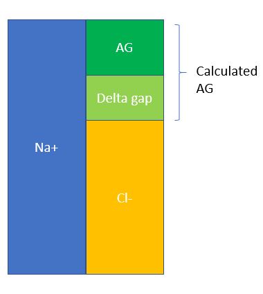
  5. Delta gap = calculated AG – normal AG = 52 – 12 = 40. Now there are several ways to do this, but one method that’s quick and simple is to simply add this delta gap to the measured bicarb and see what other metabolic acid/base disorders can be hiding beneath the anion gap. In this case, 40 + 6 = 46, which is suggestive of an underlying metabolic alkalosis.
  6. Lastly, calculate compensation. For AGMA, use Winter’s Formula = 1.5 (HCO3) + 8 +/-2. The calculated PCO2 should be 15-19. Since our measured PCO2 is 13, which is lower than expected, then this is suggestive of an underlying respiratory alkalosis as well, probably due to sepsis.
  7. Our final answer for the acid/base disorder for this patient is: AGMA with metabolic alkalosis and concurrent respiratory alkalosis!


Lactic Acidosis: Traditionally we view this as a sign of sepsis, or a marker of tissue hypoperfusion. However this is not always the case!

  • Type A: Inadequate oxygen delivery
    • Rule of thumb: Tissue hypoperfusion, tissue ischemia
    • Can also be seen with increased anaerobic muscular activity, like generalized convulsions in a seizure
    • Can also be seen in hypoxemia, severe anemia
    • Cryptic shock (elevated lactate, okay BP), similar to overt septic shock in terms of mortality in setting of severe sepsis.
  • Type B: altered physiology, others
    • B1: Underlying disease, malignancy, hepatic & renal failure
    • B2: Drugs, toxins
    • B3: Inborn errors of metabolism, enzyme deficiency
  • Management: Tx the underlying cause, bicarb controversial, studies have shown no benefits? But most experts would agree with usage in critically ill patients with profound acidemia.


MALA: Lastly, this case illustrates the dangers of metformin, in the right setting, can lead to severe acid/base disturbance (MALA).

  • Do NOT use metformin if GFR < 30, be careful in CKD patients.
  • If suspecting metformin toxicity and pt is hyperglycemic, this is a sign of severe toxicity.
  • Always do a thorough med reconciliation on every single patient!

Aplastic Anemia 9/12/2018

Today our Doctor-in -Training, Emma, presented a case of a middle age woman with no significant medical or family history coming in with a month of fatigue, menorrhagia, and gum bleeding. She was incidentally found to be pan-cytopenic. Bone marrow biopsy revealed hypocellularity with no abnormal morphologies, which is consistent with aplastic anemia!

Aplastic anemia

  • Epidemiology
    • Rare, 2 per million per year, higher incidence amongst Asians, can occur in all ages but most of the time in the first 1st-4th decades.
  • Pathophysiology
    • Big picture: Think of it this way. Aplastic anemia is similar to a factory not working due to not enough workers (stem cells). Compare and contrast this with MDS, which is basically a loss of quality control (mutation in the stem cells) leading to bad products that get worse over time (leading eventually to pancytopenia, leukemia).Capture
      • Causes
        • Primary (rare), most of the time acquired
        • Infectious: CMV, Parvo, EBV, HIV, hepatitis, TB
        • Toxins: Benzene, glue vapors, solvents, pesticides
        • Autoimmune: SLE, eosinophilic fasciitis, graft v host,
        • Other: Pregnancy, paroxysmal nocturnal hemoglobinuria, thymoma, irradiation
        • Medications: NSAIDS, Bactrim, Lasix, Gold, Allopurinol, penicillamine, phenothiazines, AEDS, anti-psychotics
      • Association
        • PNH: CD55 & CD59 abnormalities can be sen in up to 50% of patients with aplastic anemia!
      • Presentation
        • S/S of anemia
        • S/S of thrombocytopenia (menorrhagia, petechiae)
        • Recurrent infections
      • Diagnosis
        • Bone marrow biopsy most definitive, helps distinguish AA from other causes of pancytopenia
        • BM finding
          • Hypocellular, marrow space mostly fat cells and marrow stroma.
          • Remaining cells are morphologically normal
          • No fibrosis, no e/o malignancy
        • Severe AA criteria
          • BM cellularity < 25 + 2 of ANC < 500, plt < 20k, retic < 20k
        • Very Severe AA criteria
          • ANC < 200
        • Mgx: Severe AA require treatment
          • Under age 20: Early allogeneic HCT as soon as possible. If HCT not possible, immunosuppressives
          • 20 – 50: HCT also recommended if no contraindications, immunosuppressives (Anti-thymocyte globulin, cyclosporine for example) + eltrombopag (thrombopoietin agonist) otherwise.
          • > 50:
            • Transplantable: Early hematopoietic cell transplantation
            • Older, not candidates for HCT: Immunosuppressive, less intense preferred as age increases.
          • Treat other underlying/reversible causes in all cases.

Fun Fact: Blood transfusion can lead to falsely elevated iron studies within the first 24-36 hours.

Angioedema 9/11/2018

Today Arthur presented a case of a young woman with a personal and family history of facial swelling presenting with acute onset periorbital and scalp swelling, in setting of recent hair-dye exposure. No urticaria or pruritus on presentation but she did have acute pruritus after exposure to the hair-dye. She responded quickly to benadryl and steroids. The final diagnosis is allergic angioedema.

What is angioedema? It is a non-pitting swelling of the deep/subcutaneous tissues.

Angioedema

(Image adapted to JAMA)

Pathophysiology of angioedema

  • Mast cell mediated (most common)
    • Examples: Food allergies, insect stings
    • IgE -> Mast Cells -> inflammatory response
      • BeE, FoodiE = IgE
    • Findings:
      • Flushing
      • Urticaria
      • Generalized pruritus
      • +/- bronchospasm, throat tightness, hypotension
    • Onset: Minutes after exposure, develop over the next few hours
    • Recovery: 24-48 hours
    • Anaphylaxis = life threatening systemic reaction which is a severe manifestation of an allergic reaction. Angioedema is a finding.
    • Management: IM Epi is FIRST LINE, 0.3 – 0.5mg IM, repeat Q5-15min PRN. Everything else (steroids, benadryl, anti-histamines) are adjuvant only. Get that IM Epi stat.
  • Bradykinin mediated
    • Examples: Acquired angioedema (AAE, associated with underlying malignancy or autoimmune conditions), RAAS-blocker angio edema (RAE, African Americans are 3x likely vs Whites), hereditary angioedema (HAE, autosomal dominant mode of inheritance)
    • Findings:
      • Absent urticaria/pruritus, bronchospasm.
      • Minimal skin findings but can have swelling without urticaria.
    • Onset: More prolonged course, can develop over 24-36 hours
    • Recovery: 2-4 days
    • Management: depends
  • Histamine/Unclear: Seen in idiopathic angioedema (IAE)

Angioedema algorithm

Capture

HAE Management

  • C1 Inh concentrate (takes a while to obtain)
  • Icatibant (synthetic bradykinin receptor antagonist)
  • Ecallantide (recombinant plasma kallikrein inhibitor)
  • FFP (fastest to get!)
  • Supportive care, airway monitoring

Still’s Disease – 9/10/18

Thanks to Becky Lee yet again for presenting an interesting case of a young woman presenting with acute onset of fever and polyarthritis, found to have a history of similar episodes in the past together with a rash concerning for Still’s disease!


Clinical Pearls

  • Still’s disease is a diagnosis of exclusion!  Yamaguchi criteria can help with ruling in the diagnosis.
  • Still’s remains a multi-systemic disorder of unknown etiology because it’s difficult to diagnose and rare (0.16 cases per 100,000).
  • RF and ANA are generally negative but can be positive in <10% of patients with Still’s in low titers.
  • ~66% of patients present with sore throat secondary to cricothyroid perichondritis or aseptic nonexudative pharyngitis.
  • The disease is often recurrent.  Predictors of poor outcome include erosive polyarthritis on presentation and shoulder/hip involvement.

Acute polyarthritis (>5 joints involved):

Remember that for rheumatologic disorders, timing, symmetry, and number of joints involved is crucial to coming up with a differential diagnosis.  So for our patient with acute polyarthritis, consider the following:

  • Infection
    • Viral: hepatitis, HIV, parvovirus B19
    • Spontaneous bacterial endocarditis
  • Rheumatologic:
    • Rheumatoid arthritis
    • Reactive arthritis
    • SLE
    • Dermatomyositis
  • Vasculitis
    • PAN
  • Drug reactions
  • Auto-inflammatory or disease of the innate immune system (as opposed to autoimmune or diseases of the adaptive immune system):
    • Periodic fever syndromes (TRAPS, PFAPA, hyper-IgD syndrome)
    • Still’s disease
  • Schnitzler’s syndrome – chronic urticaria associated with a monoclonal gammopathy (usually IgM kappa)
  • Sweet syndrome – painful skin lesions
  • Sarcoid
  • Kikuchi disease – cervical LAD and fever (necrotizing lymphadenitis)
  • HLH/Macrophage activation syndrome – leukopenia and thrombocytopenia, elevated triglycerides, low fibrinogen and haptoglobin

Still’s disease:

  • Some clarifications on nomenclature:
    • Systemic juvenile idiopathic arthritis (sJIA): first presentation <17 years old, previously referred to as Still’s disease
    • Adult onset Still’s disease (AOSD): first presentation  > 17 years old
  • Epidemiology of AOSD:
    • 0.16 cases per 100,000
    • No sex predominance (F=M)
    • Bimodal age distribution with peak between 15-25 and another 36-46 years of age.  New diagnosis in patients >60 have been reported.
  • Clinical features ⇒ Yamaguchi criteria (need 5 total with  > 2 major)
    • Major criteria:
      • Daily fevers to 39
      • Arthritis >2 weeks
      • Non-pruritic salmon-colored macular/maculopapular rash on trunk or extremities (though cases of pruritic rash have also been reported)
      • ↑ WBC >10k, >80% neutrophils
    • Minor
      • Sore throat
      • LAD and/or splenomegaly
      • ↑ AST, ALT, or LDH
      • Negative ANA/RF
  • Treatment
    • Mild: NSAIDs
    • Moderate: NSAIDs + DMARDs
    • Severe: NSAIDs + DMARDs (IL1 receptor antagonists like anakinra appear to be more helpful than TNF inhibitors especially in sJIA)
  • Prognosis:
    • Overall good prognosis
    • Disease can be limited to one episode or recurrent over time
    • Poor prognostic indicators:
      • Hip and shoulder involvement
      • Erosive polyarthritis at initial diagnosis
  • Complications
    • Macrophage activation syndrome (ie HLH) can occur in 15% of cases
    • DIC
    • TTP
    • Diffuse alveolar hemorrhage
    • Pulmonary HTN
    • Aseptic meningitis

References: 

Great recent review article on Still’s disease (AOSD Review) and this prior post on our blog!