All posts by vmcimchiefs

Small cell bladder cancer and hematuria- 10/17/18

Thanks to Naina for presenting the case of an elderly man presenting with acute onset of n/v, and abdominal pain, found to have anemia and AKI, with work up revealing small cell cancer of the bladder causing ureteral obstruction with mets to the lymph nodes, liver, lung, and bone, hospitalization complicated by TLS prior to onset of chemo and contrast induced nephropathy.


Clinical Pearls

  • Bladder cancer is the most common malignancy of the urinary system and urothelial (transitional cell) carcinoma is the culprit >90% of the time.  Less common subtypes include squamous, adeno, small cell (our patient), and sarcoma.
  • Unexplained hematuria in anyone >40 years is bladder cancer until proven otherwise!
  • CT urography is the diagnostic imaging of choice in the work up of hematuria.
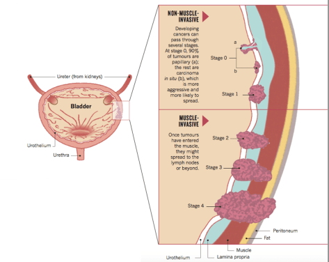
  • Diagnosis of bladder cancer is often delayed due to similarity of symptoms with other benign disorders.  However, majority of cases are still caught in stage 0-1 (muscle non-invasive disease) with overall good prognosis.

Bladder cancers:

  • Epidemiology
    • Most common malignancy of the urinary system, 3-4 x more common in men but women are usually diagnosed with more advanced disease and have a higher mortality rate.
    • Median age at diagnosis is ~70
    • Incidence has increased by more than 50% during the past 20-30 years.
  • Types:
    • Urothelial (transitional cell) carcinoma is the predominant histologic subtype in the US and Europe (>90% of all bladder cancers) and can arise in renal pelvis, ureter, or urethra
    • Other: squamous, adeno, small cell, sarcoma
  • Degree of invasion:
    • Superficial (non-muscle-invasive)
    • Muscle-invasive
    • Metastatic
  • Clinical presentation
    • Painless hematuria
    • Irritative voiding symptoms (frequency, urgency, dysuria) – only in 30% of patients
    • Sometimes metastases cause the initial symptoms that lead to diagnosis (as in our patient)
    • Most cancers eventually become symptomatic
  • Diagnosis: often delayed due to similarity of symptoms to other benign d/o
    • Urine cytology >98% specific, 12-64% sensitive based on grade of tumor
    • Imaging
      • CT favored over IVP
    • TURBT done for diagnosis and staging
    • DDx
      • Hematuria from enlarged prostate
      • Pregnancy
      • Cystitis
      • Prostatitis
      • Passage of renal calculi

Staging bladder cancer

Source: Nature Outlook.


  • Management
    • Over 50% of people diagnosed with non-invasive disease develop recurrence
    • Assess performance status with Karnofsky or Eastern Cooperative Oncology Group scales for older patients before deciding on chemotherapy
    • Chemo regimens are often cisplatin-based which carry the side effects of nephrotoxicity, ototoxicity, and neuropathy

treatmetn of bladder cancer

Source: Nature Outlook

Hematuria

Refer to this thorough algorithm on UpToDate.

  • Incidence of malignancy in microscopic hematuria is ~2-5%
  • Incidence of malignancy in macroscopic hematuria is ~20%

Extra pearls on onset of hematuria during voiding:

  • Occurs at the beginning? Urethral source
  • Discharge noted between voidings or stain on undergarment? Urethral meatus or anterior urethra
  • Terminal hematuria? Bladder neck or prostatic urethra
  • Throughout voiding? Anywhere in the GU tract

Hepatopulmonary Syndrome 10/10/2018

A 67 year old man with history of cirrhosis secondary to Hepatitis C and alcohol, hepatocellular carcinoma with recent TACE, presented with worsening dyspnea on exertion and positional shortness of breath. His breathing was worse when he sat upright, and better when he was supine. What’s going on?

Just to go over some terms:

  • Orthodoxia: Drop in PaO2 by 5mmHg or O2sat by 5% when moving from supine to upright.
  • Platypnea: Dyspnea that is induced by moving to an upright position, relieves when supine.

Hepatopulmonary syndrome

Triad

  • Chronic liver disease or portal hypertension
  • Intrapulmonary vascular dilations (IPVD)
  • Impaired oxygenation

Epidemiology:

Up to 25% of patients with chronic liver disease will have some degree of shunting, can occur at any stage (mild or severe)

Pathophysiology

  • Not well understood but the theory is due to increased nitric oxide production and reduced NO clearance, resulting in pulmonary vasodilation (IPVDs) mostly concentrated at the lung bases.
  • When upright, blood preferentially perfuse the lower lung  zones due to gravity.
  • Vasodilation leads to poor gas exchange.

Diagram

  • This leads to a VQ mismatch

Diagnosis:

  • CXR: Not helpful, might show e/o interstitial lung markings.
  • CT: Can reveal IPVDs
    • Dilated peripheral pulmonary vessels
    • Inc pulmonary artery to bronchus ratios
  • PFT: Not helpful
  • Transthoracic contrast echo (TTCE): Can be used to demonstrate presence of intrapulmonary shunts supportive of presences of IPVDs
    • Concept of bubble study: Shooting agitated saline (with bubbles into the vasculature
    • Bubbles visible in the R heart chambers, should not be visible in the left heart chambers.
    • If presence of bubbles in the left: This is indicative of a shunt:
      • Intracardiac shunt: bubbles seen within 1 beat
      • Intrapulmonary shunts: bubbles seen after 3-8 beats.


Normal Echo: Notice how the agitated saline bubbles remain on the right side of circulation and do not cross over. The bubbles were filtered out by the pulmonary vasculature.

Normal Echo


Echo demonstrating intrapulmonary shunting (see bubbles crossing over from the right to the left)

HPS


Management

  • Supplemental O2 indicated if O2 sats < 88%, PaO2 < 55mmHg
  • Mild to moderate: Monitor Q6-12 months
  • Severe to very severe: Referral for liver transplant
  • Insufficient data on other treatment options (garlic, pentoxifylline, NO synthase inhibitors, IPVD embolization, plasma exchange, oxtreotide).

HPS_diagnosis_algorithm

Image adapted from Uptodate

Check out this article if you’re interested in the data behind pentoxifylline!

On rhabdo and myopathies – 10/9/18

Thanks to Cameron and Adam for presenting the case of a middle aged man with no significant PMH who presented with diffuse myalgias and chronic progressive proximal muscle weakness, found to have a CK >12k and EMG findings concerning for an inflammatory myopathy, awaiting muscle bx for diagnosis.


Clinical Pearls

  • Rhabdomyolysis literally means dissolution of skeletal muscle and has a broad differential outside of the typical traumatic or exertional processes associated with it see below).
  • The four main inflammatory myopathies are dermatomyositis, polymyositis, inclusion body myositis, and necrotizing autoimmune myositis.
  • Polymyositis is rare and a diagnosis of exclusion after the other three main inflammatory myopathies have been investigated.
  • Overall, the prognosis of inflammatory myopathies is good with appropriate treatment.  The exception is inclusion body myositis which is a progressive disorder without any effective therapy.
  • Pigment nephropathy can occur with rhabdo regardless of the underlying etiology especially in patients with CK >5000.  Aggressive IV hydration to lower CK levels is important to reduce the risk of kidney injury.

Rhabdomyolysis:

DDx:

  • Traumatic
    • Crush injuries, surgery, prolonged compression from immobility or coma
  • Non-traumatic
    • Exertional:
      • Normal muscle: strenuous exercise, heat stroke, seizures, hyperkinetic states
      • Abnormal muscle: metabolic myopathies, mitochondrial myopathies, malignant hyperthermia, NMS
    • Non-exertional
      • Alcoholism
      • Drugs and toxins: lipid-lowering drugs (fibrates, statins), alcohol, heroin, cocaine, meth, colchicine
      • Infections: influenza, coxsackie, EBV, HIV, legionella
      • Electrolyte abnormalities: hypokalemia, hypophosphatemia, hypocalcemia
      • Endocrinopathies: DKA, HHS, hypothyroidism, vitamin D deficiency
      • Inflammatory myopathies (rare)
      • Paraneoplastic
      • Miscellaneous

Inflammatory myopathies

Largest group of potentially treatable myopathies in children and adults.

  • Four subtypes: distinguishing which process is important because each subtype has a different prognosis and response to therapy
    • DM
      • Anti-Mi-2, anti-MDA-5, anti-TIF-1, anti-NXP-2
    • PM
      • Rare, often misdiagnosed
      • Dx of exclusion
    • Necrotizing autoimmune myositis
      • More common than PM
      • Occurs alone or after viral infections or in association with cancer, CTD, or post-statin
      • Anti-SRP or anti-HMGCR
      • Highest CK level
    • Inclusion body myositis
      • Most common in people >50
      • 7.9 cases/million in the US
      • Distal muscles impacted first
      • Facial muscles impacted
      • Muscle atrophy occurs earlier than in others
      • Extramuscular manifestations are uncommon
      • Dysphagia occurs in >50%
      • Muscle atrophy is common
      • Lowest CK level
  • Up to 30% of patients with DM or PM have a constellation of clinical findings termed “antisynthetase syndrome”
    • Acute disease onset
    • Constitutional symptoms (fever, weight loss)
    • Myositis
    • Raynaud’s
    • Mechanic’s hands
    • Non-erosive arthritis
    • ILD
    • Labs show antibodies to tRNA synthetase enzymes (anti-Jo-1)
  • Extramuscular manifestations
    • systemic symptoms
    • cardiac arrhythmias or ventricular dysfunction
    • pulmonary complications (ILD)

Capture

Table above adapted from this and this review article by NEJM.

Lymphocytic hypophysitis – 10/3/18

Thanks to Sahar for presenting the interesting case of a middle-aged woman with metastatic melanoma recently started on ipilimumab who presented with a headache and fatigue, found to have hypothyroidism and adrenal insufficiency with work up consistent with hypopituitarism related to an adverse effect of ipilimumab: lymphocytic hypophysitis!


Clinical Pearls

  • Remember that adrenal insufficiency and hypothyroidism are causes of elevated ADH levels.
  • Red flags for obtaining head imaging for headache include age >55, sudden onset, positional, onset after trauma or exercise, fever, focal neuro findings, and immunosuppression.
  • Pituitary adenomas can have three manifestations: mass effect, hormonal hypersecretion, and hypopituitarism.  When imaging shows a pituitary mass, your work up should address each of these categories.
  • The most sensitive test to assess hypothalamic-pituitary access function is LH/FSH!
  • Immunotherapies are commonly associated with a flare of autoimmune diseases.  A more rare side effect of CTLA-4 inhibitors (like ipilimumab) is lymphocytic hypophysitis (inflammation of the pituitary gland)
    • This condition commonly presents with headache out of proportion to neurologic findings and preferential decline in ACTH and TSH though other hormones can also be impacted.
  • For hypopituitarism, remember to always treat adrenal insufficiency first before replacing thyroid hormone.  Failure to do so can precipitate adrenal crisis!

Indications for imaging a patient with headache:

  • Age >55
  • Sudden onset
  • Worse with lying down or wakes patient from sleep
  • Rapid onset after trauma or exercise
  • Fever
  • Focal neurologic findings
  • New headache in immunosuppressed patient

Pituitary adenoma:

  • Evaluate for the following
    • Mass effect: visual field deficit, headache
    • Hormonal hypersecretion
      • Prolactin ⇒ galactorrhea, amenorrhea, infertility
      • GH ⇒ Acromegaly
      • TSH ⇒ hyperthyroidism
      • ACTH ⇒ Cushing disease
      • ADH ⇒ SIADH
    • Hyposecretion:

Capture

Hypophysitis:

  • Inflammation of the pituitary
  • Four categories based on histologic findings:
    • Lymphocytic
      • Most common form
      • Seen in late pregnancy and post-partum period
      • Also associated with ipilimumab as our patient here!
    • Granulomatous
      • Idiopathic or secondary to GPA, sarcoid, TB
    • Plasmacytic (IgG4-related)
    • Xanthomatous (most rare)
  • Clinical presentation
    • Headache out of proportion to exam findings
    • Preferential decrease in ACTH and TSH ⇒ adrenal insufficiency and hypothyroidism
  • Prognosis:
    • Pituitary size eventually normalizes but pituitary loss of function is often permanent.

Hyponatremia:

Lastly, refer to this algorithm from our recent morning report to help you think through hyponatremia.

Acute pancreatitis – 10/2/18

Thanks to Tiffany for presenting the case of a middle-aged man presenting with acute onset of epigastric abdominal pain and nausea/vomiting, found to have a normal lipase initially which jumped up to 1150 48 hours later consistent with acute pancreatitis.


Clinical Pearls

  • Gallstones and ETOH account for the majority of cases of acute pancreatitis.
  • Up to 30% of cases of acute pancreatitis are idiopathic!  This is a diagnosis of exclusion.
  • Lipase typically rises within 4-8 hours after the onset of pancreatitis and lasts for >8 days as opposed to amylase (6-12 hours, lasts for 3-5 days).  Lipase is also more sensitive and specific than amylase.
  • Common electrolyte abnormalities associated with pancreatitis are hyperglycemia and hypocalcemia.
  • BISAP, Ranson’s, and APACHE II scores are useful for prognostication
  • In a patient with history of recurrent pancreatitis presenting with acute GI bleed, think hemosuccus pancreaticus (pseudoaneurysm between the splenic artery and pancreatic duct) which requires IR or surgical intervention.

Causes of pancreatitis: GET SMASHED

  • Gallstones
  • EtOH
  • Trauma
  • Scorpion bite (and Gila monster!)
  • Mumps, Malignancy (pancreatic adenocarcinoma)
  • Autoimmune (seen in IgG4 related disease and celiac)
  • Steroids
  • Hypertriglyceridemia and Hypercalcemia (unclear mechanism)
  • ERCP
  • Drugs (sulfas, thiazides, ACEi, lasix, ARVs are most common)

If you have ruled out the above etiologies, here is a more thorough list to sift through:

  • Infections
    • Viruses: coxsackie, CMV, HIV, VZV, HBV, HSV
    • Bacteria: salmonella, legionella, mycoplasma, leptospira
    • Fungi: aspergillus
    • Parasites: toxo, crypto, ascaris
  • Hypotension
  • Atheroembolism
  • Vasculitis (SLE or PAN)
  • Hereditary mutations
  • Cystic fibrosis

Work up for pancreatitis without identifiable cause:

  • EUS with bile sampling for microlithiasis
  • If EUS is negative or unavailable, then MRCP with secretin administration to evaluate dynamic obstruction or early chronic pancreatitis

Prognosis:

Capture

For a very thorough review of pancreatitis, please see this prior blog post.

GPA – 10/1/18

Yours truly presented a case of a middle-aged woman with a recent history of otitis, sore throat, conjunctivitis, photophobia, and arthralgias who presented with chronic and progressive decline in functional status and AMS, found to be uremic with work up revealing c-ANCA associated ESRD.


Clinical Pearls

  • Remember that oval fat bodies are specific for glomerular pathology (more commonly nephrotic syndrome but can be seen in nephritic disease as well).
  • ANCA-associated vasculitides include GPA, MPA, eGPA (and renal-limited vasculitis).
  • All have similar features on renal histology (focal necrotizing, crescentic, pauci-immune glomerulonephritis).
  • They can affect multiple organ systems (see breakdown below) which makes their clinical diagnosis challenging apart from the following differences:
    • c-ANCA is associated with GPA, p-ANCA is seen in MPA and eGPA
    • Granulomas are seen in GPA and eGPA
    • Eosinophilia and asthma are associated with eGPA

ANCA-associated vasculitides

Capture

Chart above adapted from this paper by Koldingsnes et al.

Granulomatosis with polyangiitis (GPA)

Diagnostic criteria (two or more has 88% sensitivity and 92% specificity):

  • Nasal or oral inflammation (painful/painless oral ulcers, or purulent or bloody nasal discharge)
  • Abnormal chest radiograph showing nodules, fixed infiltrates, or cavities
  • Abnormal urinary sediment (microscopic hematuria w/w/o red cell casts)
  • Granulomatous inflammation on bx of artery or perivascular area

Clinical presentation:

  • Most commonly in older adults, M=F
  • More common among white individuals (~89%)
  • S/s
    • Fatigue, fever, weight loss, arthralgias, rhinosinusitis, cough, dyspnea, urinary abnormalities, purpura, and neurologic dysfunction.
    • ENT
      • 90% of GPA cases, only 35% of MPA
      • Nasal crusting, sinusitis, otitis media, earache, polychondritis, ulcers, discharge
      • Conductive and/or sensorineural hearing loss
      • Saddle nose deformity
    • Tracheal and pulmonary disease
      • Airways or parenchyma
    • Renal
      • ~18% at presentation but subsequently develops in 77-85% of patients within the first 2 years of disease onset
      • High risk of progression to ESRD
      • Asymptomatic hematuria
      • Subnephrotic range proteinuria
      • Rapidly progressive GN
    • Cutaneous
      • ~50% of patients
      • Leukocytoclastic angiitis is most common which causes purpura of lower extremities
      • Other findings: urticarial, livedo reticularis, nodules, erythema nodosum, pyoderma gangrenosum, and Sweet syndrome
    • Ophthalmic/orbital
      • Conjunctivitis, corneal ulcers, episcleritis/scleritis, optic neuropathy, retinal vasculitis, and uveitis.
    • Other organs
      • CNS: neuropathy, CN abnormalities, mass lesions, hearing loss, granulomatous inflammation of the CNS
      • GI tract, heart, lower GU, parotids, thyroid, liver, or breast
      • High incidence of DVT (unclear mechanism)
    • Can progress slowly over months or explosively over days
    • Relapses can manifest differently than original presentation

Diagnosis requires biopsy!

Treatment:

  • Prompt initiation of therapy can be life and organ sparing
  • Induction therapy: Steroids +-Cyclophosphamide +-Rituximab
  • Maintenance therapy: multiple options-Azathioprine, MTX, Rituximab, Leflunomide

Wound botulism – 9/26/18

Thanks to Alison for presenting the case of a middle aged man who presented with acute onset of ptosis, dysphagia, and dysarthria, with an evolving exam found to have botulism secondary to IV injection of black tar heroin.


Clinical Pearls

  • Botulism is extremely rare! Only 110 cases were reported last year in the US with 70-75% of them being related to infant botulism, 20-25% foodborne, and 5-10% wound botulism.  The latter category has been on the rise especially in California.
  • Most common form of botulism is infant botulism
  • Botulism presents with bulbar symptoms; progressive, descending paralysis; absent reflexes; and autonomic dysfunction
  • Management involves the following
    • Notify the Department of Public Health ASAP if botulism is on your ddx
    • Obtain wound/blood cultures (special tube to be sent to DPH)
    • Administer antitoxin EARLY (do NOT wait for culture confirmation)

Capture

For more information, check this prior post on our blog.

Ophthalmoplegic migraine – 9/25/18

Thanks to Joe fore presenting the case of a middle aged woman with a history of migraines who presented with acute onset of painless binocular diplopia, found to have an isolated CN3 palsy concerning for ophthalmoplegic migraine after an extensive work up.


Clinical Pearls:

  • Types of diplopia:
    • Binocular diplopia refers to diplopia that is only present when both eyes are open (goes away when one eye is closed) and results from ocular misalignment.
    • Monocular diplopia refers to diplopia that is present even when one eye is closed and is more consistent with a local eye disease (globe related processes involving the cornea or the lens).
  • Approach to diplopia is similar to any neurologic deficit in which you would localize the lesion!
    • Upper motor neuron (brain)
    • Peripheral nerve (cranial nerves)
    • Neuromuscular junction
    • Muscle (extra-ocular muscles)
    • Globe (local eye disease or refractive error)
  • CN3 palsy usually presents with the eye in the “down and out” position and can also impact the levator palpebrae muscle resulting in ptosis.  These result from ischemia at the center of the nerve (secondary to diabetes/HTN).
    • In a patient with CN3 and a dilated pupil, you must rule out a PCA aneurysm!  Impingement of the CN3 by an enlarging aneurysm cuts off the parasympathetic fibers running on the outside of the nerve, resulting in a dilated pupil.  This is the only aneurysm that gives a warning sign before rupture!

Commonly tested gaze palsies:

Capture

Ophthalmoplegic migraine:

  • Rare condition, often manifests in children and young adults
  • Diagnosis of exclusion
  • Most commonly affects CN3 (but can go to CN4 and CN6 as well)
  • Can sometimes precede the headache
  • Permanent nerve damage has been reported and some believe that it is a demyelinating neuropathy (for more info, refer to this review article)


Acute Epiglottitis 9/24/2018

Yves-Paul presented a case of a middle age man with poorly controlled diabetes presenting with acute onset sore throat, which quickly progressed to dyspnea and dysphagia. He was noted to have stridor on evaluation and he was urgently intubated for airway protection. Subsequent endoscopic exam revealed a grossly purulent and inflamed epiglottis consistent with an abscess.

Stridor vs Wheezing

  • Stridor: Upper airway, inspiration, single pitch (remember the sound quiz we did?)
  • Wheezing: Relatively lower airway, expiratory musical sounds, but in bad cases can see wheezing in both

Picture1

Epiglottitis

Epidemiology

  • Kids more common (H. influenzae type B) but less common now due to vaccination.

Risk Factors

  • Kids: Immune deficiency, incomplete immunization
  • Adults: Immune deficiency, diabetes

Presentation: Drooling, dysphagia + odynophagia, dyspnea/distress

  • Acute in adults over 24-48 hours Kids can be hyperacute (< 12 hours) leading to airway compromise.
  • Obstruction less acute in adults due to larger AW diameter
  • Sore throat and odynophagia in most cases
  • Fewer have airway compromise; signs to look for are:
    • Drooling
    • Muffled voice
    • Respiratory distress
    • Stridor (impending obstruction)
    • Tripoding, extended neck (maximizes airway diameter)

Diagnosis:

  • Lateral X-ray with thumb sign demonstrates an enlarged epiglottis.

Picture2

  • Direct visualization (fiber optic), beefy red, stiff, edematous epiglottis is diagnostic.

Picture3

As you can see, the surrounding structures were grossly edematous and inflamed to the point we cannot identify the vocal cords at all.

Pathophysiology

  • Organisms: Strep, staph, non-type H.influenzae, beta hemolytic strep, Klebsiella
  • Viral: HSV, EBV, Para/influenza, VZV
  • Non-infectious: Foreign body

Management:

  • Secure airway FIRST. No not manipulate or remotely touch. Can be nasotracheal or orotracheal. If unable to intubate, tracheostomy can be done.
  • Abx: Beta lactamase resistant class (usually 3rd gen cephalosporin) in general unless also suspecting staph/strep, then add vancomycin as well
  • I&D if abscess

Picture4


Elevated Alk Phos… Only Clue to Breast Cancer 9/20/2018

Sarah presented a middle age woman with a history of schizophrenia, HFpEF, possibly COPD, who presented from her Board and Care facility due to inability to ambulate. She was able to provide much of a history but her exam was normal. Her labs were notable for alk phos in the 700-1000 range, and mildly elevated AST/ALT in the 60-70s. An abdominal US revealed hepatic steatosis but really nothing else… Her medical history was also obscure since she receives her care from multiple institutions.

She was incidentally found to have a breast mass on exam, and subsequent work up for her elevated alk phos unfortunately revealed metastatic breast cancer.

Elevated Alk Phos

Background: Alk Phos is derived from mainly bone and liver, higher in men, varies with age (higher in kids, thought to be due to physiologic osteoblastic activity)

GGT (gamma glutamyl transpeptidase): liver specific, can be used to verify if alk phos elevation is due to biliary disease if GGT is also elevated.

Alk Phos


Breast Cancer

Most common tumor in women

Risk

  • > 50
  • Personal Hx
  • Strong family Hx of pre-menopausal breast cancer
  • Genetic BRCA 1 & 21
  • Personal hx or ovarian or endometrial cancer
  • Dense breasts
  • OCP use for > 15 years
  • Late menopause

Screening:

  • Screen F > 50 or < 5-10 yrs prior to age in 1st degree relative with breast cancer, and then Q2yr
  • More frequent screening recommended for specific mutations, i.e. BRCA, TP53, then MRI Q year
  • How about F with breast implants? MRI, CT, or mammogram? The recommendation is still mammogram but with multiple views


Common Scenarios:

Breast

Local disease in situ No malignancy beyond basement membrane Lumpectomy + RT, or mastectomy. If ER +, use tamoxifen/aromatase inhibitors
Lobular carcinoma in situ (LCIS) Isolated to lobule, within basement membrane, not exactly cancer yet but high risk ER+ use tamoxifen/AI to dec risk of development into invasive breast cancer.

NSABP-P1 trial: Pt with LCIS tx with tamoxifen dec risk of invasive breast cancer, but inc risk of endometrial cancer esp in > 50yo

Infiltrating ductal carcinoma, LN negative Spread beyond BM, need to sample sentinel LN, if negative, no further need for dissection. Wide excision of mass with free margins + RT, adjuvant chemo for size > 1cm. Tamoxifen/AI if ER +
Infiltrating ductal carcinoma with LN positive Same as above but LN +, warrants further LN dissection, automatic adjuvant chemo Wide excision (modified radical mastectomy), RT + adjuvant chemo + Tamoxifen/AI if ER+.
Local invasive dz involving skin or chest wall   Chemo followed by mastectomy, + tamox/AI if ER+

Lymph node positivity is the most important prognostic factor, followed by tumor size, then receptors, and then grade.

For diagnosis, always get excision biopsy for dx, FNA has low sensitivity, hence even if negative, always follow by excisional bx.

Receptors and Management

  ER+ ER- HERR2+
Pre-menopausal Chemo + tamoxifen Chemo + trastuzumab
Post-menopausal Aromatase inhibitors + chemo Chemo + trastuzumab

Tamoxifen: Use for 10 years if ER/PR+. Dec risk of new breast cancer and effective for metastatic dz of receptor positive.

Down side: inc risk of endometrial cancer 3x, inc thromboembolic risk.

Aromatase inhibitors: Watch out for osteoporosis