Category Archives: Morning Report

Stanford Type B Dissection in a Young Patient! 3/4/2019

A 41 (below 45 is young in my book) man with infrequent medical care, but otherwise no medical history other than obesity, presenting with acute onset chest pressure when he was showering. He initially tried to walk it off, but the pressure became sharp, and it started to radiate from the anterior chest to his shoulder blades. At the same time, he started feeling short of breath, dizzy, and diaphoretic. CTA revealed a Stanford Type B aortic dissection extending all the way to the R renal artery!


Chest pain: This is a topic we deal with almost every single day. In general, I like to classify chest pain in two categories:

Can’t Miss!

  • ACS & associated complications
  • Pulmonary emboli
  • Pneumothorax
  • Aortic dissection
  • Tamponade
  • Esophageal rupture

Everything Else!

  • Infection (abscess, pneumonia, etc), arguably also belongs in the Can’t Miss category
  • Peri/myocarditis
  • Pleuritis
  • Mass
  • Effusion
  • MSK
  • Trauma
  • etc

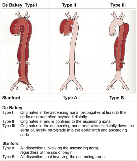
When it comes to dissection, there are two classification systems that describes the nature of the dissection (source: Grepmed). The Stanford classification is used more commonly.

classification-dissection-diagnosis-stanford-typeiii-original

Epidemiology

  • Men > women
  • Age 60-80 most common, mean age 63, 65% men
  • Younger < 40: 34% with HTN, 8.5% with h/o Marfan

Other Risk Factors

  • Trauma
  • Hypertension (most important predisposing factor of acute aortic dissection, up to 72% of pts will have underlying HTN. HTN is more common in those with type B vs type A.
  • Pheochromocytoma (very rare, case reports, thanks Alexa for pointing this out!)
  • Connective tissue disorders
  • Turner Syndrome
  • Vasculitis
  • Pregnancy
  • Crack cocaine, meth
  • Biscuspid aortic valve (9% of pts under age 40 have biscuspid AV)\
  • Pre-existing aortic aneurysm
  • Heavy lifting
  • Cardiac cath

Pathophysiology:

  • In a nut shell, due to a tear in the aortic intima, blood flow creates a false lumen separating the intima and the media. The higher the blood pressure, the higher the size of the false lumen due to shear force.
  • 50-65% originate in the ascending aorta, while 20-30% originate near the left subclavian artery and extend distally.

Presentation

  • Typical Triad
    • Acute chest or abdominal pain, can be sharp, tearing, or ripping
    • Pulse/pressure deficit between extremities
    • Mediastinal/aortic widening on CXR
  • Ascending aortic dissections are 2x as common as descending, 30% involves aortic arch
  • Acute onset chest pain that radiates to the back is the typical illness script. Pain can be the classic tearing or sharp.
    • Type A: more likely to experience anterior chest pain
    • Type B: more likely to experience back pain or abd pain > anterior chest pain
  • Propagation leads to acute AR, tamponade, stroke, shock, can propagate proximal or distally

Physical Exam Highlights

  • You might have heard of a term called pulse/pressure deficit when it comes to aortic dissections. In a nut shell, if there is a difference in SBP of greater > 20mmHg between right and left, then there is a deficit. This deficit is only found in 30% of cases of its presence can be very useful. High specific but poor sensitivity.
  • Findings Sensitivity Specificity LR if Present LR if Absent
    Pulse Deficit 12-49% 82-99% 4.2 0.8
  • Presence of an aortic regurg murmur has a LR of 1.5
  • The presence of 3 findings increase likelihood of a Type A dissection:
    • SBP < 100
    • AR
    • Pulse deficit
  • Presence of mediastinal or aortic widening on CXR increases probability of dissection somewhat, with LR 2.0

Diagnosis

  • CTA is fastest and easiest to get, 90+ % sensitivity and specificity
  • Can also do MRA or echo (TEE preferred) but might get longer to arrange in our hospital
  • EKG won’t have any specific signs but it can help you rule out other things.

Management/Prognosis

  • Stanford Type A
    • Surgical emergency, CT Surgery should be consulted ASAP.
    • High mortality, 2% per hour if left untreated.
    • Surgical mortality: up to 30%
    • Typically open repair, role of endovascular repair is not well studied.
  • Stanford Type B
    • Uncomplicated:
      • Medical management, goal is to reduce BP < 120/80 and HR < 60
      • Medical management:
        • Beta blocker: Esmolol or labetalol or propranolol, first line
        • Vasodilator: Nicardipine or nitroprusside, can cause reflexive tachycardia, hence do NOT use alone and only add if BP remains elevated with beta blocker
          • Nitroprusside over time can lead to cyanide toxicity
        • CCB: Non-DHP i.e. verapamil or diltiazem if there is a contraindication to beta blockers
      • Complicated:
        • If impending aortic rupture, end organ damage, continuing pain and hypertension despite medical therapy, false lumen extension, or large area of involvement, Type B is now considered complicated.
        • Endovascular intervention with Vascular Surgery is recommended (INSTEAD-XL study)
  • Long term management
    • Identify associated genetics factors
    • HR, HTN control
    • Serial imaging: 3, 6, and 12 months after event, and then annually after initial episode

Sarcoidosis

Today, we talked about the fascinating case of a middle-aged man presenting with subacute cough, night sweats, and 15 pound weight loss, found to have bilateral hilar LAD on CXR and CT concerning for pulmonary sarcoidosis.  While awaiting LN biopsy, he developed L sided Bell’s palsy with MRI showing inflammation of CN5 and CN7 as well as nodular dural thickening of the trigeminal cave concerning for neurosarcoidosis.  LN biopsy showed non-caseating granulomas and he was subsequently started on high dose steroids for neurosarcoidosis.


Clinical Pearls

  • Sarcoidosis is a multisystem granulomatous disorder of unknown etiology.
  • The most common clinical presentation of sarcoidosis is pulmonary related.
  • In asymptomatic individuals with incidental hilar LAD, there is no indication for LN bx to diagnose sarcoidosis because 2/3 of cases resolve spontaneously without treatment.  Close follow up is needed to ensure that patients do not develop symptoms.
  • Similarly, Lofgren syndrome (arthritis, erythema nodosum, hilar LAD, and fevers) and Heerfordt’s syndrome (uveoparotid fever) are clinically consistent with sarcoidosis and there is no indication for LN biopsy.

DDx for unilateral facial droop

  • CNS lesion
    • Especially if forehead is spared.  However, keep in mind that a stroke involving the region of pons which houses the nucleus of CN7 would mimic Bell’s palsy!
  • PNS lesion
    • Infections
      • Zoster
      • HIV
      • Lyme
      • Syphilis
    • Malignancy
      • Parotid tumors
      • Acoustic neuroma/schwannoma
      • Lymphoma or other mass compressing CN7
    • Infiltrative/autoimmune
      • Sjogren’s syndrome
      • Sarcoidosis

Sarcoidosis

  • Multisystem granulomatous disorder of unknown etiology
  • 3-4 x more common in blacks
  • Overall prevalence is 10-20 per 100,000 people
  • Pathophys
    • Aberrant formation/accumulation of non-caseating granulomas
  • Clinical presentation
    • Age of onset is 20-60 years
    • Often discovered incidentally on a CXR
    • Most common organ involved is the lung (cough, SOB, CP) with ILD being the most common type of lung involvement. 30% of patients present with extrathoracic manifestations.
    • Fatigue, malaise, fever, and weight loss are common
    • Lofgren’s syndrome: arthritis, erythema nodosum, b/l hilar LAD
    • Heerfordt’s syndrome: parotid gland enlargement, facial palsy, fever, anterior uveitits
  • Diagnosis
    • No biopsy needed in the following
      • Asymptomatic with hilar LAD
      • Lofgren’s syndrome
      • Heerfordt’s syndrome
    • Otherwise, biopsy the easiest site to access.  Keep in mind that erythema nodosum does not have granulomas and would not help in diagnosing sarcoidosis.
    • Lab abnormalities that may be present:
      • Anemia
      • Leukopenia, eosinophilia, thrombocytopenia
      • ESR and CRP may be elevated
      • Hypercalciuria is more common than hypercalcemia
      • Moderate elevation in ALP suggests diffuse granulomatous hepatic involvement
      • Hypergammaglobulinemia and a positive RF (not usually part of routine work up)
      • ACE levels are elevated in 75% of untreated patients with sarcoid but has poor sensitivity and insufficient specificity (10% false positive rate with cocci, DM2, TB, hyperthyroidism, lung cancer, pneumoconiosis, etc.)
  • Organs that may be impacted in sarcoid
    • Pulmonary: occurs in over 90% of patients. Bilateral hilar LAD as well parenchymal disease. Can present with a restrictive spirometry pattern due to underlying fibrosis, pulmonary HTN
    • Cutaneous: can be disfiguring can be macules, papules, plaques and erythema nodosum
    • Liver/Spleen: a high alkaline phosphatase level suggests granulomatous liver disease
    • Neurologic: Noted in 5% of cases. MRI with contrast can help with diagnosis. Complications:
      • Cranial-nerve palsies (20-50%) → most common
      • Headache
      • Ataxia
      • Encephalopathy
      • Granulomatous meningitis
      • Weakness
      • Seizures
      • Neuroendocrine dysfunction
    • Optho: anterior uveitis most common manifestation
    • Cardiac: Typically cardiomyopathy, can also see arrhythmias (tachy or brady).
    • Renal: Can have hypercaliuria (more common than hypercalcemia) and renal calculi
    • Bone: chronic arthritis and cysts resembling rheumatoid, and diffuse granulomatous myositis.
  • Treatment/monitoring:
    • Asymptomatic? Follow up outpatient q3-4 months and annually thereafter to monitor for development of symptoms.
    • Symptomatic? Start steroids, re-evaluate q1-2 months
    • Refer to this NEJM article for organ specific treatments.
  • Chronic complications of pulmonary sarcoid
    • VTE is more common than the average patient population
    • Chronic pulmonary aspergillosis
    • Pulmonary HTN due to advanced fibrosis
  • Prognosis
    • Up to 80% of patients with hilar LAD spontaneously improve on their own!
    • With more symptomatic disease or more extrapulmonary manifestations, prognosis declines to less than 30% remission.
    • Overall mortality is <5%

ECG Report!

Clinical Pearls from our first ever (and hopefully monthly) ECG Report!

  • Make sure to always work through an ECG in a systematic way in order to avoid missing key information.
  • Remember that you might not see any p waves (or retrograde P waves) in AVRT and AVNRT.  Both of these rhythms would be fast and regular.  A fib, by contrast, would be irregular.
  • Hypercalcemia presents with a shortened QT and a loss of the ST segment on ECG.  But make sure you are ruling out an MI because often times the loss of ST segment resembles a STEMI.
  • Whenever you see a downward p wave in lead I, think of two diagnoses:
    • Dextrocardia
    • Limb lead reversal
  • Easiest way to distinguish between dextrocardia and limb lead reversal is to look at the QRS amplitude as you move across the precordium.  If the amplitude is decreasing as you advance from V1 to V6, then the diagnosis is dextrocardia because you are moving away from the heart.  If the amplitude is not changing or increasing, then the diagnosis is limb lead reversal.
  • The presence of > 2 mm coved precordial ST-segment elevation (leads V1through V3) with T wave inversions is suggestive of Brugada morphology.  In a patient with history of syncope, ventricular arrhythmias, or family history of Brugada syndrome, this is consistent with a diagnosis of Brugada syndrome and would require ICD placement.Capture

Have an interesting ECG?  Save them/send them our way for our next ECG report!

Multiple Myeloma

Thanks to Erica for presenting the case of a middle aged man who presented with acute back pain and B symptoms after trauma to his back, found to have stage 3B multiple myeloma.


Clinical Pearls 

  • Remember that majority of cases of acute low back pain (<6 weeks) is due to musculoskeletal etiologies that spontaneously improve on their own.  Imaging and further diagnostic work up is not indicated unless there are red flags (see below).
  • A straight leg test is more useful when negative as it has a high negative predictive value for ruling out radiculopathy.  False positive rates are quite high.
  • Unexplained anemia and worsening renal function in the outpatient setting should trigger a work up for multiple myeloma.
  • The most common presenting symptoms for MM are anemia (73%), bone pain (58%), and renal insufficiency (48%).
  • In diagnosing MM, sensitivity increases with each added test: SPEP (82%) → IFE (93%) → FLC/UPEP (97%).  The other 3% that would not be diagnosed with these tests have a non-secretory MM (monoclonal increase in plasma cells in bone marrow that do not produce immunoglobulins or light chains).

Red flags for acute low back pain:

  • Focal neurologic complaints/deficits
  • History of cancer
  • Age >50 years
  • Fever not explained by another cause
  • History of recent bacteremia or IVDU
  • Steroid use
  • Weight loss
  • Pain that is worse at night
  • No relief with bed rest or pain lasting >1 month

Multiple Myeloma: refer to this prior blog post.  Other info below:

Diagnostics:

  • SPEP: picks up M protein or elevated immunoglobulins (heavy + light chain) in the serum.  You can diagnose over 80% of patients with MM using an SPEP.
  • IFE: identifies the specific type of immunoglobulin that is elevated with its light chain.
  • Free light chains (FLC): measures the amount of free light chains not bound to a heavy chain floating around in the blood.  Normally people have about a 2:1 ratio of kappa to lambda chains.  In light chain only multiple myeloma, there is a disproportionate increase in one type over the other and the ratio will be off.  If there is an increase in both light chains but the ratio is normal, think kidney disease!
    • Keep in mind that the reason to check FLC when you suspect MM is to diagnose those people who are only producing light chains and not whole immunoglobulins that would have been picked up by SPEP/IFE.
  • UPEP: measures light chains dumped in the urine (Bence Jones protein)

Capture

Thrombocytopenia and ITP

Thanks to Connie for presenting the case of an elderly woman who presented with petechial bleeding, found to have a platelet count of 15, with work up revealing possible Evans’ Syndrome as well as bladder cancer!


Clinical Pearls

  • Think of thrombocytopenia as a problem with platelet production, platelet destruction, sequestration, or lab problem.
  • ITP is a diagnosis of exclusion. It can be a primary process due to autoimmune platelet destruction, or triggered by an associated condition (secondary ITP).
  • Remember that decadron is superior to prednisone in treatment of ITP based on this meta-analysis.
  • TPO mimetics are best as a last resort and more useful in maintenance of remission rather than inducing remission.

Thrombocytopenia DDx

  • Decreased production
    • Aplastic anemia
    • MDS
    • Drugs
    • Alcohol
    • Cirrhosis
    • Leukemia
    • Severe megaloblastic anemia (folate/B12 deficiency)
    • Myelofibrosis
    • Granulomas
    • Solid malignancy mets to the bone
  • Increased destruction
    • Immune mediated
      • Primary ITP
      • Secondary
        • Infection: HIV
        • Rheum d/o: SLE, APLS
        • Lymphoproliferative process: CLL, lymphomas
        • Drugs
        • Post-transfusion purpura (alloimmune process)
    • Non-immune mediated
      • MAHA (DIC, HUS/TTP)
      • Plavix
      • Vasculitis
      • HELLP
      • Infections: sepsis, CMV, EBV, malaria, rickettsia, ehrlichiosis, dengue, Hanta virus, etc.
      • CVVH
  • Sequestration due to hypersplenism
  • Pseudo-thrombocytopenia: clumping due to EDTA anticoagulated tubes (confirm by request blood draw in citrate coated tubes)

Thrombocytopenia work up

  • Good history and review of meds, infections, prior history of autoimmune disorders
  • Check CBC, coags and peripheral smear!
    • Spherocytes ⇒ AIHA, hypersplenism
    • Schistocytes ⇒ MAHA
    • Pancytopenia ⇒ aplastic anemia, MDS, leukemias, etc.

Immune Thrombocytopenic Purpura (ITP)

Check out our thorough post on this topic here.

Status Epilepticus & Serotonin Syndrome

Today we talked about the case of a young woman who presented after suicide attempt by ingestion of multiple prescription medications, found to be obtunded, initially in status epilepticus and later with exam findings concerning for serotonin syndrome.


Clinical Pearls

  • Continuous EEG is indicated if a patient is not returning to baseline mentation 15 mins after a seizure to rule out non-convulsive status epilepticus.
  • Status epilepticus is defined as a seizure lasting > 5 mins or > 2 discrete seizures between which there is incomplete recovery of consciousness.
  • Treatment of status epilepticus involves acute management with IV/IM benzos, urgent long-term control with IV fosphenytoin (preferred), phenytoin, or valproic acid.  Remember that keppra is more useful in suppressing future seizures than treating an acute episode.
  • Serotonin syndrome is a clinical diagnosis and manifests with neuromuscular activation like tremors, hyperreflexia, and clonus (generally worse in lower extremities).  Use Hunter’s criteria to help with diagnosis.

Status Epilepticus

  • When is continuous EEG needed?
    • If patient is not returning toward baseline in 15 mins after a seizure, goal is to rule out nonconvulsive seizures
  • How to define status?
    • > 5 mins OR
    • > 2 discrete seizures between which there is incomplete recovery of consciousness
  • Non-convulsive status epilepticus
    • Suspect if LOC not improving by 10 mins after cessation of movement
    • Mental status remains abnormal for 30-60 mins after movement cessation
    • Dx requires a 24-hour EEG (we don’t have one ☹)
    • Treatment is the same as generalized status epilepticus, but prognosis is worse (mortality 65% vs 27%)
  • Treatment of status
    • Assessment and supportive treatment
    • Initial pharmacologic therapy
      • Ativan (2 mg IV q1-2 mins), total dose 0.1mg/kg
        • Watch out for respiratory depression and hypotension
        • Alternatives: versed 0.2mg/kg IM, valium 0.2mg/kg IV
    • Urgent long-term control
      • Fosphenytoin (preferred): 20 mg/kg at 150mg/min
        • Give extra 10 mg/kg if not responding
        • Dephosphorylates into phenytoin.  It’s more soluble in water and less likely to precipitate in the skin and vessels.
      • Phenytoin: 20mg/kg at 50mg/min (slower than fospheny)
        • If infused too fast, can irritate skin/vessels causing skin necrosis
      • Valproic acid: 20mg/kg at 5 mg/kg/min
        • Sometimes the preferred choice in patients with known generalized epilepsy b/c phenytoin can provoke absence seizures in that population.
      • What about Keppra?
        • Technically not FDA approved for status.  It has weak evidence to support its use.  More useful in suppressing subsequent seizures after status has been controlled.

Serotonin syndrome:

  • Clinical diagnosis. Serum serotonin concentrations do not correlate with clinical findings.
  • Severe disease can lead to DIC, rhabdo, renal failure, and ARDS.
  • Diagnostic criteria: use Hunter’s (84% sensitive, 97% specific)Hunter's decision rule
  • DDx
    • NMS
    • Anticholinergic toxicity
    • Malignant hyperthermia
    • Sympathomimetic intoxication
    • Sedative-hypnotic withdrawal
    • Meningitis
    • Encephalitis
  • Serotonin syndrome may be distinguished from other causes of agitated delirium on the basis of neuromuscular findings. Whereas patients with serotonin syndrome show signs of neuromuscular activation (eg, tremor, hyperreflexia and clonus that are greater in the lower extremities, ocular clonus, and increased muscle tone), patients with sympathomimetic toxicity or infections of the central nervous system lack these findings.
  • Treatment
    • Supportive care and sedation
      • Chemical sedation preferred over physical restraint
    • Autonomic instability
      • High BP: esmolol or nitroprusside (Short acting). Avoid longer acting agents
      • Low BP: neo or epi. Avoid idirect agents like dopamine because they are converted to epi and norepi.  When monoamine oxidase is inhibited, epi and norepi production at the cellular level is not controlled and could lead to an exaggerated HD response.
    • Hyperthermia
      • No benefit to Tylenol b/c increase in body temp is due to increased muscular activity rather than alteration in the hypothalamic temperature setpoint.
      • If temp >41.1, then sedate, paralyze, intubate
    • Antidote: cyproheptadine
      • Histamine receptor antagonist. Also has weak anticholinergic activity
      • 12 mg loading dose, then 2 mg q2h until clinical response is seen.
      • Is sedating (good) and can also cause transient hypotension.

Capture

Hyponatremia due to secondary adrenal insufficiency

Thanks to Jess for presenting the fascinating case of a middle-aged woman with family history of autoimmune disease who presented with acute onset of fatigue and abdominal pain, found to have vitiligo on exam.  Work up revealed hyponatremia due to a secondary adrenal insufficiency, pancytopenia, and panhypopituitarism possibly due to a yet to be diagnosed autoimmune disorder!


Clinical pearls

  • Remember that hyponatremia is a problem of water regulation that can be compounded by low solute intake.
  • Primary adrenal insufficiency is disorder at the level of the adrenal glands and manifests with low sodium and high serum potassium levels.
  • Secondary adrenal insufficiency is disorder at the level of the pituitary and manifests with low/normal sodium and normal potassium levels (because low cortisol leads to high ADH levels and hyponatremia).
    • Make sure to do work up to rule out panhypopituitarism.  Keep in mind that the most sensitive test for HPA access integrity is LH/FSH.
  • Tertiary adrenal insufficiency is disorder at the level of the hypothalamus and presents similarly to secondary AI.
  • Test for adrenal insufficiency with a cort-stim test and/or AM cortisol and ACTH levels.

Hyponatremia

Remember these three steps to working up hyponatremia:

  1. Is there a sodium problem? check serum osm
  2. Are the kidneys responding appropriately? check urine osm
  3. Is ADH revved up for a hemodynamic reason? check urine Na 

Capture

Adrenal insufficiency

Adrenal insufficiency
Source: NIDDK.gov

Primary AI:

  • Failure of adrenal glands
  • Causes: Addison’s (most common in the US), infiltrative processes (TB, sarcoid), hemorrhage, toxins
  • Labs would show ↓Na and ↑potassium (b/c aldosterone is gone)

Secondary AI:

  • Failure of pituitary (low ACTH)
  • Causes: pituitary lesions, surgeries, TBI, drugs
  • Clinically may present with loss of other anterior pituitary hormones
  • Labs would show ↓Na (because low cortisol leads to high ADH levels) but normal potassium levels (b/c aldosterone is active)

Tertiary AI:

  • Failure of hypothalamus (low CRH)
  • Causes: more commonly iatrogenic (cessation of high dose glucocorticoid therapy without taper) or post surgical interventions.

Hypopituitarism

Capture

Hypophysitis:

  • Inflammation of the pituitary
  • Four categories based on histologic findings:
    • Lymphocytic
      • Most common form
      • Seen in late pregnancy and post-partum period
      • Also associated with CTLA4 inhibitors like ipilimumab
    • Granulomatous
      • Idiopathic or secondary to GPA, sarcoid, TB
    • Plasmacytic (IgG4-related)
    • Xanthomatous (most rare)
  • Clinical presentation
    • Headache out of proportion to exam findings
    • Preferential decrease in ACTH and TSH ⇒ adrenal insufficiency and hypothyroidism
  • Prognosis:
    • Pituitary size eventually normalizes but pituitary loss of function is often permanent.


Angioedema and hyperkalemia management

Thanks to Audris for presenting the case of a middle-aged man with vasculopathy on ACEi who presented with angioedema requiring intubation!  We discussed the  diagnostic work up and management of angioedema as well as hyperkalemia!


Clinical Pearls

  • First order of business when suspecting angioedema is the ABCs!
  • Treat angioedema in the acute setting with H1 blockers and steroids, even if you are suspicious of a non-histaminergic pathway.
  • Always assess for concurrent anaphylaxis (hypotension or bronchospasm in addition to hives or angioedema).  If anaphylaxis is present, then treatment involves IM 0.3-0.5 mg of 1:1000 dilution epinephrine (1mg/mL), repeat every 20 minutes until symptoms resolve (max 3 doses)
  • If you have access to a functioning kidney, favor loop diuretics over cation exchange binders (i.e. kayexalate) to lower serum potassium!
  • Patiramer is much better tolerated than kayexalate and has a more favorable side effect profile.
  • Calcium gluconate has a role in the treatment of hyperkalemia when EKG changes are present. Give a dose and repeat the EKG.  If no improvement, repeat to a maximum of 3 doses until EKG has normalized.

Angioedema 

  • 3 pathophysiologic subtypes:
    • Mast cell/histamine mediated
      • Etiologies:
        • Allergic reactions: food/insect stings, latex, drugs. Can also be idiopathic. IgE type 1 hypersensitivity
        • Direct mast cell release: drugs (opiates, contrast). IgE is not involved.
        • ASA/NSAIDs: via IgE or direct mast cell release
        • Chronic urticaria w/w/o angioedema
      • S/sx affecting organ systems other than the skin? Suspicious for anaphylaxis ⇒ give epi
      • Treatment: H1 blockers, glucocorticoids. 
    • Bradykinin mediated
      • Inhibition of enzymes involved in the degradation of bradykinin, or deficiency/dysfunction of complement C1 inh
      • More prolonged time course, develops over 24-46 hours and resolves within 2-4 days
      • Relationship between trigger and onset of symptom is not as apparent
      • Not associated with other s/sx. More common to have abdominal pain due to bowel wall involvement.
      • Treatment: bradykinin pathway mediators (ecallantide, icatibant), C1 inhibitor concentrate, or plasma replacement.
    • Unknown mechanism
      • Idiopathic angioedema
      • Infections (in children)
      • CCBs
      • Other drugs: sirolimus, everolimus, amiodarone, metoprolol, risperidone, paroxetine, and etanercept, inhaled cocaine.
      • Herbal meds
      • Urticarial vasculitis
      • Hypereosinophilic syndrome and Gleich syndrome


Capture

Hyperkalemia

Agents that reduce serum potassium via transient intracellular shift:

  • Insulin: give with D50 if normoglycemic to avoid hypoglycemia and be sure to check FSG hourly for 4 hours after to ensure no hypoglycemia develops
  • Albuterol (10-20 mg) nebs: this is significantly higher than the dose we give in COPD (2.5 mg) and is equal to ~8 treatments! So make sure to continue the nebs when the patient arrives on the floor from the ER if they are still hyperkalemic.
  • NaHCO3: best for management of chronic hyperkalemia in the outpatient setting.  In the acute management of hyperkalemia, alkalinization of serum with a large bicarb load can lead to a reduction in serum calcium levels.  Lower serum calcium can lead to more cardiac membrane instability and fatal arrhythmias!

Agents that eliminate potassium from the body:

  • Loop diuretics: first choice if a functioning kidney is available!
  • Cation exchange binders: preferred when kidneys are not available
    • Patiramer (available at VMC), much more tolerable than kayexalate and highly effective at lowering serum potassium.  Like kayexalate, it works over hours to days.
    • Sodium zirconium: similar to patiramer but not currently available
    • Kayexalate: not pleasant to take orally. Also carries with it the slight risk of colonic ischemia especially in post renal transplant patients and those with baseline colonic dysfunction (due to infection or inflammation).
  • Dialysis

Indication for using calcium gluconate: when EKG changes are noted.  Repeat doses (maximum 3) until EKG changes have resolved.

Stroke in a 23 year old… secondary to a neck massage 2/13/2019

We have a very bizarre case of a 23 year old woman, with a history of hypertension, presenting as a stroke alert. She has dysarthria and focal right-sided deficits, and she required intubation for airway protection. She has no prior stroke or clotting history, no illicit drug use (other than marijuana), and no significant family history. CT of the head was unremarkable, but CTA revealed a… vertebral artery dissection! We later learned that the patient was in the middle of getting a massage when the stroke occurred!

That’s right, this is a case of cervical artery (more specifically the vertebral artery) dissection secondary to traumatic massage!


Stroke typically is due to atherosclerotic disease in most cases, but if you need a case that occurred in a young patient, and you’ve ruled out everything else that would present similar, consider the following:

Stroke in young patients 

  • Coagulopathy
    • APLS (one of the most common)
    • DIC
    • HIT
    • Hypercoagulable/hyperviscosity states
  • Vascular
    • Vasculitis
      • Primary: Takayasu, GCS, Kawasaki, PAN, ANCA
      • Secondary: TB, HIV, syphilis, fungi, SLE
    • Dissection (most common in young patients)
    • Trauma
    • Structural malformation i.e. AVM, aneurysms
    • Cerebral venous sinus thrombosis
  • Metabolic
    • CADASIL
    • MELAS
    • Fabry’s disease
    • Homocysteinuria
  • Drugs
    • Cocaine
    • Meth
  • Cardiac
    • Congenital defects
    • PFO/ASD leading to paraoxysmal stroke
    • Arrhythmias
    • Endocarditis
    • Rheumatic valve disease

Vertebral artery dissection: 

Epidemiology

  • Can occur at any age and usually infrequent, but vertebral arterial dissection in general is a common cause of stroke in the young.
  • Carotid artery dissection is much more common than vertebral.
  • Incidence: 1-1.5 per 100,000.
  • Spontaneous dissections of the carotid and vertebral artery account for ~ 2% of ischemia strokes but in young and middle age patients, they can account up to 10-25%.
  • Peak incidence at 5th decade of life, occurs earlier in F > M
  • Most of these patients likely have underlying connective tissue pathology leading to weakening of artery structural integrity but this is a theory.

Pathophysiology

  • False lumen expansion leading to cerebral ischemia from hypoperfusion or thromboembolism, also can cause neurological sx from compression of adjacent nerves/vessels leading to CN involvement, Horner syndrome, pain, etc.
  • Main mechanism, as revealed by angiography studies, point toward a thromboembolic mechanism from the dissection leading to stroke.
  • Hemorrhage due to rupture, expanding hematoma can also lead to focal neurological signs.

Risk Factors

  • Connective tissue disorders (i.e. Ehlers Danlos, Marfans, ADPKD, osteogenesis imperfecta, subclinical/unnamed)
  • Yoga
  • Migraine
  • Coughing
  • Painting a ceiling
  • Vomiting
  • Sneezing
  • Sudden neck movement
  • Trauma
  • Chiropractic manipulation, estimated 1 in 20000 spinal manipulations lead to a stroke.

Presentation

  • Most cases have a precipitating event
  • Intracranial dissection
    • 50% of cases result in subarachnoid hemorrhage
  • Extracranial dissection
    • Associated with trauma, vertebral artery is most mobile and vulnerable to mechanical injury at C1 to C2 as it leaves the transverse foramen of the axis vertebra and enters the intracranial space.
    • Severe neck pain, dizziness, vertigo, double vision, ataxis, dysarthria are common symptoms.
    • Lateral medullary and cerebellar infarctions are common
    • Typical: Local pain, HA, ipsilateral Horners, CN palsies, hemiparesis typically ipsilateral
    • Exam: Hematoma, vascular bruit (1/3 in carotid dissections, usually absent on vertebral), Horner’s syndrome, neck tenderness

Diagnosis

  • Catheter angiography used to be the gold standard, nowadays moving toward…
  • MRA
  • CTA

Management

  • Anticoagulation for at least 3-6 months to prevent thromboembolic complications unless there is hemorrhagic transformation of the infarct, an intracranial aneurysm, or intracranial extension.
    • Most ischemic damage is actually due to thromboembolic effect, not hypoperfusion.
  • Surgical: May be required for SAH/intracranial VAD or if the defect fails to heal on its own
    • Endovascular stenting used for extracranial carotid or vertebral artery dissection when medical mgx fails, anticoag or thromboiysis does not exclude subsequent endovascular therapy but ideally endovascular intervention should be done within 6 hours.
  • Thrombolytics: Consider in extracranial dissection, contraindicated in intracranial dissections or aorta involvement.

Prognosis

  • Extracranial VAD: Generally has good prognosis, estimated 50% of patients will recover with no neurological deficit and 25% with moderate to severe deficits.
  • Intracranial VAD: Prognosis is generally pretty poor.

Take Home Points:

  • Cervical (Carotid more common than vertebral) artery dissection is a common cause of strokes among young patients.
  • Chiropractic manipulation, even massages, are risk factors for cervical artery dissection!
  • A good portion of patients with cervical artery dissection might have underlying, sometimes subclinical, connective tissue abnormalities.

Lupus Nephritis

Shout out to Paige for admitting the interesting young woman with no medical history who presented with blurry vision, found to be in hypertensive emergency and nephrotic syndrome.  Her work up revealed diffuse proliferative lupus nephritis (class IV) without any other clinical features of lupus!


Clinical Pearls

  • Proteinuria is primarily caused by three processes: 1) overflow (due to elevated paraprotein levels), 2) glomerular disease, 3) tubular disease (rarely reaches nephrotic range)
  • Renal involvement is noted in ~50% of patients with SLE and can present as nephrotic and/or nephritic syndromes.
  • The most common and severe form is diffuse proliferative lupus nephritis (class IV)
  • Keep in mind that SLE flare is associated with a normal/low WBC, normal/low CRP, and absence of fever.  Infection, which can instigate a lupus nephritis flare, would cause an elevated WBC, elevated CRP, and fever.
  • Lab findings suggestive of SLE flare also include an elevated anti-dsDNA (correlates with disease activity), low complement levels (especially C3), worsening proteinuria, and elevated creatinine.
  • Treatment involves an induction phase with cytoxan or MMF plus solumedrol followed by a maintenance phase with the goal of reducing urine protein excretion to <0.33 g/day.

Hypertension urgency/emergency:

  • Urgency: BP>180/120 without end organ damage
    • Manage with orals, goal to lower (not necessarily correct) over 24-48 hours
  • Emergency: elevated BP + end organ damage (brain, eye, heart, kidney)
    • Manage with IV meds, reduce DBP by 10-15% over the first hour, then by 25% over the next 6-12 hours.
    • Special situations:
      • Lower rapidly in acute aortic dissection (goal SBP <110 ASAP)
      • Recent ischemic stroke: do not lower BP unless > 220/120 (> 185/110 if received reperfusion therapy)

Lupus

  • Female to male prevalence is 9:1
  • Diagnose using Systemic Lupus International Collaborating Clinics (SLICC) criteria.  Must have > 4 (at least 1 clinical and 1 laboratory) OR biopsy proven lupus nephritis with a positive ANA or dsDNA
  • Autoantibodies:
    • dsDNA is 50% sensitive but 97% specific –> can monitor disease activity
    • Smith is 10-20% sensitive but >90% specific
  • Work up of presumed flare:
    • Important to distinguish flare from acute infection but keep in mind that infection can trigger flare
    • SLE flare:
      • Low WBC and normal CRP (except when serositis is present)
      • Fever is unusual
    • Infection:
      • High WBC and high CRP
      • Fever is common 
  • Treatment:
    • Plaquenil for all unless contraindicated
    • Mild/mod:
      • plaquenil, NSAIDs, low dose steroids (pred 5 daily)
    • Severe
      • Acute: high dose steroids
      • Chronic: Immunosuppressive agents (MTX, cyclosporine, cyclophosphamide, azathioprine, mycophenolate, belimumab)
  • Prognosis
    • Worse with pediatric onset, ethnic minority, renal involvement, increased number of diagnostic criteria, low complement, e/o end organ damage
    • High association of CVD and SLE

SLE and renal disease

  • Renal involvement is common and eventually occurs in ~50% of SLE patients
  • 10% progress to ESRD
  • High mortality compared to SLE without nephritis
  • More common and severe in African Americans, Hispanics, Asians
  • Classifications of GN: can evolve from one to another
    • Minimal mesangial lupus nephritis (class I)
      • Earliest and mildest form
      • Rarely diagnosed b/c pts have a normal U/A, no or  minimal proteinuria, and normal Cr
    • Mesangial proliferative lupus nephritis (class II)
      • Microscopic hematuria and/or proteinuria
      • Light microscopy would show mesangial hypercellularity or mesangial matrix expansion
    • Focal lupus nephritis (class III)
      • Hematuria, proteinuria, some HTN, decreased GFR
      • Less than 50% glomeruli affected by light microscopy
      • Segmental glomerulonephritis
        • A: active lesions –> focal proliferative
        • A/C: active and chronic lesions –> focal proliferative and sclerosing
        • C: chronic inactive lesions and scarring –> sclerosing
    • Diffuse lupus nephritis (class IV)
      • Most common and most severe
      • Hematuria, proteinuria, nephrotic syndrome, HTN, reduced GFR
      • Hypocomplementemia (esp C3) and elevated anti-dsDNA during active disease
      • >50% of glomeruli are affected
    • Lupus membranous nephropathy (class V)
      • nephrotic syndrome, Cr normal or slightly elevated
      • Diffuse thickening of the glomerular capillary wall and subepithelial deposits
      • Can present without any other clinical or serologic manifestations of SLE
    • Advanced sclerosing lupus nephritis (class VI)
      • Slow, progressive renal dysfunction with proteinuria and relatively bland urine sediment
      • Global sclerosis >90% of glomeruli
      • Active GN no longer observed
  • Treatment:
    • Best to initiate early
    • Aimed at proliferative lupus nephritis
    • Induction
      • 3 – 12 months: goal is to obtain renal response.
      • Cytoxan or MMF PLUS solumedrol 250-1 g/day x 3 days (former takes 10-14 days to have an effect so the latter is much faster) or prednisone 60 mg/day
    • Maintenance
    • Response:
      • substantial reduction in urine protein excretion to <0.33 g/day
      • improvement or stabilization of serum creatinine
      • improvement of urinary sediment

Approach to glomerular disease:

Capture
* can be nephritic or nephrotic