Category Archives: Morning Report

Necrotizing Fasciitis & Ludwig’s Angina

Thanks to Joe for presenting the case of an elderly man with no known medical history who presented with acute AMS, found to have L facial swelling and crepitus, eventually diagnosed with necrotizing Ludwig’s angina!


Clinical Pearls

  • Necrotizing fasciitis (NF) is a surgical diagnosis and involves infection of muscle and subcutaneous fat.
  • CT is a useful tool to help with diagnosis and in one case series had a 100% sensitivity and >80% specificity for diagnosing NF.
  • LRINEC or Laboratory Risk Indicator for NF is a lab-based risk assessment tool to help risk stratify patients with possible NF.  It has a sensitivity of 80% and specificity of 67%.  It should NOT supplant your clinical judgement.
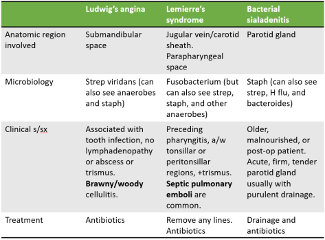
  • Ludwig’s angina refers to any infection of the submandibular space (not just NF).  Normally the treatment for Ludwig’s angina is antibiotics.  In the case of NF, urgent surgical debridement is necessary.  In spite of antibiotics and debridement, head and neck necrotizing infections are associated with a high mortality rate (~40%).
    • In patients with Ludwig’s angina, always involve anesthesia AND ENT to help secure airway. Oral intubation is associated with higher rates of laryngospasm in these patients so oftentimes nasal intubation is preferred.

Deep neck infections:

Capture

Necrotizing fasciitis

  • Background
    • Infection of deep tissues, specifically muscle fascia and subcutaneous fat.
    • Two main types
      • Type 1: polymicrobial
        • More common in elderly and those with significant comorbidities including diabetes, immunocompromised states, PVD, etc.
        • Blood cultures are positive in ~20% of patients.
      • Type 2: monomicrobial (usually GAS but can be other beta-hemolytic strep and MRSA)
        • Can be seen in any age group and without any underlying disease.
  • Clinical manifestations
    • Remember that you do not need to have a penetrating injury for NF.  Oftentimes, blunt trauma is the preceding history and overlying tissue does not show any signs of infection, leading to the “pain out of proportion to exam” finding.
    • Systemic signs of toxicity (including hypotension and shock), rapid progression, crepitus.
    • LRINEC or the Laboratory Risk Indicator for NF is a lab-based risk assessment tool to help risk stratify patients with possible NF.
      • It has a sensitivity of 80% and specificity of 67%.  It should NOT supplant your clinical judgement.
    • CT scan is highly sensitive (100% in one case series of 67 patients) and specific for differentiating NF from celllulitis.
    • Ultimately, NF is a surgical diagnosis so consult surgery early if you are concerned about the diagnosis and before waiting for imaging in an unstable patient!
  • Treatment
    • Early surgical intervention and debridement
    • Empiric antibiotics
      • Beta lactam/beta lactamase inhibitor or carbapenem PLUS
      • vancomycin or other similar drug for MRSA coverage PLUS
      • clindamycin
        • Eagle effect: at high bacterial loads, there is reduced efficacy of beta-lactam antibiotics for strep pyogenes infections due to reduced exposure of penicillin binding protein on the bacteria.  Clindamycin works better in these situations and does not rely on the penicillin binding protein site.
        • Toxin neutralization: clindamycin has the ability to suppress synthesis of bacterial toxins that cause systemic symptoms in patients with NF.
    • Other therapies such as hyperbaric oxygen and IVIG have not shown reliable evidence of benefit in studies and are not currently recommended by the IDSA.
  • Prognosis
    • Mortality is high even with appropriate treatment (up to 45%).

References: 

Refer to this amazing review by NEJM for more info on NF.


Acute Rheumatic Fever

Today, we talked about the very interesting case of a middle-aged man who presented with acute migrating oligoarthritis, found to be febrile with an inflammatory synovial fluid and elevated ASO titers consistent with acute rheumatic fever!


Clinical Pearls

  • Nonsuppurative manifestations of GAS infection include acute rheumatic fever (ARF), acute GN, and Scarlet fever.
  • Use the modified Jones Criteria to help you diagnose ARF and treat early if high suspicion for the disease (do not wait for titers to come back).
  • Late complications of ARF include rheumatic heart disease (10-20 years after infection) and Jaccoud arthropathy.
  • Treatment of ARF involves NSAIDs for arthritis, PCN G IM x 1 dose for acute presentation and then monthly for prophylaxis, and patient education about oral hygiene to prevent endocarditis and need for prophylaxis before invasive procedures.

Differential diagnosis for a migratory arthritis

  • Rheumatic fever
  • Infective endocarditis
  • Vasculitis (IgA, cryo, ANCA associated)
  • SLE 
  • Acute leukemia
  • Serum sickness
  • Viral arthritis
  • Bacteremia (staph, strep, mening/gonococcal)
  • Pulmonary infections (mycoplasma, histoplasma)
  • Lyme
  • Whipple’s

Nonsuppurative complications of GAS infection

  • ARF
  • Scarlet fever
  • Acute GN

Rheumatic fever 

  • Nonsuppurative sequela that occurs 2-4 weeks after GAS pharyngitis
  • Epi
    • More common in children 5-15 years of age
    • More common in resource limited settings
  • Pathogenesis:
    • Poorly understood, ?molecular mimicry
  • Clinical manifestations:
    • Two primary manifestations of disease

Two manifestations of ARF

(Table above from UpToDate)

  • Late sequelae
    • Rheumatic heart disease (10-20 years after infection), primary involves the mitral valve >aortic valve.
      • Leading cause of cardiovascular death in the first 5 decades of life in resource limited settings
    • Jaccoud arthropathy
  • Diagnosis:
    • Revised Jones criteria (joint and cardiac manifestations can only be counted once).
      • Major
        • Carditis and valvulitis (clinical or subclinical) – 50-70%
          • Usually pancarditis. Valvulitis especially of mitral and aortic valves, shown as regurg on echo.
          • Carey Coombs murmur: short mid-diastolic murmur heard loudest at the apex
        • Arthritis (migratory, involving large joints) – 35-66%, earliest symptom
          • Several joints affected in quick succession, each inflamed for a day or two to one week. Most common are knees, ankles, elbows, and wrists.
        • CNS involvement (Sydenham chorea) – 10-30%
        • Subcutaneous nodules – 0-10%
        • Erythema marginatum – <6% 
      • Minor
        • Arthralgia
        • Fever >38.5
        • Elevated acute phase reactants (ESR, CRP)
        • Prolonged PR interval on EKG
      • Diagnosis requires evidence of prior GAS infection plus:
        • 2 major OR
        • 1 major + 2 minor criteria OR
        • 3 minor criteria (only if patient has history of prior episode of ARF)
      • In a high prevalence setting, slightly modified criteria are used.
    • Labs:
      • Prior GAS infection through either
        • Throat culture
        • Positive rapid strep antigen test
        • Elevated or rising ASO titers
      • Treatment
        • Goals
          • Symptomatic relief of acute disease manifestations
            • Arthritis: NSAIDs
            • Carditis: if severe, heart failure treatments
          • Eradication of GAS
            • IM PCN G benzathine x 1
            • Contacts (throat culture test and treat if positive)
          • Ppx against future GAS infection to prevent progression of cardiac disease
            • PCN G IM once a month
            • For 5 years or until 21 years of age (whichever is longer)
            • If ARF with carditis and residual heart disease
              • 10 years or until 40 years, sometimes even lifelong
            • Education
              • Oral health
              • Ppx before any invasive procedures

Stroke from CNS TB induced vasculitis!

Thanks to Katie for presenting the interesting case of a young man with history of disseminated TB with TB meningitis and hydrocephalus requiring VP shunts, admitted for acute LUE weakness, L homonymous hemianopsia, and memory impairment, found to have acute strokes in multiple vascular territories due to TB related CNS vasculitis!


Clinical Pearls

  • Remember that arterial dissection is the most common cause of stroke in a young patient.
  • CNS vasculitis can be primary or secondary to a systemic illness.  It typically presents with infarcts in multiple vascular territories.  Treatment involves immunosuppression with high dose steroids + cytoxan/rituxan.
  • CNS vasculitis is the most common cause of severe neurologic deficit in patients with TB meningitis.
  • Vasculitis in CNS TB is the result of a hypersensitivity reaction to proteins released from the bacteria.
  • TB meningitis requires an extended course of anti-TB treatment, generally up to 1 year or more.  Serial LPs are obtained to monitor adequate response to therapy.

Etiologies of stroke in a young adult

CNS TB:

Three main manifestations:

  1. TB meningitis (most common presentation in low incidence settings like the US)
  2. Intracranial tuberculoma
  3. Spinal tuberculous arachnoiditis

Spillage of tubercular protein into the subarachnoid space results in an intense hypersensitivity reaction and inflammation resulting in

  • Proliferative arachnoiditis (fibrous mass encasing cranial nerves and vessels adjacent to it)
  • Vasculitis with resultant aneurysm, thrombosis, and infarction
  • Communicating hydrocephalus 

TB Meningitis

  • 1% of all TB cases, 5% of all extrapulmonary TB cases
  • 15-40% mortality rate
  • Clinical manifestations
    • 3 stages:
      • Prodromal phase: malaise, headache, low grade fever, personality changes
      • Meningitic phase: meningismus, headache, vomiting, lethargy, confusion, CNS signs, some motor deficits
      • Paralytic phase: stupor, coma, seizures, hemiparesis (death within 5-8 weeks)
  • Diagnosis:
    • Characteristic CSF findings of low glucose, elevated protein, lymphocytic pleocytosis 
    • CSF AFB smear and culture: in general, a minimum of 3 serial LPs should be performed, as diagnostic yield increases f
    • Nucleic acid tests: Xpert MTB/RIF assay should be submitted in the setting of high clinical suspicion and negative AFB staining.
  • Treatment
    • Intensive phase (2 months): four drugs RIPE. Ethambutol has poor CNS penetration so some use fluoroquinolones instead.
    • Continuation phase (7-10 months)
    • Steroids
      • A review of 9 trials on 1337 patients found that use of steroids reduced death and disability by ~25%.
      • Benefit higher if started earlier in disease process.
      • Treat for 8 weeks, slow taper.
    • Stroke
      • A retrospective study in Stroke 2018 on patients with TB meningitis found that those >40, with concurrent HTN, dysplipidemia, and DM were more likely to have this complication. Some small case series showing benefit in reducing future strokes with the use of Aspirin.
      • No role for tPA.


Acute encephalopathy… found to have thrombocytopenia and evidence of microangiopathic hemolytic anemia… A case of TTP! 11/19/2018

Bri presented a case of a gentleman with multiple medical comorbidities with a recent lap chole presenting with confusion. His labs were significant for anemia, thrombocytopenia, elevated indirect bilirubin, elevated LDH, and undetectable haptoglobin. A smear revealed numerous schistocytes concerning for MAHA. ADAMTS13 levels were found to be very low, and the presence of an ADAMTS13 inhibitor was detected as well. This presentation is consistent with thrombotic thrombocytopenic purpura (TTP)!


Common differential for microangiopathic hemolytic anemia (MAHA):

  • DIC
  • HELLP/Eclampsia
  • TTP/HUS, atypical HUS
  • Mechanical heart valves
  • Severe B12 deficiency

TTP

Epidemiology

  • Rare, most often > 40 in adults, congenital ADAMTS13 deficiencies can be seen in kids (Upshaw-Schulman Syndrome, autosomal recessive)
  • 2:1 female to male predominance

Pathophysiology

  • Non-immune mediated platelet and RBC destruction due to mechanical shearing of platelets and RBC when they pass through platelet/fibrin deposits on small vessel walls in absence of ADAMTS13 activity.
  • Further consumption of plts via formation of microthrombi in small arterioles/capillaries, brain/heart/kidneys are especially affected.
  • ADAMTS13 cleaves VWF, preventing large multimer formation on vessel walls

ADAMTS

J Evan Saldler. Von Willebrand factor, ADAMTS13, and thrombotic thrombocytopenic purpura. Blood 2008 112:11-18; doi: https://doi.org/10.1182/blood-2008-02-078170

Causes

  • Idiopathic
  • Drug-induced (Immunosuppressants, chemo)
  • Pregnancy (preeclampsia/eclampsia)
  • Hemorrhagic colitis
  • HUS: more likely in kids, more commonly presents with AKI and in higher severity. Associated with E.coli O157:H7 infection and some strains of Shigella
  • Atypical HUS: very similar to TTP but differnet pathophys (congenital complementary activation defect)

Presentation

  • Pentad of FATRN: < 1/3, can be indolent (days to weeks of malaise)
    • Fever( 10%)
    • Anemia (100%)
    • Thrombocytopenia (100%)
    • Renal dysfunction, more common in HUS
    • Neuro (encephalopathy): More common in TTP (53%), less in HUS
  • Triad that’s almost always present:
    • LDH elevation
    • Schistocytes
    • Thrombocytopenia
  • Sx: Non-specific, encephalopathy, abd pain, N/V, diarrhea, arrhythmia.
  • Exam: SICK compared to pts with ITP.

Diagnosis

  • Thrombocytopenia
  • E/O hemolysis: Anemia, polychromasia, elevated retic, reduced hepato, elevated LDH, elevated indirect bilirubin
  • Fibrinogen is normal (although early DIC can also be relatively normal)
  • PT/PTT are normal (vs elevated in DIC!)
  • Low ADAMTS13 level (<10%) is highly specific for TTP
  • ADAMTS13 inhibitor usually seen in adults, suggestive of autoimmune related deficiency of ADAMTS13. Generally responsive to immune suppression.

Management

  • Emergent consultation with specialists, coordinate with MICU, Heme/Onc, and Renal! 
  • FFP can be given to temporize things, fastest treatment option
  • DO NOT TRANSFUSE PLATELETS
  • 90% mortality without tx
  • Emergent PLEX: reduces mortality to 20-30%, but those who survive the initial episode can have relapses 20-50% of the time.
    • Low ADAMTS13 activity and higher titers of ADAMTS13 inhibitor are associated with worse prognosis.
    • Plasma exchange usually continued until e/o dz activity has decreased (nrl plt, nrl LDH)
  • Immune suppression with corticosteroids, rituximab can be considered in refractory cases.
  • For HUS, tx is mainly supportive +/- dialysis but Eculizumab can be used.


Neurosyphilis? Wait… reactive arthritis!

Thanks to Tim for presenting the interesting case of a middle-aged man with h/o inadequately treated syphilis who presented with neck stiffness worse in the mornings, back pain, and blurry vision, admitted for presumed neurosyphilis.  Exam revealed inflammation of T2/T3 joints, L SI joint tenderness, and an inflamed R foot with dactylitis of the 3rd and 4th digits.  Further history revealed a recent gonorrhea/chlamydia for which he was treated and HLA B27 positivity consistent with reactive arthritis!  He was started on NSAIDs with significant improvement of symptoms.


Clinical Pearls:

  • Neurosyphilis is most commonly seen in HIV positive patients and can present at any time after infection.
  • Early neurosyphilis occurs within the first year after infection and involves the CNS, meninges, and vasculature
    • Neurosyphilis presents with posterior uveitis or pan-uveitis whereas reactive arthritis presents with anterior uveitis
  • Late neurosyphilis occurs >10 years after infection and involves the brain and spinal cord parenchyma
  • The four main spondyloarthropathies are ankylosing spondylitis, psoriatic arthritis, reactive arthritis, and IBD-related arthritis.
  • The genital pathogen most commonly associated with reactive arthritis is chlamydia trachomatis.
    • HLA B27 is positive in 30-50% of patients
    • Mainstay of treatment is NSAIDs
    • Disease typically lasts 3-5 months.


Syphilis

Clinical manifestations and treatment of different stages of syphilis

Neurosyphilis manifestations

  • Refer to this prior post
  • Early (w/n first year of infection)
    • CSF, meninges, vasculature
    • Symptomatic meningitis
    • Ocular syphilis (posterior uveitis, panuveitis)
    • Meningovascular syphilis
      • Arteritis of any sized vessel which can lead tostroke or spinal cord infarction
  • Late
    • Brain and spinal cord parenchyma
      • General paresis (10-25 years after initialinfection)
        • Progressive dementia
        • Psychiatric symptoms
      • Tabes dorsalis (>20 years after initialinfection)
        • CSF may be completely normal
        • Affects dorsal columns
        • Symptoms
          • Sensory ataxia
          • Argyll-Robertson pupil
          • Lancinating pains
  • Diagnosis
    • Non-treponemal tests (poor sensitivity but highspecificity)
      • VDRL
      • RPR
    • Treponemal tests
      • FTA-ABS
      • Syphilis EIA
    • In an HIV negative patient with suspectedneurosyphilis and a non-reactive CSF-VDRL, one can establish the diagnosis with
      • CSF lymphocytes >5 cells/microL
      • CSF protein concentration >45

Reactive Arthritis

  • Epimiology
    • Young adults, M:F equal
  • Typically follows GI or urogenital infections (several days to weeks after infection)
    • Chlamydia trachomatis (most common genital infection associated)
    • Yersinia
    • Salmonella
    • Shigella
    • Campylobacter
    • E coli
    • C diff
    • Chlamydia pneumoniae
  • Manifestations
    • Mono- or oligoarticular pattern of arthritis,often involving the lower extremities, sometimes associated with dactylitis and enthesitis
    • The triad of arthritis, urethritis, andconjunctivitis is only present in a subset of patients (formerly called Reiter’s syndrome)
    • Ocular manifestions: conjunctivitis, less frequently anterior uveitis, episcleritis, and keratitis.
    • Other: 
      • Skin: keratoderma blennorhagica, erythema nodosum
      • Circinate balanitis 
      • Nail changes resembling psoriatic arthritis
  • Lab
    • E/o of antecedent or concomitant infection
    • Elevated acute phase reactants
    • Positive HLA-B27 (present in 30-50% of patients)
    • Inflammatory synovitis
    • Imaging consistent with enthesitis or arthritis
  • Treatment
    1. Treat any ongoing concurrent infection
    2. NSAIDs (first line)
    3. Steroids (if refractory to NSAIDs)
    4. DMARDS (for chronic reactive arthritis)
    5. Anti-TNF (last resort)
  • Prognosis
    • Duration is typically 3-5 months
    • >6 months duration is considered chronic reactive arthritis
    • Most remit completely or have little active disease w/n 6-12 months after presentation
    • 15-20% may experience more chronic persistent arthritis

Spontaneous Bacterial Peritonitis secondary to… Acinetobacter? 11/14/2018

Our doctor-in-training Jacqueline presented a middle man with infrequent medical care, with a history of heavy alcohol use, who presents with generalized swelling and anorexia. He was septic on presentation with a distended abdomen. Ascitic fluid anlysis was concerning for bacterial peritonitis, and blood cultures (4/4 bottles) were positive for acinetobacter with OXA resistance marker!


Spontaneous bacterial peritonitis

Important to distinguish between Spontaneous bacterial peritonitis (SBP) vs Secondary bacterial peritonitis (also SBP but for the sake of clarity, SBP from this point on will refer to spontaneous bacterial peritonitis)

  • Secondary: Bacterial peritonitis secondary to something else besides spontaneous, i.e. bowel perforation, surgery.
    • 100% mortality without surgical intervention. Surgical risk is high but patient mortality is almost guaranteed without surgery!
    • If Spontaneous BP, mortality can approach 80% with abdominal surgery.
  • Diagnosis: history, fluid analysis
  • Cultures from peritoneal fluid usually polymicrobial (gut flora)
  • Tertiary bacterial peritonitis: Persistence of peritonitis or abscess following adequate treatment of primary or secondary peritonitis

Epidemiology

  • Pts with cirrhotic ascites, suspect SBP in all these patients, and also pts with ascites suffering from a GIB.
  • Organisms: E.coli, Klebsiella, strep pneumo are most common, usually single organism
  • Less common: Acinetobacter, pseudomonas, proteus
  • If polymicrobial of anaerobes, suspect secondary bacterial peritonitis
  • Rarely fungal but they have been described, poor prognosis.

Presentation

  • S/S of ascites
  • May have fever, malaise, encephalopathy, decompensated liver cirrhosis, peritoneal signs sometimes.
  • Frequently an instigator for hepatorenal syndrome in cirrhotic patients.

Diagnosis:

  • PMN > 250 cells/mL
  • Positive cultures/Gram-stain
  • Absence of secondary causes

Management:

  • Antibiotics:
    • Cefotaxime 2g Q8H
    • Ceftriaxone once daily is an alterative with some evidence trending toward improved survival and less ICU stay with 2g daily dosing vs 1g.
    • Cefepime 1-2g Q8H is an alternative as well esp for resistant pathogens.
    • Fluoroquinolones: Consider alternative if pt already on a quinolone for prophylaxis prior to developing SBP. Can use Cipro, Levo, or Moxi.
    • Carbapenems
    • Beta lactam/Beta lactamase inhibitors i.e. Zosyn
    • Duration: At least 5 days
  • Albumin: Recommended, RCT published in NEJM in 1999 established the administration of albumin decreases the incidence of renal failure with albumin + antibiotics as well as decrease in mortality.
    • Patients in the study who were most likely to benefit from albumin had:
      • Bili > 4
      • Cr > 1
      • BUN > 30
    • 1.5g albumin /kg on day 1, the 1.0g/kg on day 3. Dose limited to max of 100g
  • HRS (Hepatorenal syndrome): 1.0g/kg albumin days 1 & 2 and see if renal function improves (albumin challenge)

Prognosis

  • Renal failure can be seen in 30-40% of patients with SBP
  • Prognosis tends to be poor once HRS sets in
  • HRS
    • Type 1: Rapid progressive decline, 50% 1 month mortality
    • Type 2: More subacute/chronic, not associated with an inciting event, median survival 6 months

 Prophylaxis

  • Primary
    • Cirrhotics presenting with GIB should get primary prophylaxis, total duration of therapy x 7 days
    • Ascitic protein < 1.0 g/dL can also be considered (RCT published in Journal of Hepatology in 2008)
    • Ascitic protein <1 and Childs Pugh > 9 or T.bili > 3 or renal dysfunction: can also consider long-term primary prophylaxis, based on an RCT from Gastroenterology in 2007, drug of study was norfloxacin.
  • Secondary
    • Indicated after first episode of SBP, one year recurrence rate of 40-70%, mortality rate of 50-70%
    • Meds: Norfloxacin or cipro daily, Bactrim also an equivocal alternative
    • Life-long or until transplant

Please refer to this article for an overview of SBP.


Acinetobacter

Epidemiology:

  • Nosocomial, ICU
  • Tropical/humid environments
  • Water and soil
  • Certain strains can survive in a desiccated environment for weeks.

Presentation

  • Most commonly in ventilator associated pneumonia and blood stream infection (1.5% – 2.4%)
  • Others: Central line, catheters, surgical site infection
  • Can be contamination, pts and health care workers can be colonized
  • Can also present as endocarditis or meningitis or ocular infection (contact lens)
  • Peritonitis secondary to acinetobacter usually more common in peritoneal dialysis patients.

Risk Factors

  • Prior antibiotics, especially beta lactams, carbapenems, fluoroquinolones
  • Presence of catheters, ICU
  • PD (especially in setting of peritonitis secondary to actinobacteria)
  • Trauma, burn, immunosuppression

Resistance

  • Increasingly resistant, both acquired and inherent
  • ESBL phenotype also emerging

Management

  • 1st line: cephalosporin (ceftaz, cefepime), beta-lactam/beta lactamase inhibitor, carbapenems are highly effective, ampicillin0sulbactam is also very effective.
  • Sometimes combination therapy is used i.e. with a fluoroquinolone or aminoglycoside due to concerns of emerging and acquired resistance but limited data on combo therapy vs emergences of resistance or whether clinical outcome is improved.
  • Other possible options: minocycline, tigecycline, polymyxins

Prognosis

  • 2x more likely to die from a carbapenem resistant strain

Hypothermia and Myxedema Coma – 11/13/18

Thanks to Austin for presenting the case of an elderly woman with h/o psychiatric disorder who presented with acute/subacute onset of AMS, severe hypothermia, sinus bradycardia, and hypotension with work up revealing hypothyroidism suspicious for myxedema coma!


Clinical Pearls

  • Exam findings for hypothermia change depending on severity of hypothermia (see below).
  • It is crucial to measure core body temperature for accuracy especially when you are rewarming the patient (esophageal is the best, rectal/bladder are ok prior to rewarming but can remain low in spite of increasing core body temp so do not rely on these metrics alone)
  • Think of etiologies of hypothermia broadly within the categories of increased loss or decreased heat generation.
  • The most common causes of hypothermia are sepsis, exposure, and hypoglycemia.
  • The hallmarks of myxedema coma are AMS, hypothermia, and a precipitating event (i.e. infection, exposure, meds, etc.)
  • Myxedema coma is a medical emergency with a high mortality rate.  So consult endocrine early when you are suspecting it.
  • Always treat myxedema coma with levothyroxine AND steroids until you have ruled out a concurrent adrenal insufficiency.

HYPOTHERMIA

Types:

  • Mild (32-35°C)
    • Clinical manifestations
      • Shivering, rigors
      • ↑ HR, ↑ RR, ↑ BP, hyperventilation
      • Cold diuresis
        • Body’s attempt to preserve heat.  When peripheral vasoconstriction occurs to keep blood closer to vital organs, BP rises.  Kidneys see this rise in BP and act to correct it by dumping fluid! (Oh kidneys…)
    • Treatment
      • Passive, external
        • Blankets
        • Humidified inspired air
  • Moderate (28-32°C)
    • Clinical manifestations
      • ↓ shivering
      • Confusion, slurred speech
      • ↓ HR, hypoventilation
      • Can also start to notice other cardiac manifestations such as prolonged QTc, QRS, osborn (J) waves, ST elevations/depressions.
      • ↓ renal blood flow
    • Treatment
      • Passive, external (see above) PLUS
      • Active external
        • Forced heated air
        • Warm blankets
        • Warm water immersion
      • Active internal
        • Warm humidified air (42°C)
        • Warm IV fluids (42°C)
        • Body cavity lavage (in trauma patients only)
  • Severe (<28°C)
    • Clinical manifestations
      • NO shivering
      • Edema (due to poor renal blood flow) of extremities and lung
      • ↓ HR, ↓ BP (due to drop in cardiac output), hypoventilation, ventricular arrhythmias
      • Cardiac manifestations more common as with moderate hypothermia
      • AMS
      • Paradoxical undressing
        • mechanism is poorly understood but thought to be due to paralysis of the nerves regular vascular muscle tone leading to vasodilation and sensation of a heat flush which results in the patient wanting to take their clothes off.
    •  Treatment
      • Any of the above (passive external, active external, active internal) and/or
      • Extracorporeal
        • HD
        • ECMO

Etiologies of hypothermia:

Capture

Items in red above are the most common causes of hypothermia.

Lab findings:

  • Less reliable since labs have to be warmed prior to processing
    • ABG is often inaccurate
    • Coagulopathy may be masked
    • Hyperkalemia due to rewarming

Complications of rewarming:

  • Hypotension due to peripheral vasodilation
  • Ileus and urinary retention
  • Worsening coagulopathy
  • Arrhythmias
  • Hyperkalemia
  • Core temperature after-drop (a condition in which cold peripheral blood gets shunted to the core and results in further decline in temperature.  You can avoid this by active internal rewarming like warmed IV fluids)

MYXEDEMA COMA 

Learn all about it from our prior blog post here.

Metastatic… Insulinoma? 11/7/2018

Yonglu presented a middle age man with no medical history presenting with syncope. In the preceding months, he has been having non-specific fatigue, decreased exercise tolerance, dizziness, and diaphoresis. He was found to be hypoglycemic after this syncopal episode, and in the hospital his labs were consistent with hyperinsulinism when he was in a hypoglycemic state. CT revealed diffuse liver masses concerning for HCC, as well as a lesion on his left iliac crest appearing to be an osteosarcoma. He was also found to have a pancreatic mass as well…

Three malignant processes? Octreotide scan revealed increased uptake at these regions, and biopsy of the liver revealed a diagnosis of a neuroendocrine tumor!


Hypoglycemia

When we think about hypoglycemia, its pattern can actually give us a clue.

  • Fasting: Most common
  • Post-prandial: non-insulinoma pancreatogenous hypoglycemia syndrome (NIPHS), post-bariatric surgery hyperinsulinemic hypoglycemia
  • Both: Insulin autoantibody, insulinoma

hypoglycemic-differential-hypoglycemia-malignancy-diagnosis-original

Source: grepmed


Insulinoma

Epidemiology

  • Rare, not enough data
  • Small cohort: median age 48 years, 77% men
  • MEN Type 1: Younger presentation, 20s

Pathophysiology

  • Pancreatic islet cell origin
  • Generally benign, single vs multiple
  • Rare to be malignant (10%)

Presentation

  • Pattern: fasting hypoglycemia mainly but can be both
  • May have some sympathoadrenal sx i.e. palpitations, diaphoresis (seen in this patient), tremulousness
  • Likes to spread to liver, rarely can have bony mets (~13%)

Whipple’s Triad: Presence of all three demonstrates “true” hypoglycemia

  • Symptoms of hypoglycemia
  • Low plasma glucose at time of symptoms
  • Relief of symptoms when glucose is back to normal

Diagnosis

  • Evidence of inappropriately high serum insulin during episode of hypoglycemia
  • 72 hour fasting plasma glucose test: Supervised fast in order to bring on hypoglycemia in order to evaluate etiology. If pt has underlying hyperinsulinism, 95-99% of the time they will be hypoglycemia within 48 hours of fasting.
    • Blood test is drawn when pt has sx of hypoglycemia
    • Test: Glucose, insulin, proinsulin, and c-peptide level.
    • Normal: suppression of endogenous insulin
    • Abnormal: Inappropriately elevated insulin, pro-insulin, and c-peptide in setting of hypoglycemia.
  • Octreotide scan: Increased uptake seen in tumors of neuroendocrine etiology, more sensitive than US, CT, or MRI for detection of somatostatin receptor positive tumors
  • Evidence of hyperinsulinism
    • Low BHB
    • High insulin level
    • High C-peptide
    • High pro-insulin
  • Chromogranin A: used to help diagnose carcinoid tumors (NET of the digestive tract and lungs). Nowadays carcinoid is generally used to refer to well differentiated NETs originating in the lungs. GI tract tumors are now termed NET.

Management

  • Localized lesion: Surgical resection is curative
  • Hypoglycemia
    • Somatostatin analogy
      • Octreotide: Inhibits growth hormone secretion, can switch to Q-monthly formulation
      • Lanreotide
    • Diazoxide: Diminishes insulin secretion, side effects include hirsutism and edema
  • Radiation therapy: Data also limited in utility but can be consider if evidence of bony mets (which is also rare for NET)
  • Chemo:
    • Minority of NET, namely high-grade, well differentiated with Ki67 index > 20%, are rare and there is no consensus on how to treat these patients. These patients generally respond poorly to platinum/etoposide based regimens used to treat most NETs.
    • Other options: Temozolomide, Sunitinib (RTK inhibitor), Everolimus (mTOR inhibitor)

Helpful table for hypoglycemia work up.

Beta-hydroxybutyrate (BHB) is by product of alternate metabolism (more specifically ketone bodies) in a fasting state, so it can be elevated in setting of prolonged fasting (not just DKA).

Also thanks to Arathi for pointing out that insulin has a negative feedback on this process, hence in a hyperinsulinemic state (despite concurrent hypoglycemia), beta hydroxybutyrate would be very low!

Insulinomas can appear like hypoglycemia secondary to oral glycemic agents, but the key is the oral glycemic agent screen would be positive in the latter case!

IGF-omas can cause s/sx hypoglycemia due to similarity with insulin. Expect IGF2 levels to be elevated in such cases and elevated BHB.

Capture.JPG

Please refer to this helpful review article if you want to know more about NETs!

Also please refer to this paper for a case report on AFP-producing pancreatic NET (AFP elevated in this patient!)

Abdominal Pain Secondary to… Lots of Foreign Bodies 11/6/2018

Our doctor-in-training, Jacqueline, presented a case of a 46yo man with a complicated abdominal surgical history, as well as schizophrenia, who presents with acute onset vague abdominal pain. He could not provide any remarkable history (other than abd pain and losing a bag of coins), and his exam was otherwise benign except for mild diffused abdominal pain…

The mystery was resolved on a radiography.

Picture1


Foreign Body Ingestion

Epidemiology

  • Mostly in kids, peaks 1-2 years of age
  • Adults: Typically, accidental (95% of cases) usually related to fish, chicken bones, or toothpicks. More common in older adults, pts with mental illnesses, intoxicated, or inmates (drug trafficking, packers vs stuffers).
  • Most frequent cause of esophageal obstruction = food bolus on existing stricture

Presentation

  • Asymptomatic
  • Stridor/airway compromise/aspiration
  • Chest pain/abdominal pain
  • Fever, shock (perforation)
  • Hemoptysis, hematemesis

Diagnosis

  • Imaging, clinical history

Management:

  • Will depend on stability, the location, nature of the objects ingested, and progression.
  • Expectant management for most blunt objects, ~ 70-80% of objects will pass by day 4. Consider surgical/endoscopic intervention if failure to progress

Battery

  • Presentation
    • Local necrosis secondary to pressure, electrical current, or caustic chemicals.
    • Ulceration can occur within 2-4 hours
    • Perforation can be seen as early as 4-8 hours
    • VERY IMPORTANT to distinguish between coin batteries (thicker, concentric circles) vs coins (thinner, confluent)!
    • Picture2.jpg
  • Complications
    • Vocal cord paralysis, esophageal perf, stricture, tracheal/esophageal fistula, aspiration pneumonia, mediastinitis, erosions into arteries, gastric hemorrhage, intestinal perf
  • Management
    • Esophagus: Emergent removal
    • Beyond esophagus: Depends, most (89%) will pass within 7 days
      • Surgical/Endoscopic option: consider if co-ingestion of magnets, or if remained in stomach for more than 48 hours.
      • GI symptoms
    • Cylindrical batteries: Relatively harmless and usually pass through GI tract without issues, but if stuck in stomach or esophagus, endoscopic removal is recommended

Magnets

  • Presentation
    • Fistula, perforation, volvulus, obstruction, localized necrosis (pressure)
    • Higher chance of complications if multiple magnets and/or metallic objects were ingested.
    • Can react with metal external of the body and cause injury
  • Complications
    • Localized bowel necrosis, obstruction
  • Management
    • Prompt removal endoscopically if in esophagus or stomach.
    • Beyond stomach: Surgery if symptomatic or failure to progress
    • Single magnet: Expectant management, serial XR, monitor progress, don’t be around anything ferromagnetic

Sharp

  • Presentation
    • High risk of perforation/injury if in esophagus, medical emergency
  • Complications
    • Esophageal perforation
    • Intestinal perforation
  • Management
    • Immediately endoscopic removal if in esophagus
    • Beyond:
      • Stomach/proximal duodenum: still consider urgent endoscopic removal, complication risk varies from as low as 10% to 40%
      • Beyond and failure to progress: Surgical intervention recommended.

Packers vs Stuffers

  • Packers: Carefully PACKING illicit substances into packages, lower chance of leakage (image adapted from Vectortoons.com)
    • Picture3.png
  • Stuffers: Hastily STUFFING illicit substances to hide evidence from law enforcement (image adapted from Family Guy), higher chance of content leakage.
    • Picture4
  • Management:
    • Decontamination:
      • Packers: Whole-bowel irrigation safe and feasible
      • Stuffers: Controversial
    • Symptomatic:
      • Opioid (CNS depression, hypoventilation, pinpoint pupils): IV Naloxone 0.05 in nonapneic patients, 0.2 – 1mg in apneic patients. Larger doses may be required if pt ingested a large amount of heroin.
      • Sympathomimetic (agitation, hypertension, hyperthermia): Symptomatic management, airway monitoring, temperature control. AVOID pure beta blockers. Can consider GI decontamination but consult Poison Control.

If suspecting ingestion of potentially toxic substance, don’t hesitate to call Poison Control!

Picture5


Endobronchial Tuberculosis 11/5/2018

Sarasa presented a case of a young woman with recently diagnosed pulmonary TB on HREZ presenting with worsening dyspnea and voice changes. Her fiberoptic endoscopy of the upper airway was normal. She was found on CT to have tissue thickening and stranding in the mid/lower trachea as well as a small tracheal diverticula, very suspicious for endobronchial TB!


Endobronchial tuberculosis

Definition: TB that involves the tracheobronchial tree

Epidemiology

  • More common among patients with extensive pulmonary TB, especially with cavitary lesions, can occur in 10-40% of patients but less common now with anti-TB therapy.
  • For some reason more likely to occur in women in second to third decades of life.
  • Usually seen in main and upper bronchi, but in 5 % of cases: involves the lower trachea

Pathophysiology

  • Unclear but thought to be by either direct extension of pulmonary disease into the bronchi, spread via infected sputum, or hematogenous/lymphatic spread.

Presentation:

  • Cough, CP, hemoptysis, wheezing (low pitch), fatigue, fever, dyspnea. Can mimic foreign body aspiration, non-resolving pneumonia, or malignancy.
  • Can have significant sputum production, leading to bronchorrhea (> 500mL/day of sputum)
  • Complications: Atelectasis, obstruction, bronchiectasis, tracheal stenosis, fistula, hilar lymph node rupture

Diagnosis: CT and bronchoscopy are methods of choice for dx, with bronch being the most validated for confirming the diagnosis.

  • XR: Can be normal in 10-20% of cases
  • CT: Can demonstrate endobronchial lesions, stenosis (up to 2/3 of patients), or fistulas.
  • Bronchoscopy: able to visualize stenosis and biopsy. Can interview if severe symptomatic stenosis.

Management

  • Same as for other forms of TB but also prevent tracheobronchial stenosis
  • Medical Therapy
    • Intensive Phase: HREZ (aka RIPE) x 2 months
      • R: Rifampin
      • I: Isoniazid
      • P: Pyrazinamide
      • E: Ethambutol
    • Continuation Phase
    • Ex: 2HREZ/4HR = standard regimen, 2 months of HREZ (RIPE), followed by 4 months of isoniazid and Rifampin
  • Specific for endobronchial TB
    • Corticosteroids: Controversial.
      • Shown improvement in outcome (prevention of bronchial compression) in children.
      • Some data on shortening healing time and decrease severity of bronchial stenosis
    • Nebulized INH or streptomycin, mixed data
    • Surgery: Usually indicated for stenosis or stricture. Balloon dilatation, stenting, ablation, resection, cryosurgery.
    • Severe tracheobronchial stenosis sometimes requires pneumonectomy or lobectomy

Laryngeal TB:

Distinctive entity, also more prevalence in younger patients, most commonly presents with dysphonia (96%), odynophagia (25%), and stridor (9%). True vocal cords, epiglottis, and false vocal cords are most commonly involved.


Drug Resistance and TB

Definition:

  • Drug-resistance TB: resistant to one or more anti-TB drugs
  • Mono-resistant: single agent
  • Poly-resistant: resistant to multiple drugs, but susceptible to either INH or rifampin but not both
  • MDR: R to INH and rifampin + others.
  • XDR-TB: Extensively drug resistant TB: resistant to INH, rifampin, fluoroquinolones + either aminoglycosides or capreomycin or both.

1st Line Agents

  • Rifampin
  • Isoniazid
  • Pyrazinamide
  • Ethambutol
  • Streptomycin

2nd Line Agents

  • Fluoroquinolones (Levofloxacin, Moxifloxacin)
  • Injectable: Amikacin, Capromycin, Kanamycin
  • Other: Cycloserine, Linezolid, Ethionamide

Addon Agents/Tertiary

Bedaquiline, Para-aminosalicyclic acid, imipenem, meropenem + Augmentin, thioacetazone.

If suspecting resistant, strategy usually is to add at least 2 additional drugs. Adding single drug inc risk for resistance.

Management of drug-resistant TB:

Tx of MTB PCR will be based on susceptibility data

  • Conventional: 20-26 months treatment, with an intensive phase with at least 5 effective drugs for at least 6 months after negative sputum, followed by a continuation phase of at least 4 drugs for 18-24 months
  • Shorten version for 9-12 months if no extra-pulmonary manifestation and susceptible to quinolones.

Please refer to this informative article on a review of endobronchial TB.